Glaucoma
Dr.Adnan ShallalAnatomy:
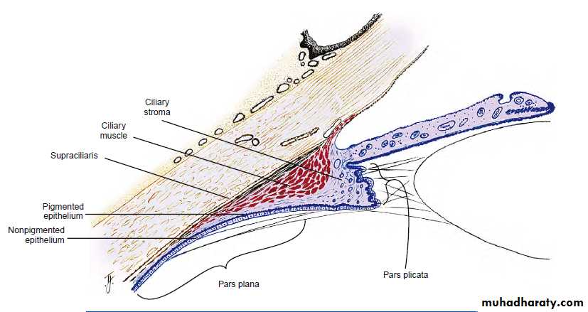
Ciliary body: The ciliary body is 6- 7 mm wide and consists of 2 parts: the pars plana and the pars plicata. The pars plana is a relatively avascular, smooth pigmented zone; it is 4 mm wide and extends from the ora serrata to the ciliary processes. The pars plicata is richly vascularized and consists of approximately 70 radial folds, or ciliary processes. The zonular fibers of the lens attach primarily in the valleys of the ciliary processes but also along the pars plana. The ciliary body has 2 principal functions: aqueous humor formation and lens accommodation The ciliary body is lined by a double layer of epithelial cells. The inner nonpigmented epith.which is the main part secreting aqueous and the outer pigmented epithelium .Ciliary muscle lie within ciliary stroma & is responsible for accommodation &also increases drainage rate
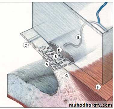
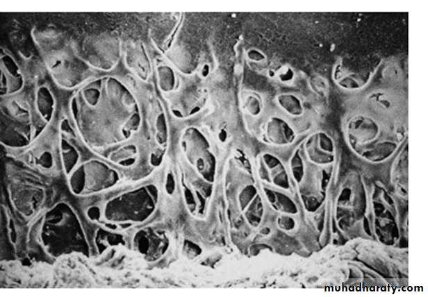
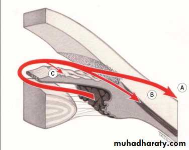
The trabecular meshwork: is a sieve-like structure at the angle of the anterior chamber, through which 90% of the aqueous humour leaves the eye . It is made of the following three portions:
a
The uveal meshwork is the innermost portion and consists of cord-like endothelial cell-covered strands arising from the iris and ciliary body stroma, and extending from the root of the iris to Schwalbe line. The intertrabecular spaces are relatively large and offer little resistance to the passage of aqueous.b
The corneoscleral meshwork forms the larger middle portion which extends from the scleral spur to Schwalbe line. The layers are sheet-like and composed of connective tissue strands also with overlying endothelial-type cells. The intertrabecular spaces are smaller than those of the uveal meshwork, conferring greater resistance to flow.
c
The juxtacanalicular (cribriform) meshwork is the outer part of the trabeculum, and links the corneoscleral meshwork with the endothelium of the inner wall of the Schlemm canal. This offers the major proportion of normal resistance to aqueous outflow,
Schlemm canal is a circumferential channel in the perilimbal sclera, bridged by septa. The inner wall is lined by irregular spindle-shaped endothelial cells containing infoldings (giant vacuoles) which are thought to convey aqueous via the formation of transcellular pores. The outer wall is lined by smooth flat cells and contains the openings of the collector channels which leave the canal at oblique angles and connect directly or indirectly with episcleral veins.
Physiology
Aqueous secretion Two mechanisms are involved: 1 Active secretion(80-90%) by the non-pigmented ciliary epithelium accounts for the vast majority, and involves a metabolic process that depends on several enzyme systems, especially the Na+/K+ ATPase pump which secretes sodium ions into the posterior chamber.
2 Passive secretion by ultrafiltration and diffusion, which are dependent on the capillary hydrostatic pressure and the level of IOP.This play a minor role in the genesis of aqueous humour under normal conditions
Aqueous outflow: Aqueous flows from the posterior chamber via the pupil into the anterior chamber, from where it exits the eye by two different routes: 1 Trabecular (conventional) route accounts for approximately 90% of aqueous outflow. The aqueous flows through the trabeculum into the Schlemm canal and is then drained by the episcleral veins
2 Uveoscleral (unconventional) route accounts for the remaining 10% in which aqueous passes across the face of the ciliary body into the suprachoroidal space and is drained by the venous circulation in the ciliary body, choroid and sclera. Some aqueous also drains via the iris.
Ocular hypertension:
In the general population the mean IOP is 16 mm Hg; two standard deviations on either side of this gives a 'normal' IOP range of 11-21 mm Hg. In the elderly, the mean IOP is higher, particularly in women, and the standard deviation greater than in younger individuals. This means that 'normal' IOP in elderly women may range up to 24 mm Hg and not 21 mm Hg. It is estimated that about 7% of the population over age of 40 years have IOPs > 21 mm Hg without glaucomatous damage on standard clinical tests. These individuals are referred to as ocular hypertensives or glaucoma suspects.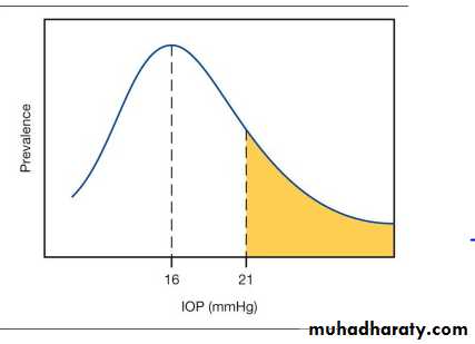
Untreated patients with ocular hypertension have only a 9.5% cumulative risk of developing primary open angle glaucoma (POAG) after 5 years, therefore do not require treatment and only those at risk should be treated in order to delay or prevent the development of POAG. It should be remembered that once treatment is instituted, it is continued throughout the patient's lifetime and may have significant side-effects.
Risky patients are those with:
1- IOP 30 mm Hg or more.
2- C/D ratio 0.4 or more.
3- Family history of glaucoma in a first degree relative.
4- High myopia.
Glaucoma:
It is an optic neuropathy with characteristic appearance of the optic disc and specific pattern of visual field defect that is associated frequently but not invariably with raised intraocular pressure (IOP).
It is a potentially blinding disease.
Classification of glaucoma:
1- Congenital (developmental).
2- Acquired: divided into:
a- Open angle: this is either primary (raised IOP not associated with other ocular disorders) or secondary.
b- Angle closure: also either primary or secondary.
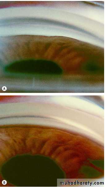
* We use gonioscopy to determine whether it is open angle or angle closure glaucoma, so if we see the structures of the angle, then it is open angle glaucoma, while if we do not see some or all of these structures, then the angle is closed or occludable.
* The structures that we can see normally by gonioscopy (using goniolens) in the angle are; (anterio-posteriorly) schwalbe's line, trabecular meshwork,
scleral spur and anterior face of ciliary body.
The optic nerve head:
It represents the collection of 1.2 million ganglion cell axons as they pass across the retina to enter the scleral canal to form the optic nerve head (optic disc)Here there is no photoreceptors (blind spot). The optic cup:Is a pale depression in the center of the optic nerve head which is not occupied by neural tissue, but it is occupied by glial tissue made mainly of astrocytes.
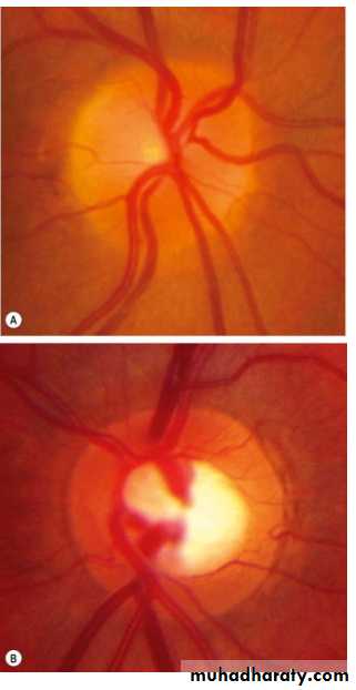
The cup:disc ratio (C/D ratio):
Indicates the diameter of the cup expressed as fraction of the diameter of the disc and should be measured in both vertical (which is more important) and horizontal meridian.* Most normal eyes have a vertical C/D ratio of 0.3 or less (<30%). So C/D ratio more than 0.3 or difference of 0.2 (20%) between the two eyes should be regarded with suspicion.
* About 2% of population have CD ratio more than 0.3 and may even reach 0.7 and they are really normal, and to consider it glaucoma we have to have either raised IOP or optic neuropathy (we must have two of three of the diagnostic criteria of glaucoma to say this is glaucoma).
C/D ratio assessed and calculated by:
1- Clinical fundal examination and fundus photograph. (crude method)
2- Now a day we use very sophisticated method of imaging technique called Heidelberg retinal tomography (HRT). The imaging technique is used in diagnosis and follows up of patients with glaucoma.
3- Ocular coherent tomography (OCT) which works in the similar way of ultrasonography but instead of using reflected sound, reflected light is used. It gives accurate measurement of nerve fibers layer changes and C\D ratio assessment.
Primary Open Angle Glaucoma (POAG)
Definition: a bilateral disease (in general) although not necessarily a symmetrical disease, characterized by:1- Adult onset.
2- IOP >21 mmHg.
3- An open angle of normal appearance.
4- Glaucomatous optic nerve damage.
5- Visual field loss typical of glaucoma.
* In POAG, the trabecular meshwork is exposed (angle is open) but the pores are occluded (sclerosis of trabecular meshwork for unknown etiology).
Pathogenesis of optic disc cupping:
1- Ischemic theory (indirect): compromised microvasculature of optic nerve axons duo to rising IOP.
2- Direct mechanical theory: chronically elevated IOP directly damages optic head nerve fibers at lamina cribrosa duo to mechanical stretching of nerve fibers.
Risk factors and associations:
1- Age: older people, after age of 65 years due to aging process and sclerosis of trabecular meshwork.
2- Race: more common and amore severe in Blacks and Asians.
3- Family history and inheritance.
4- Myopia: increased incidence of POAG.
Clinical features: (of POAG)
Insidious (gradual), asymptomatic until late in the course of the disease due to significant loss of visual field (i.e. it presents with advanced optic nerve damage and visual field impairment, but with preserved visual acuity as it is not involved in the course of disease except in very late stages).
Premature presbyopia (usually presbyopia seen clinically at age of 45-46 years, but here occurs earlier at about 38-40 years and its symptoms are difficulty in reading and visualization of near objects.
Rapid increase in hypermetropia (same mechanism of presbyopia, and also it is early and rapid) in patients over the age of 40 years.
Rarely: eye ache (as the elevation of IOP is gradual over many years), headache, halos (due to transient corneal epithelial oedema, due to raised IOP that damages the endothelium of cornea).
Signs of POAG:
1- Raised IOP.
2- Fluctuation in IOP (normally there is diurnal variation of IOP according to circadian rhythm of hormones but not more than 5 mmHg). In glaucoma the fluctuation is more than 5 mm Hg.
3- Optic disc changes, increased C/D ratio more than 0.3 or asymmetry of it (difference more than 0.2 between both eyes).
4- Glaucomatous field change.
5- Gonioscopy, show open angle with no any abnormality (e.g. no new blood vessels or inflammatory cells occluding the angle)
Visual field changes:
1- Generalize constriction of peripheral VF (earliest field defect but not specific sign for POAG and occurs in many other diseases).2- Paracentral scotoma: (central VF change which is specific sign).
3- Arcuate scotoma (central VF change).
4- Temporal (crescent) or central island of vision only (both are end- stage glaucoma).
Management of POAG:
Treatment Aim: to preserve visual function by controlling IOP and so preventing or retarding further optic nerve damage, but there is no improvement.Treatment goals:
1- Target pressure: it is assumed that the pre-treatment level of IOP has damaged the optic nerve and will continue to do so. An IOP level below which further damage is considered unlikely called target pressure.
2- Monitoring: is performed of the optic nerve and visual fields. In the event of further damage the target IOP is reset at a lower level. Although, there is no safe level, progression is uncommon if the IOP <16 mmHg.
Medical treatment:
1- Any chosen drug should be used in its lower concentration and as infrequently as possible consistent with the desired therapeutic effect.
2- Ideally the drug with the fewest potential side-effects should be used.
3- Initial treatment is usually with one drug, usually a beta-blocker or prostaglandin analogue.
4- Follow up after 4 weeks should be performed after initiation of treatment and fall in IOP of >4 mm Hg is usually considered significant.
5- If the response is satisfactory subsequent assessment is after 2 months and at 3-4 monthly intervals thereafter for long life.
6- If the response is unsatisfactory the initial drug is withdrawn and another substituted.
7- If the response is still unsatisfactory yet another drug is added or a combined preparation (e.g. Timolol & Dorzolamide) substituted.
8- Perimetry: assessment of VF annually is sufficient if the IOP is satisfactory and the appearance of optic disc is stable.
9- Gonioscopy: should also perform annually because the AC gradually shallows with age.
Drugs used in treatment of POAG:
* Beta blockers are taken twice daily, they act by decreasing the secretion, they are contraindicated for patients with heart problems or asthmatics, but betaxolol is cardio-selective and can be used for asthmatics.
* Dorzolamide is a carbonic anhydrase inhibitor and acts by decreasing secretion. If used alone is given 3 times / day.
* Brimonidine is an alpha-2 agonist, and acts by decreasing secretion and has slight effect to increase drainage. Used 3 times / day.
* Latanoprost is a prostaglandin agonist, and acts by increasing drainage through uveoscleral outflow. Used once daily at night.
* Parasympathomimetics (e.g. Pilocarbine) act on ciliary and specifically on its longitudinal muscles which are inserted in scleral spur, so on contraction, the trabecular meshwork will be stretched and the size of pores will increase leading to increment of drainage through the conventional way.
2- Surgical treatment:
Indications:a- Failure of medical treatment.
b- Poor compliance.
c- Primary treatment (some doctors consider it primarily).
d- Unavailability of drugs or their expensiveness.
Types:
a- Laser trabeculoplasty (Argon) "ALT": here we use the laser to produce a thermal effect on the trabecular meshwork and not to perforate it, so the thermal effect will lead to shrinkage of the sites treated by laser due to fibrosis and these shrunken sites will pull the neighboring parts leading to opening of pores.
b- Cyclo-destructive: diode or YAG laser therapy and cyclocryotherapy, we destruct the ciliary process, which concerned with production of aqueous humour.
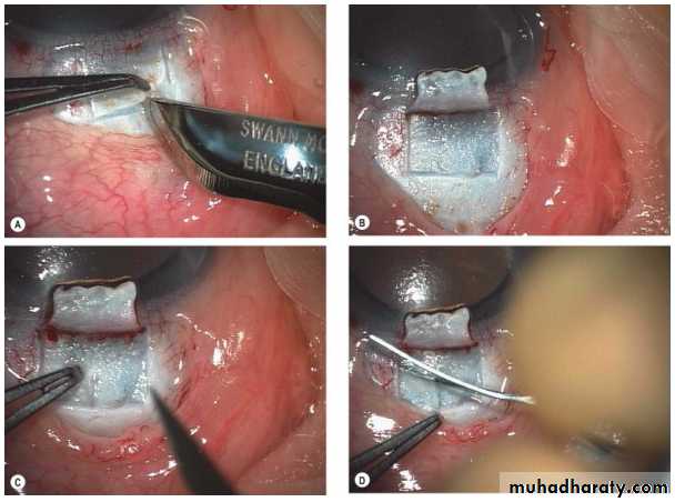
c- Trabeculectomy with or without antimetabolites (5-FU, mitomycine): we create surgical fistula between AC and subconjunctival space for drainage of excess aqueous humour and some time we use antimetabolites at site of surgery to diminish fibroblast activity and preventing risk of occlusion of this fistula and failure of surgery.
d- Antiglaucoma devices (Molteno, Ahmed): a similar operation to the previous one, but here we use a tube to connect the anterior chamber with the Subconjunctival space. It is used for resistant cases of trabeculectomy.
Corticosteroid responsiveness:
Normal population is divided into 3 groups on the basis of their IOP response to a six week course of topical betamethasone (potent steroid):
1- High responders: IOP >30mmHg.
2- Moderate responders: IOP 22-30 mmHg.
3- Non responders: no change in IOP.
* Incidence of steroid responsiveness %:
High Moderate Non
General population 5 35 60
The tendency is more marked in patients with POAG and their close relatives. Intra- and periocular steroid administration, including periocular application of steroid skin cream and nasal administration, are also prone to elevate IOP. Systemic steroids are much less prone to cause elevation of IOP, but some authorities have advocated screening for all patients on systemic steroids, perhaps those on dexamethasone in particular N.B: fluromethelone is the weakest glucomatogenic steroid.
Normal tension glaucoma (NTG)
Definition:
NTG also referred to us low tension glaucoma, is variant of POAG, it is characterized by:
1- A mean IOP ≤ 21 mm Hg on diurnal testing.
2- Glaucomatous optic disc damage and VF loss.
3- Open drainage angle on Gonioscopy.
4- Absence of secondary causes.
Pathogenesis:
The exact cause of NTG has not been conclusively determined although various mechanisms has been postulated such as:
1- Vascular insufficiency.
2- Decrease optic disc resistance.
3- IOP effects.
4- Optic nerve compression by normal carotid arteries.
Diagnosis and treatment:
Is the same as in POAG except that IOP is normal. We need here to decrease the IOP even more to reach the target pressure which is safe and causing no more optic disc damage and no more VF defects.
Primary Angle Closure Glaucoma (PACG)
Definition: is a condition in which there is obstruction to aqueous outflow due to partial or complete closure of the angle by peripheral iris.Unlike POAG, the diagnosis is by examination of anterior segment and gonioscopy. Presence of a normal optic disc and absence of visual field loss does not preclude (exclude) diagnosis of PACG (as it is acute so the damage doesn’t occur unless the condition left without treatment and blindness happen within few days or even hours). In addition, there are shallow AC and convex iris-lens diaphragm.
Risk factors:
1- Age: average age 60 years, it does not occur before this age, as the AC is still deep.2- Gender: ♀/♂ = 4/1.
3- Race: whites 6% of glaucomas, more common in South East Asians, Chinese and Eskimos, uncommon in Blacks.
4- Family history: ocular anatomical features are inherited, 1st degree relatives are at increased risk.
Anatomical predisposing factors:
1- Relative anterior location of iris-lens diaphragm.
2- Shallow anterior chamber (AC).
3- Narrow entrance to anterior chamber angle
- Acute congestive angle-closure glaucoma:
This condition is caused by a sudden total closure of the angle.
Clinical features:
Symptoms: a- Rapidly progressive impairment of visual acuity, due to corneal oedema.
b- Periocular pain + congestion.
c- Nausea and vomiting in severe cases.
Signs seen with slit-lamp:a- Ciliary flush due to injection of limbal and conjunctival blood vessels.
b- IOP severely elevated (>50mmHg).
c- Corneal oedema with epithelial vesicles.
d- Shallow AC + peripheral iridocorneal touch.
e- Aqueous humour flare (protein) + cells.
f- Pupil vertically oval + fixed in mid-dilated position, not reactive to light and accommodation.
Treatment is surgical
but initially we start medical treatment to control elevated IOP. Supine position to encourage the lens to shift posteriorly under the influence of gravity.
a- IV acetazolamide: 500mg followed by 250mg qid orally.
b- Hyperosmotic agents: - IV Mannitol 1g/kg of 20% or
- Oral glycerol 1g/kg of 50% with orange juice.
c- Topical therapy:
i- Pilocarpine 2% x4 to affected eye, it causes miosis and pulls iris periphery away from cornea.
ii- Pilocarpine 1% x4 to unaffected eye (as prophylaxis).
iii- Beta-blocker x2, only for the affected eye.
iv- Topical steroid: to avoid permanent adhesion between the periphery of iris and the cornea (peripheral anterior synechia)( PAS).
Surgical treatment:
a- We do bilateral YAG PI, then we stop pilocarpine to fellow eye (unaffected) and we decrease medication of affected eye, 1st we stop IV mannitol, then oral acetazolamide, if IOP is elevated again (due to peripheral anterior synechiae (PAS) formation) go to:
b- Trabeculectomy is indicated, which means that 50% of the angle is already closed by permanent anterior synechia and YAG laser PI with topical medications alone cannot control IOP.
secondary glaucomas
Classification:Open-angle
Secondary open-angle glaucoma can be subdivided on the basis of the site of aqueous outflow obstruction as follows:
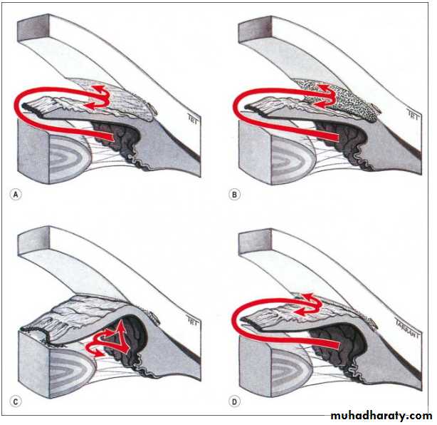
1 Pre-trabecular glaucoma in which aqueous outflow is obstructed by a membrane covering the trabeculum (Fig. 10.48A), which may consist of: • Fibrovascular tissue (neovascular glaucoma).
•Endothelial cellular membranous proliferation (iridocorneal endothelialsyndrome). • Epithelial cellular membranous proliferation (epithelial ingrowth).
2 Trabecular glaucoma in which the obstruction occurs as a result of ‘clogging up’ of the meshwork by the following:• Pigment particles (pigmentary glaucoma)•Red blood cells (red cell glaucoma).• Degenerate red cells (ghost cell glaucoma).•Macrophages and lens proteins (phacolytic glaucoma).•Proteins• Pseudoexfoliative material (pseudoexfoliation glaucoma).Trabecular glaucomas may also be caused by alteration of the trabecular fibres themselves by• Oedema (herpes zoster iritis/trabeculitis).• Scarring (post-traumatic angle recession glaucoma
3 Post-trabecular glaucoma in which the trabeculum itself is normal but aqueous outflow is impaired as a result of elevated episcleral venous pressure due to conditions such as: • Carotid-cavernous fistula.•Obstruction of the superior vena cava
Angle-closure
Secondary angle-closure is caused by impairment of aqueous outflow secondary to apposition between the peripheral iris and the trabeculum. Classification is based according to the presence or absence of pupillary block:1 With pupillary block • Seclusio pupillae (360° ‘ring’ posterior synechiae), usually secondary to recurrent iridocyclitis• Subluxated lens • Phacomorphic glaucoma• Aphakic pupillary block.• Anterior chamber lens implant without a patent iridotomy
2 Without pupillary block • Secondary causes of PAS such as advanced neovascular glaucoma and chronic anterior uveitis
Important examples 2dary glaucomas
1- Lens-related glaucoma:
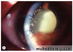
a- Phacolytic glaucoma (lens protein glaucoma)Is occur in association with hypermature cataract (leakage of lens materials and shrinkage of lens). Leaked lens material is engulfed by microphages. The trabecular obstruction is caused by high molecular weight lens proteins which have leaked through the intact capsule into the aqueous humour or by microphages laden with these proteins.Treatment: control IOP medically, then surgery (cataract extraction).
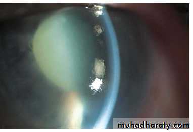
b- Phacomorphic (Intumescent) glaucoma:Is acute secondary angle-closure glaucoma precipitated by an intumescent cataractous lens. Swelling of lens pushes the lens-iris diaphragm forward to occlude the angle of the anterior chamber. Presentation is the same like that of PACG but with cataract.Treatment: surgery (cataract extraction).
c- Phacoanaphylactic (phacoantigenic) glaucoma:Is an autoimmune reaction to lens proteins occurring in an eye with a traumatic ruptured anterior capsule (large lens matter passing through a ruptured capsule, this matter is regarded as foreign body and antibodies against it is produced). Therefore, there will be occlusion of the pores of trabecular meshwork by immune complexes and cells.Treatment: cataract extraction
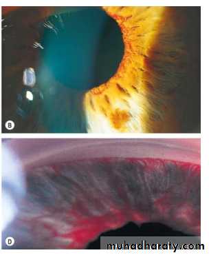
2- Neovascular glaucoma (NVG):Is a relatively common and serious condition, which occurs as a result of iris neovascularization (Rubeosis iridis).The common aetiopathogenic factor is severe, diffuse and chronic retinal ischaemia.Retinal ischaemia leads to hypoxic retina which tend to produces vasoproliferative growth factors in an attempt to revascularize hypoxic areas (form new blood vessels to compensate for hypoxia), but unfortunately these blood vessels are very fragile, so they may rupture suddenly and patient get sudden blindness due to retinal or vitreous hemorrhage. These vessels may rupture spontaneously or during valsalva manoeuvre. However, these factors also diffuse in to the anterior segment and initiate Rubeosis iridis (neovascularization of iris) and neovascularization in the angle of the anterior chamber (leading to occlusion of trabecular meshwork).* If you see rubeosis iridis in any person, you can say without any doubt that there is retinal ischaemia.
Causes of retinal ischaemia (causes of NVG):a- Central retinal vein occlusion. (commonest cause)b- Diabetes mellitus (proliferative diabetic retinopathy).c- miscellaneous, e.g.: - Carotid obstructive disease. Central retinal artery occlusion. Intraocular tumours. Long standing retinal detachment.Chronic intraocular inflammation.
Treatment:
1- Medical treatment is initially with topical beta-blockers and acetazolamide. Topical atropine and steroids may decrease inflammation and make the eye more comfortable and less congested, even if IOP remains high.
2- Panretinal photocoagulation with laser: We destruct retinal tissue in order to decrease O2 consumption and control hypoxia. We photcoagulate the mid periphery and the periphery of retina and we preserve the macula only.
3-Intravitreal Anti VEGF(vascular endothelial growth factor) injection
4- Filteration surgery may be consider if still there is useful vision either by trabeculectomy with anti metabolite or by use artificial filtering shunts.
5- Cyclodestruction by trans scleral diode laser or trans scleral cyclocryotherapy which lead to destructs part of Ciliary body and decrease aqueous production.
6- Retrobulbar alcohol injection is useful in relieving pain by destruction of sensory enervation.
7- Enucleation may be considered if all else fails
3- Inflammatory glaucomas:
In acute anterior uveitis, the IOP is usually normal or subnormal as a result of concomitant ciliary shutdown. Occasionally, however, a trabecular-block open angle glaucoma develops secondary to obstruction of aqueous outflow, most commonly just as the acute inflammation is and ciliary body function returning to normal. The block may be caused by either inflammatory cells and debris or acute trabeculitis. The IOP usually returns to normal once the inflammation has subsided.
Secondary angle closure is caused by 360° iridolenticular adhesions (seclusio pupillae), The pupil block obstructs the passage of aqueous humour from the posterior to the anterior chamber, and the increased pressure in the posterior chamber causes an anterior bowing of the peripheral iris (iris bombé), If severe, iris bombé is associated with a shallowing of the anterior chamber, and apposition of the peripheral iris to the trabeculum and peripheral cornea. If this occurs in an eye with active inflammation, the iris sticks to the trabeculum and the iridocorneal contact becomes permanent with the development of peripheral anterior synechiae (PAS).
Slit lamp examination shows seclusio pupillae, iris bombé and a shallow anterior chamber.
Gonioscopy shows angle closure from iridotrabecular contact.
Treatment involves the following measures:
1. Prevention of synechial angle closure can be effective by a reduction in the 'stickiness' of the peripheral iris using a combination of intensive topical steroids and anterior sub-Tenon's injection of a long-acting depot steroid preparation.
2. Lowering of IOP with topical ß-blockers and/or sympathomimetics may be effective in relatively mild cases where the IOP is <30 mmHg. Carbonic anhydrase inhibitors are usually required if the IOP is >30mmHg.
3. Nd-YAG laser iridotomy may be required if medical therapy fails, It will only eliminate the element of pupil block and will therefore only be effective in lowering IOP, if at least 25% of the angle is still open
Primary congenital glaucomas
They are uncommon (not rare), sever and potentially blinding disease.* in general, the most common type of glaucoma is POAG 65%, followed by secondary glaucomas 20%, then comes PACG 12% (especially the acute one) then and the least common ones are the congenital glaucomas 3%.
Types:
1- True congenital glaucoma: represents (40%) of all congenital glaucoma, the IOP is elevated during intra-uterine life.
2- Infantile glaucoma: represents (55%) of congenital glaucomas, the disease is manifested before 3 years age, but the patient was born with normal IOP.
3- Juvenile glaucoma: represents (5%) of congenital glaucomas, least common, present after 3 years but before reaching 16 years (>16 → adult glaucoma).
In general, congenital glaucoma:
- Occurs 1 in 10,000 births.
- Most cases are sporadic, some are inherited as autosomal recessive trait.
- 65% in boys and 35% in girls.
- Bilateral in 75% and 25% unilateral.
Impaired aqueous outflow in PCG is caused by maldevelopment of the angle of the anterior chamber, unassociated with any other major ocular anomalies (isolated trabeculodysgenesis). Clinically, trabeculodysgenesis is characterized by absence of the ciliary body band due to translucent amorphous material that obscures the trabeculum
Clinical Features:
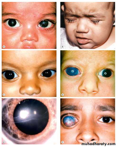
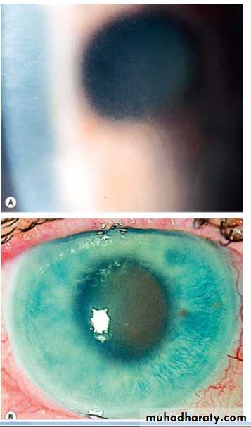
The first sign noticed by parents is the corneal haze:
1- Corneal haze (or opacity): caused by epithelial &stromal oedema secondary to elevated IOP.2- Photophobia, lacrimation and blepharospasm:
3- Buphthalmus: a large eye due to elevated IOP prior to the age of 3 years, so we should expect elevated IOP in every child with large eye below age of 3 years.
- Sclera also enlarges becoming stretched and takes blue appearance due to the enhanced visualization of underlying uvea.
- Corneal enlargement leading to deep AC (Anterior chamber), zonule becomes stretched, rarely lens becomes subluxated.
- Ocular enlargement leads to axial myopia.
4- Breaks in descemet's membrane (due to stretching).
5- Optic disc cupping, C/D >0.3, it is not a reliable sign, as stretching of sclera will cause enlargement of scleral canal and cupping due to separation of nerve fibers, and as soon as we control IOP, C/D will return to normal.
6- Visual field: cannot be done as majority of cases are under 3 years and the diagnosis is straight forward by clinical signs.
Management:
Initial evaluation
The initial evaluation should be performed under general anaesthesia with intravenous ketamine, since this lowers IOP less than other agents. Examination of the optic discs should be undertaken first, followed by measurement of IOP and corneal diameters, and finally gonioscopy.
IOP is measured with Perkins tonometer or schiotz tonometer
Corneal diameter is measured in both the vertical and horizontal meridian with callipers. A diameter >11 mm prior to the age of one year or >13 mm at any age should be viewed with suspicion. Diameters of 14 mm are typical of advanced buphthalmos.
Gonioscopy is performed with a direct goniolens.
Surgery:
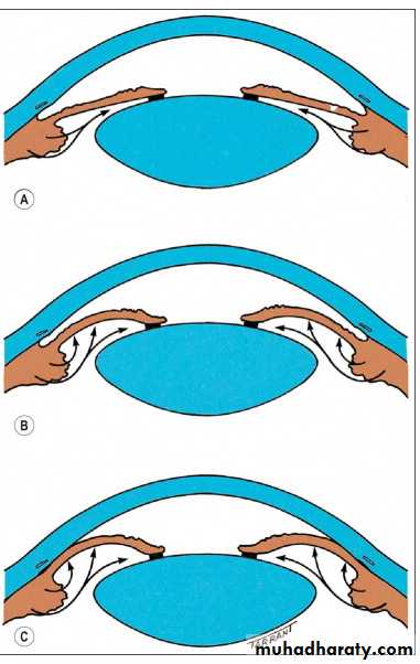
1- Goniotomy. We incised the trabecular meshwork to create a direct communication between the anterior chamber and schlemm's canal bypassing the trabecular meshwork, this operation needs clean cornea to use the instruments and visualized the structures of the angle of the AC. Successful rate is 80%.
2- Trabeculotomy. From the limbus, we introduce a probe inside the schlemm's canal and then we open the inner wall of it toward the AC. Also here there is a direct communication between the anterior chamber and schlemm's canal bypassing the trabecular meshwork
3- Trabeculectomy: same operation discussed in previous lecture .It is often successful, particularly when Combined with adjunctive antimetabolites