Copyright 2009 John Wiley & Sons, Inc.Chapter 8 The Skeletal System: The Appendicular Skeleton

Copyright 2009 John Wiley & Sons, Inc.Appendicular SkeletonThe primary function is movementIt includes bones of the upper and lower limbsGirdles attach the limbs to the axial skeleton
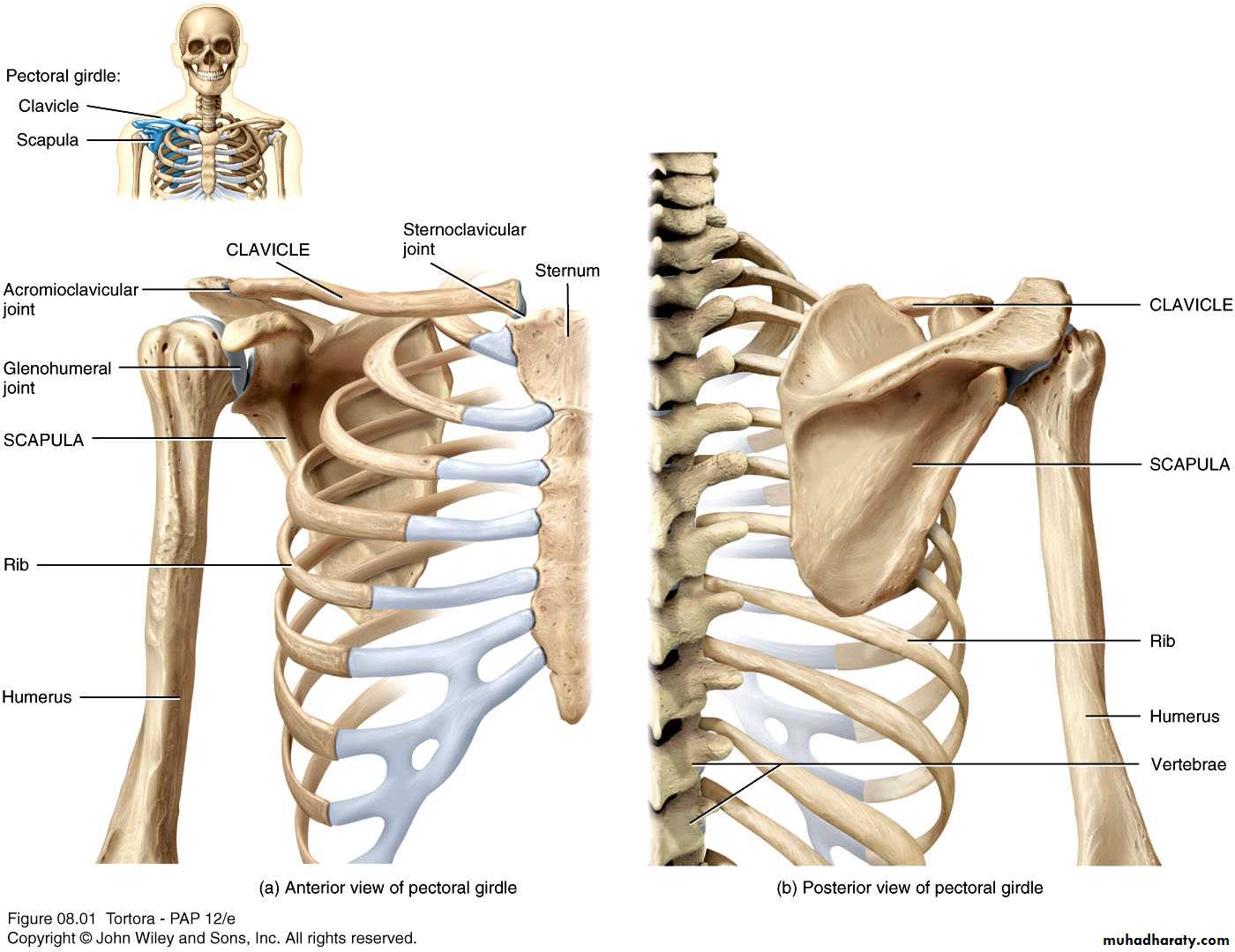
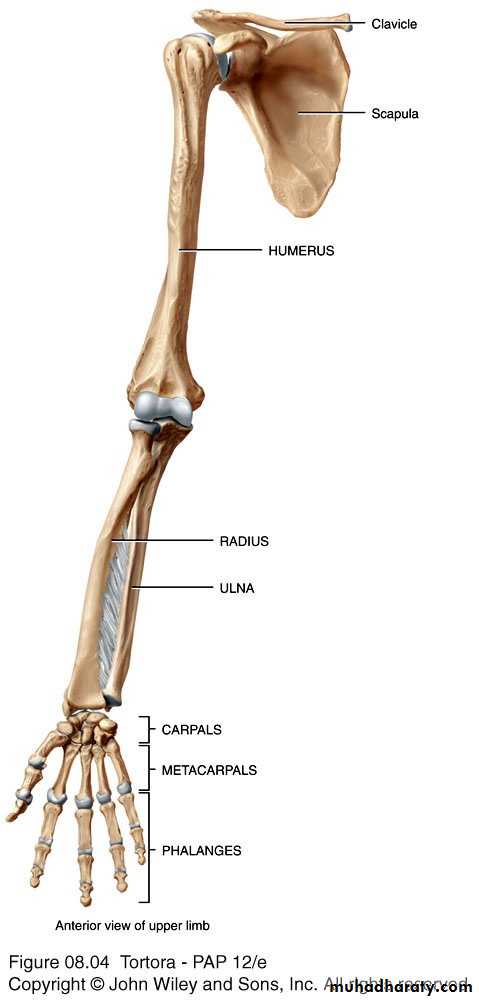
Copyright 2009 John Wiley & Sons, Inc.The Pectoral (or Shoulder) Girdle Figure 8.1Copyright 2009 John Wiley & Sons, Inc.Skeleton of the Upper LimbEach upper limb has 32 bonesTwo separate regions1. The pectoral (shoulder) girdle (2 bones)2. The free part (30 bones)
Copyright 2009 John Wiley & Sons, Inc.Upper LimbThe pectoral girdle consists of two bones, the scapula and the clavicleThe free part has 30 bones1 humerus (arm)1 ulna (forearm)1 radius (forearm)8 carpals (wrist)19 metacarpal and phalanges (hand)Copyright 2009 John Wiley & Sons, Inc.Pectoral Girdle - ClavicleThe clavicle is “S” shapedThe medial end articulates with the manubrium of the sternum forming the sternoclavicular jointThe lateral end articulates with the acromion forming the acromioclavicular joint
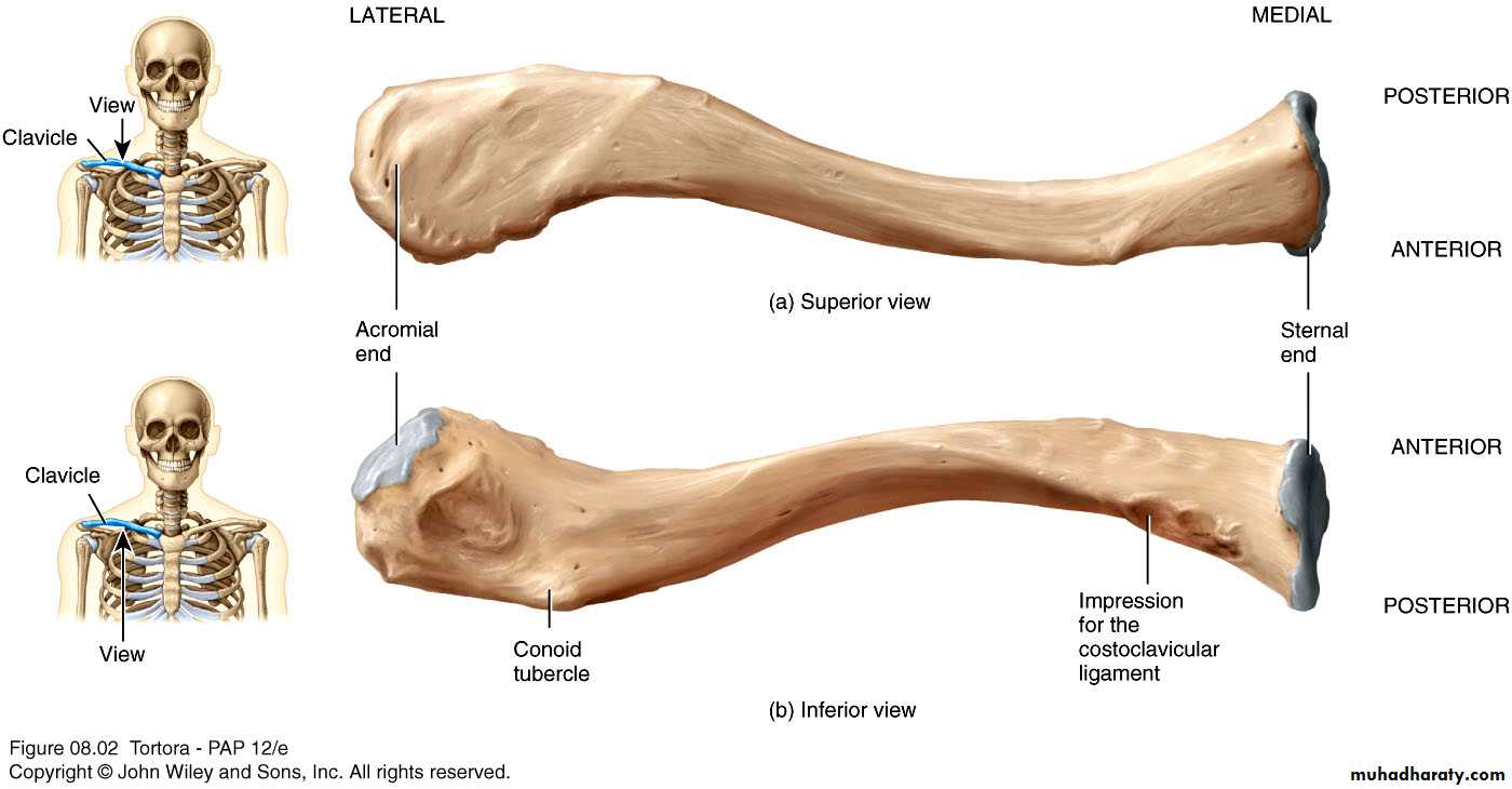
Copyright 2009 John Wiley & Sons, Inc.The Clavicle Figure 8.2Copyright 2009 John Wiley & Sons, Inc.Pectoral Girdle - ClavicleThe clavicle is convex in shape anteriorly near the sternal junctionThe clavicle is concave anteriorly on its lateral edge near the acromion
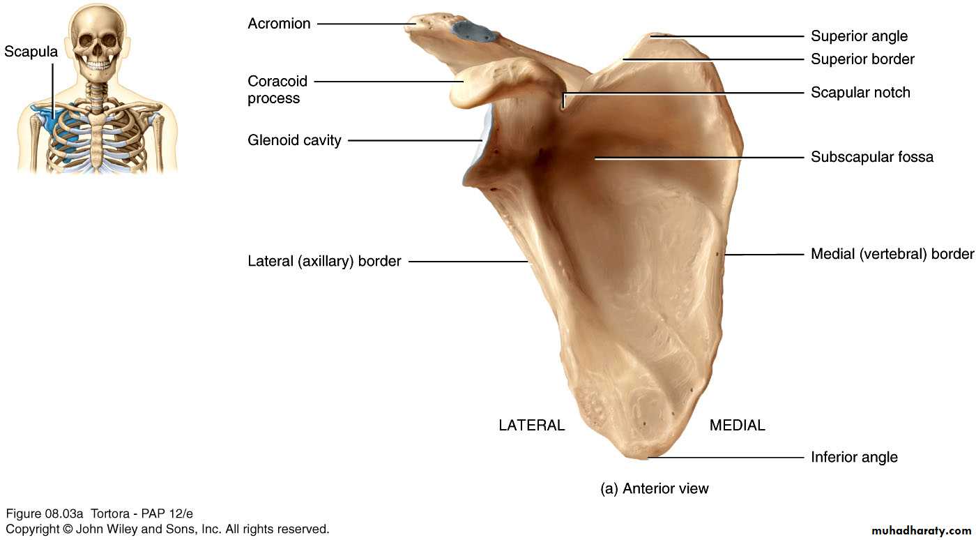
Copyright 2009 John Wiley & Sons, Inc.Clinical Connection - Fractured ClavicleA fall on an outstretched arm (F.O.O.S.H.) injury can lead to a fractured clavicleThe clavicle is weakest at the junction of the two curvesForces are generated through the upper limb to the trunk during a fallTherefore, most breaks occur approximately in the middle of the clavicleCopyright 2009 John Wiley & Sons, Inc.Pectoral Girdle - ScapulaAlso called the shoulder bladeTriangular in shapeMost notable features include the spine, acromion, coracoid process and the glenoid cavity
Copyright 2009 John Wiley & Sons, Inc.Features on the ScapulaSpine - a large process on the posterior of the scapula that ends laterally as the acromion Acromion - the flattened lateral portion of the spine of the scapulaCoracoid process - a protruding projection on the anterior surface just inferior to the lateral aspect of the clavicleGlenoid cavity - shallow concavity that articulates with the head of the humerus
Copyright 2009 John Wiley & Sons, Inc.Scapula Figure 8.3
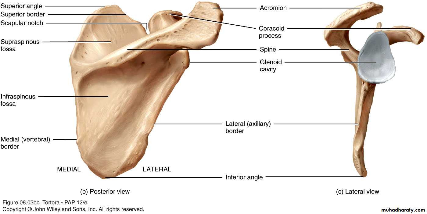
Copyright 2009 John Wiley & Sons, Inc.Scapula Figure 8.3Copyright 2009 John Wiley & Sons, Inc.Scapula - FeaturesThe medial (vertebral) border - closest to the vertebral spineLateral border - closest to the armSuperior border - superior edgeInferior angle - where medial and lateral borders meet inferiorlySuperior angle - uppermost aspect of scapula where medial border meets superior border
Copyright 2009 John Wiley & Sons, Inc.Scapula - FeaturesSubscapular fossa - anterior concavity where the subscapularis muscle attachesSupraspinous fossa - posterior concavity superior to the scapular spine, attachment site for supraspinatus muscleInfraspinous fossa - posterior concavity inferior to the scapular spine, site of infraspinatus muscleCopyright 2009 John Wiley & Sons, Inc.Skeleton of the Arm - HumerusLongest and largest bone of the free part of the upper limbThe proximal ball-shaped end articulates with the glenoid cavity of the scapulaThe distal end articulates at the elbow with the radius and ulna
Copyright 2009 John Wiley & Sons, Inc.Humerus - Surface FeaturesThe head of the humerus has two unequal-sized projectionsThe greater tubercle lies more laterallyThe lesser tubercle lies more anteriorlyBetween the tubercles lies the intertubercular groove or sulcus (bicipital groove) where the long head of the biceps brachii tendon is locatedCopyright 2009 John Wiley & Sons, Inc.Humerus - Surface FeaturesJust distal to the head is the anatomical neckThe surgical neck is where the tubular shaft begins and is a common area of fractureAbout mid-shaft on the lateral aspect is a roughened area, the deltoid tuberosity where the deltoid tendon attachesCapitulum - a round knob-like process on the lateral distal humerusTrochlea - medial to the capitulum, is a spool-shaped projection on the distal humerus
Copyright 2009 John Wiley & Sons, Inc.Humerus - Surface FeaturesCoronoid fossa - anterior depression that receives the coronoid process of the ulna during forearm flexionOlecranon fossa - posterior depression that receives the olecranon of the ulna during forearm extensionThe medial and lateral epicondyles are bony projections to which the forearm muscles attach
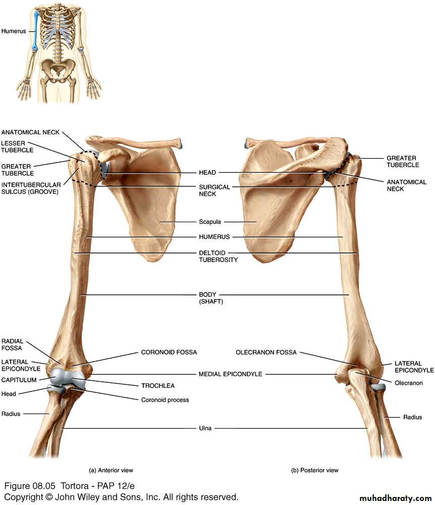
Copyright 2009 John Wiley & Sons, Inc.Humerus and Glenohumeral JointFigure 8.4
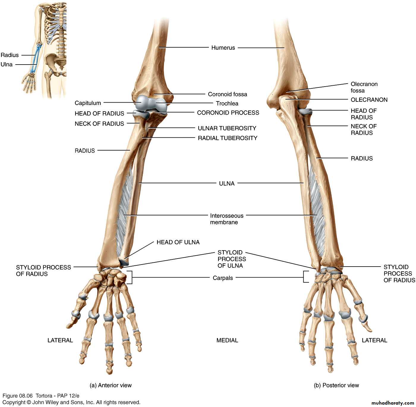
Copyright 2009 John Wiley & Sons, Inc.Skeleton of the Forearm - UlnaThe longer of the two forearm bonesLocated medial to the radiusOlecranon - the large, prominent proximal end, the “tip of your elbow”Coronoid process - the anterior “lip” of the proximal ulnaTrochlear notch - the deep fossa that receives the trochlea of the humerus during elbow flexionStyloid process - the thin cylindrical projection on the posterior side of the ulna’s head
Copyright 2009 John Wiley & Sons, Inc.
Copyright 2009 John Wiley & Sons, Inc.Right humerus in relation to scapula, ulna, and radius-- Figure 8.5
Copyright 2009 John Wiley & Sons, Inc.
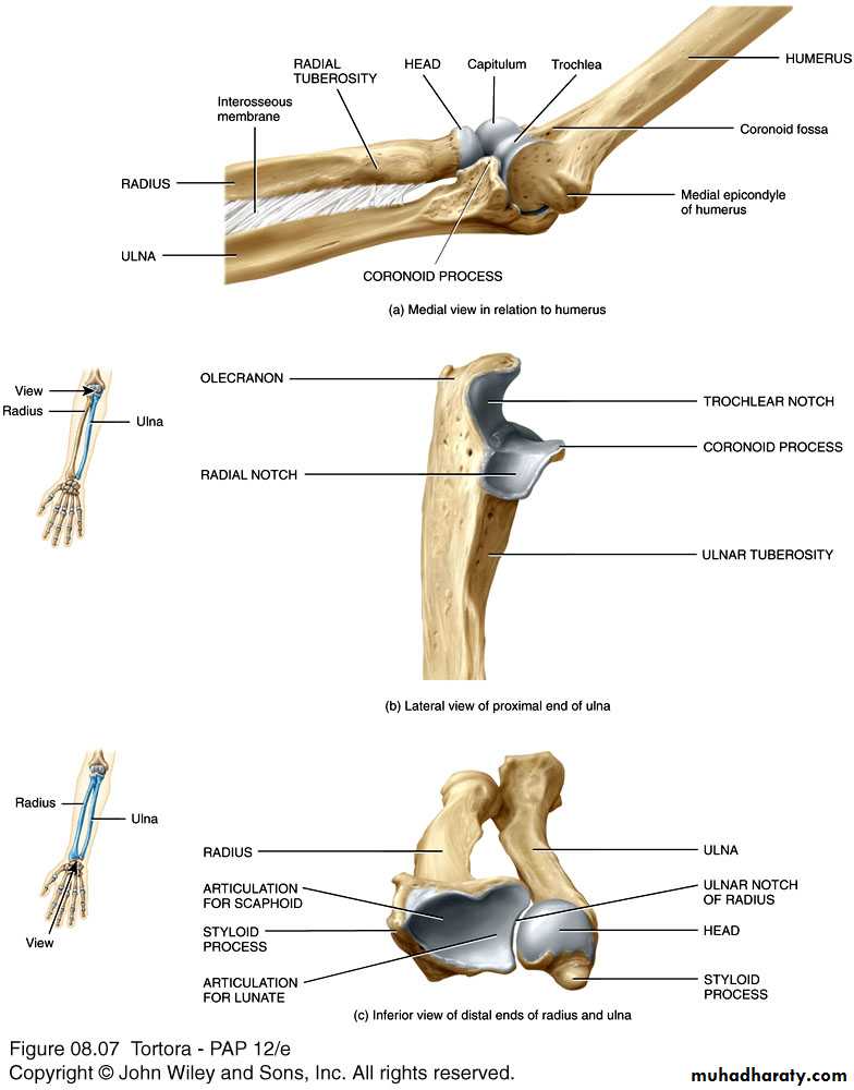
Articulations formed by the ulna and radius -- Figure 8.7Copyright 2009 John Wiley & Sons, Inc.RadiusLies lateral to the ulna (thumb side of the forearm)The head (disc-shaped) and neck are at the proximal endThe head articulates with the capitulum of the humerus and the radial notch of the ulnaRadial tuberosity - medial and inferior to neck, attachment site for biceps brachii muscleStyloid process - large distal projection on lateral side of radius
Copyright 2009 John Wiley & Sons, Inc.Ulna and RadiusThe shaft of these bones are connected by an interosseus membraneThere is a proximal radioulnar joint and a distal radioulnar jointProximally, the head of the radius articulates with the radial notch of the ulnaDistally, the head of the ulna articulates with the ulnar notch of the radiusCopyright 2009 John Wiley & Sons, Inc.
Right ulna and radius in relation to the humerus and carpals -- Figure 8.6
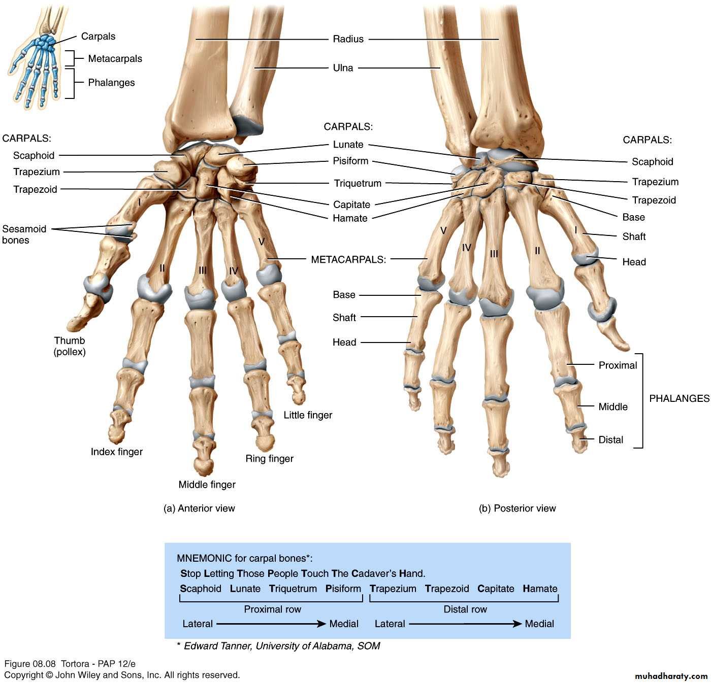
Copyright 2009 John Wiley & Sons, Inc.Skeleton of the HandThe carpus (wrist) consists of 8 small bones (carpals)Two rows of carpal bonesProximal row - scaphoid, lunate, triquetrum, pisiformDistal row - trapezium, trapezoid, capitate, hamateScaphoid - most commonly fracturedCarpal tunnel - space between carpal bones and flexor retinaculumCopyright 2009 John Wiley & Sons, Inc.Articulations formed by the ulna and radius -- Figure 8.7
Copyright 2009 John Wiley & Sons, Inc.Metacarpals and PhalangesFive metacarpals - numbered I-V, lateral to medial14 phalanges - two in the thumb (pollex) and three in each of the other fingersEach phalanx has a base, shaft, and headJoints - carpometacarpal, metacarpophalangeal, interphalangeal Copyright 2009 John Wiley & Sons, Inc.Right wrist and hand in relation to ulna and radius -- Figure 8.8