Object Localization
(Parallax technique)To obtain three-dimensional information of location of an object such as:
Foreign bodiesImpacted, unerupted teeth
Supernumerary teeth
Broken needles
Retained roots
Fractures of maxillofacial structures
Soft tissue calcifications
Intra-osseous tumors
Calculi in gland/duct of gland
Filling materials in alveolar process
Object Localization
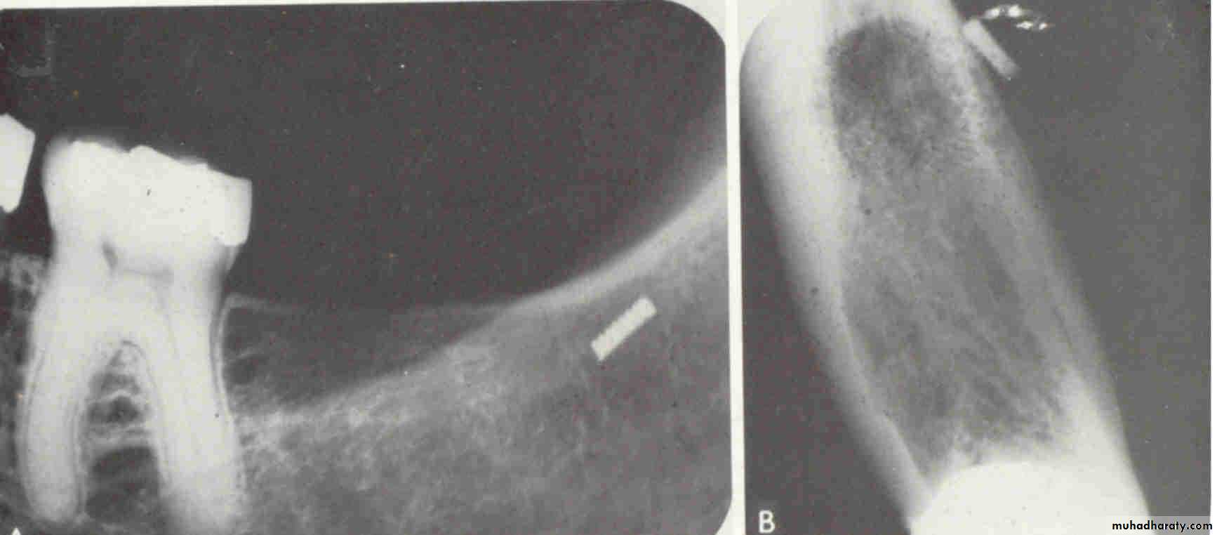
FISSURE BUR
FOREIGN BODY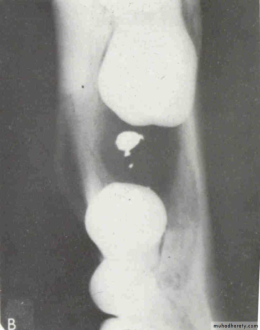
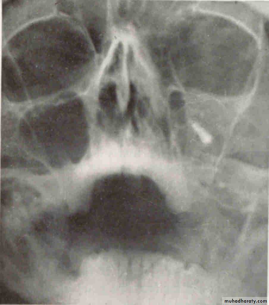
WATERS VIEW(Occipitomental )
LATERAL VIEW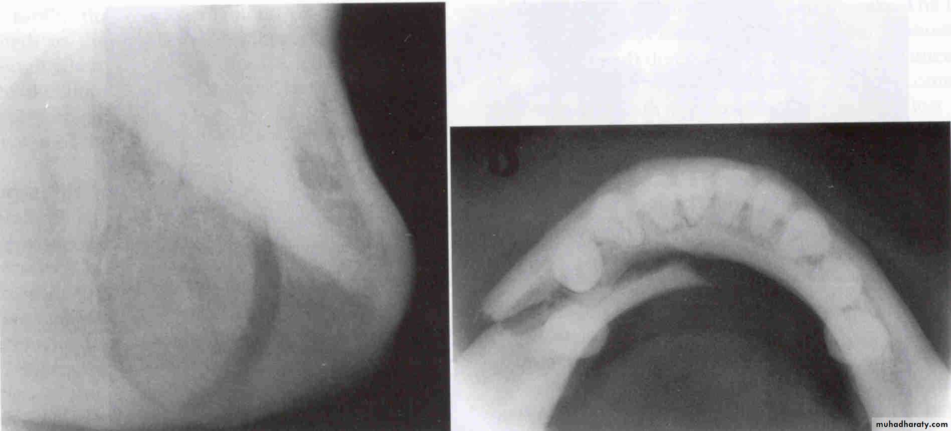
Lateral oblique
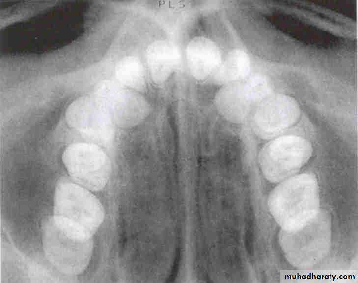
Occlusal view
A PA. film identify the location of an object vertically and in a horizontal (mesiodistal) direction, since it is a 2D. Therefore we need another method for locating objects in a buccolingual direction. The two primary methods of determining the buccolingual location of objects are:
Right-Angle Technique (Miller’s Technique) (Occlusal projection)
Primarily identifies buccolingual location, but mayalso confirm mesiodistal location seen on periapical
Tube-shift Technique (SLOB rule, Clark’s rule)
Utilizes two films with different horizontal or vertical
angulations
Object Localization (Parallax technique)
Miller’s TechniqueUsually Indicated for Mandibular Third molar area.
Two radiographs made are positioned at right angles to each other.
LOCALIZES in 3 Dimensions
DISADVANTAGESCan be used in mandible but not that useful in maxilla due to superimpositions.
(Tube shift technique (Clark’s rule
PRINCIPLE
Relative positions of radiographic images of two separate objects changes ,when projection angle at which images were made, is changed.
The image of tooth that is farther away from X-ray tube(lingual) moves in same direction as tube and image of tooth closer to X-ray tube (Buccal) moves in opposite direction.
SLOB
Same Lingual Opposite Buccal
Lingual body moves in same direction
SLOB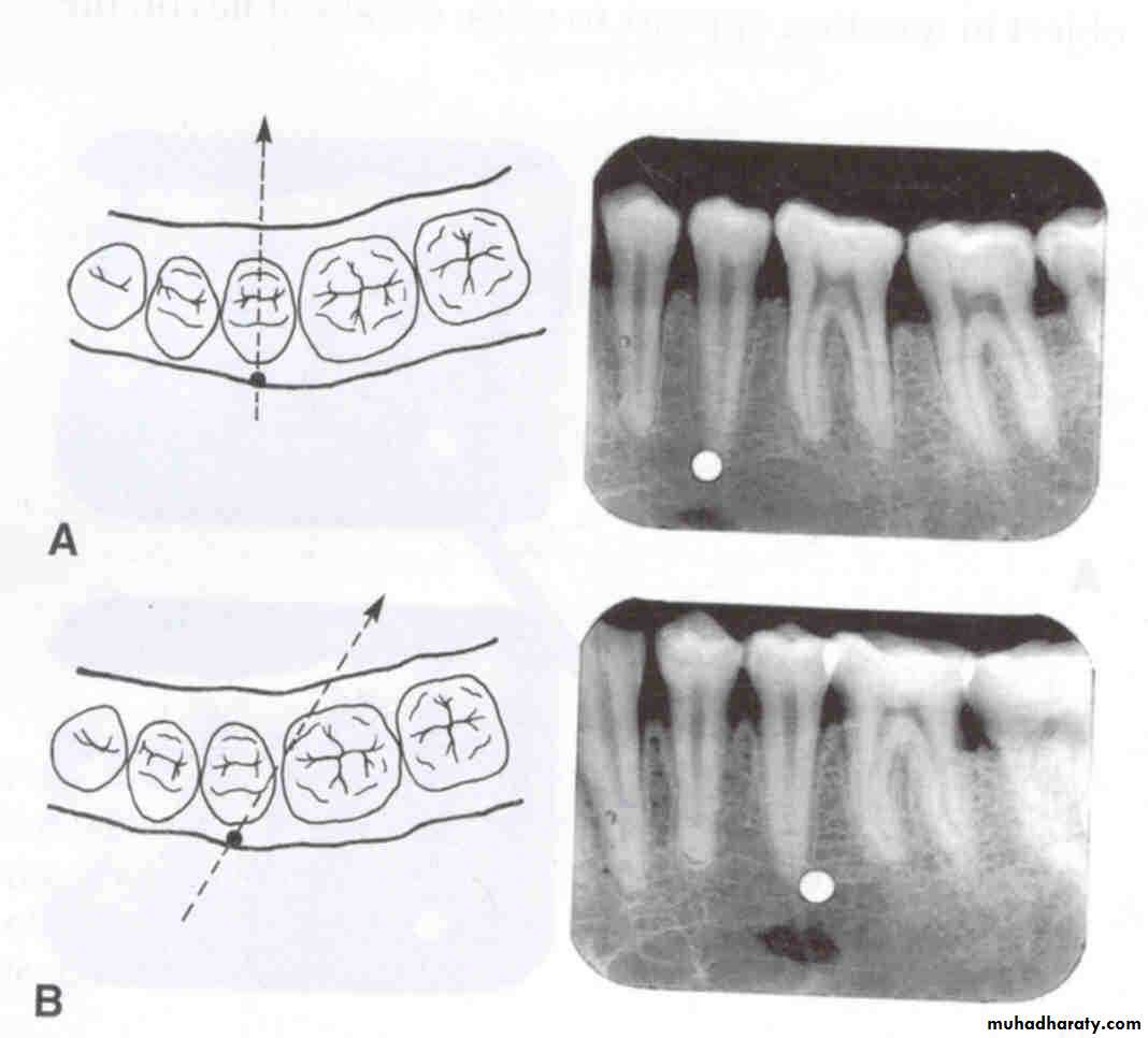
Buccal body moves in opposite direction
SLOBFor the SLOB rule to work, there must be a change in the horizontal or vertical angulation of the x-ray beam as the tube head is moved. This change in angulation will alter the relationship between the object of interest and the reference object, allowing you to determine the buccal or lingual location.
In the diagram at right, the tube head is moved, but there is no change in direction of the x-ray beam, which results in no change in location of the object of interest in relation to reference object (see below). Moving the tube head without changing the beam direction would often result in a cone cut , depending on how far the tube head is moved (see below right).
When using the SLOB rule, the direction of the beam must be opposite to the way the tube head is moved.
Horizontal Tube Shift: When the tube head is moved mesially, the beam must be directed more distally (from the mesial). If the tube head is moved distally, the direction of the beam must be more towards the mesial (from the distal).
Vertical Tube Shift: The SLOB rule also works for movement of the tube head in a vertical direction. Downward movement of the tube head requires that the beam be directed upward and when the tube head is moved upward, the beam must be directed downward.
Moving the tube head mesially or distally and changing the direction of the x-ray beam (as described in the previous slide) will result in the movement of the object of interest on the film in relation to the reference object. In the diagram below, the tube head is moved distally with the x-ray beam directed more mesially (from the distal). The object of interest, located lingual to the first molar, moves distally, in the same direction as the tube head movement.
incisors
caninepremolar
molar
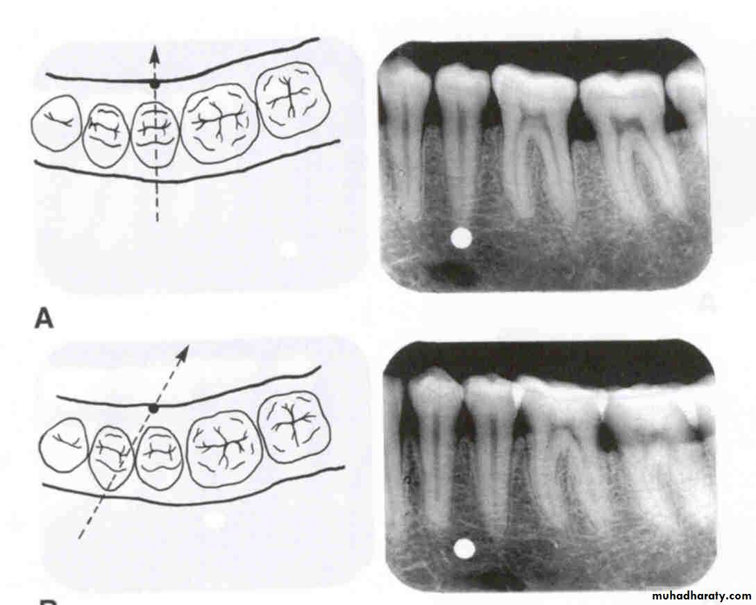
Horizontal movement of the tube head and x-ray beam
In moving from the incisor film to the canine film, the canine film to the premolar film and the premolar film to the molar film, the tube head moves distally and the beam is directed more mesially. There is not much change in angulation from the premolar to the molar film; the normal situation would be that the beam is directed slightly more from the distal (or to the mesial) as the tube head is moved distally for the molar projection.
In the diagram at left, the buccal (yellow) and lingual (red) objects of interest are superimposed on each other because the beam is directed perpendicular to both of them and they are in the same relative position mesiodistally and vertically. Both images are located above the second molar.
mesial
distal
mesial
distal
Horizontal movement
In the diagram at left, the tube head is moved distally and the beam is directed mesially. On the radiograph, the buccal object of interest (yellow) moves mesially (opposite to tube head movement) in relation to the second molar and the lingual object of interest (red) moves distally (same direction as tube head) in relation to the second molar.
mesial
distal
mesial
distalHorizontal movement
In the diagram at right, the tube head is moved mesially and the beam is directed distally. On the radiograph, the buccal object of interest (yellow) moves distally (opposite to tube head movement) in relation to the second molar and the lingual object of interest (red) moves mesially (same direction as tube head) in relation to the second molar.
mesial
distal
mesial
distal
Horizontal movement
Maxillary PA
BWMandibular PA
Vertical movement of the tube head and x-ray beam
In moving from the maxillary periapical to the bitewing and from the bitewing to the mandibular periapical, the tube head moves down and the beam is redirected upward (opposite direction; decreased vertical angulation).
In the diagram at left, the buccal (yellow) and lingual (red) objects of interest are superimposed on each other because the beam is directed perpendicular to both of them and they are in the same relative position mesiodistally and vertically. Both images are superimposed over the mandibular second premolar.
Vertical movement
In the diagram at left, the tube head is moved upward and the beam is directed downward. On the radiograph, the buccal object of interest (yellow) moves down (opposite to tube head movement) in relation to the second premolar and the lingual object of interest (red) moves up (same direction as tube head) in relation to the second premolar.
Vertical movement
In the diagram at left, the tube head is moved downward and the beam is directed upward. On the radiograph, the buccal object of interest (yellow) moves up (opposite to tube head movement) in relation to the second premolar and the lingual object of interest (red) moves down (same direction as tube head) in relation to the second premolar.
Vertical movement

TEST
Is the composite restoration on tooth # 8 (arrows) located on the buccal or lingual?
canine filmincisor film
1
The restoration is located on the buccal. The tube head moves mesially from the canine film to the incisor film (x-ray beam projected more distally) and the composite moves distally, which is the opposite direction.
0
canine film
premolar film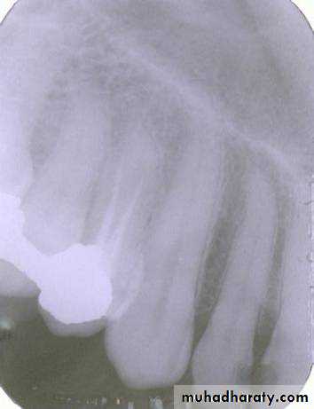
The arrow in the canine film is pointing to the gutta percha in which canal of the maxillary first premolar?
2
The arrow identifies the lingual canal. The tube head moves mesially from the premolar film to the canine film (beam directed more distally) and the gutta percha indicated by the arrow also moves mesially. (See following slide).
0
PID
PID
lingual
buccalWhen the tube head is moved mesially, with the beam directed distally, the two canals, which are initially superimposed (premolar periapical above) will separate. The lingual canal (red arrow) will follow the tube head movement and the buccal canal (blue arrow) will move in the opposite direction, as seen on the canine film.
0
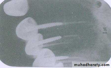
The red arrow is pointing to the gutta percha in which canal of this maxillary left first premolar?
This is the buccal canal. The tube head goes distally from the canine film to the premolar film and the gutta percha moves mesially to be positioned over the lingual canal which has the threaded post.
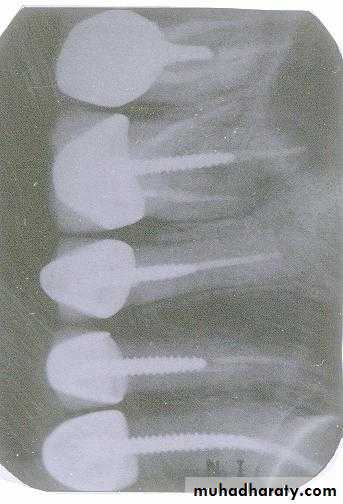
The pink arrow points to a threaded post. In which canal of this maxillary left second premolar is the post located?
The post is located in the lingual canal. As the tube head moves distally from the canine film to the premolar film, the post also moves distally to cover the canal that has all gutta percha.
0
3

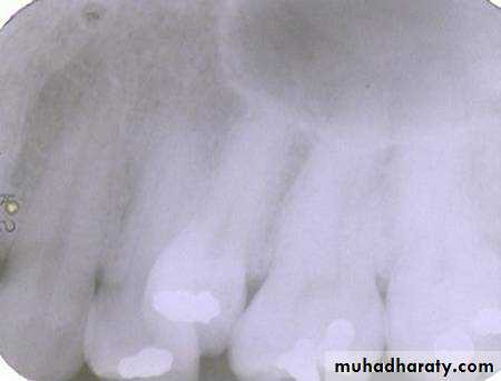
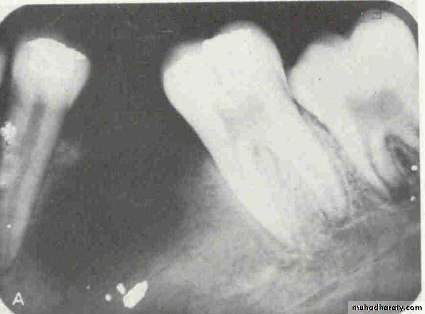
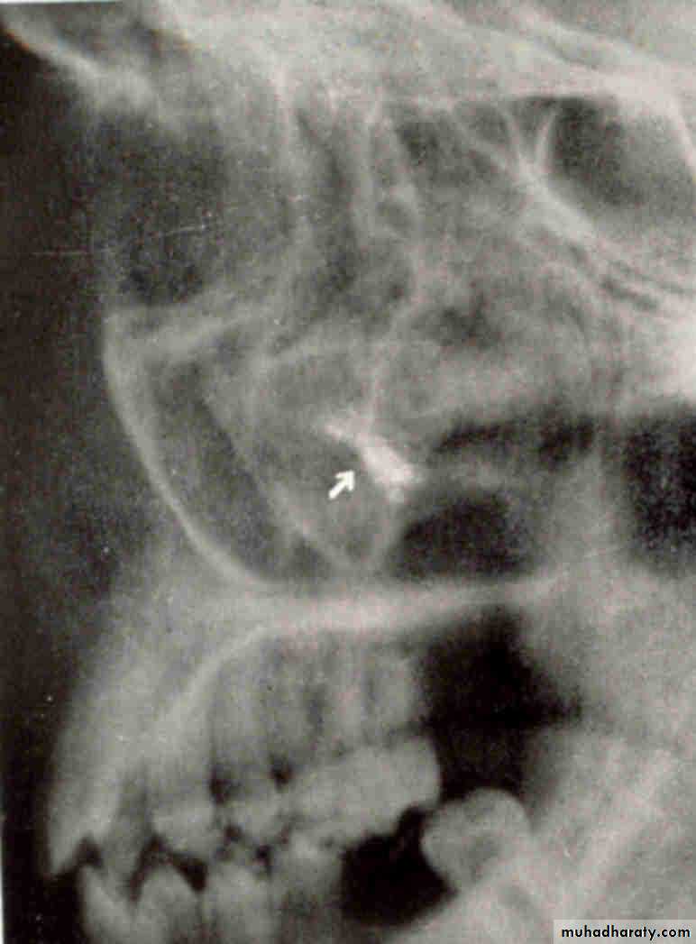
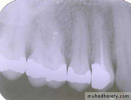
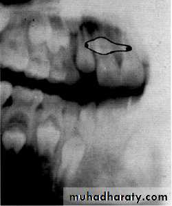
Is the maxillary second premolar (arrows) displaced to the buccal or the lingual?
premolar filmmolar film
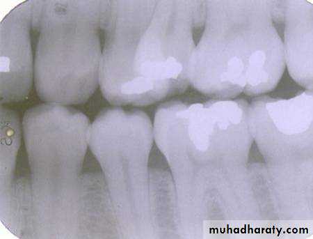
premolar bitewing
4The tube head moves distally from the premolar film to the molar film. The second premolar also moves distally, overlapping the first molar more in the molar film. In moving from the premolar periapical to the bitewing, the tube head moves down and the premolar also moves down. The displacement is to the lingual.
0
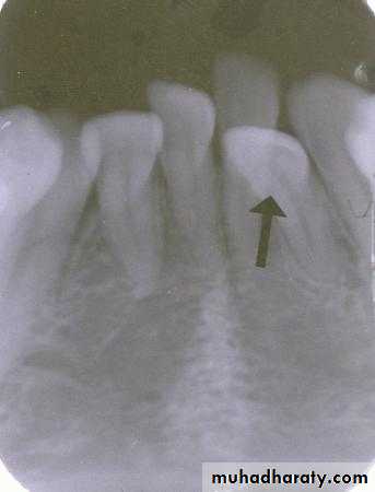
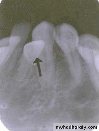
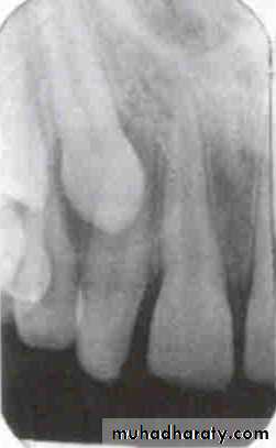
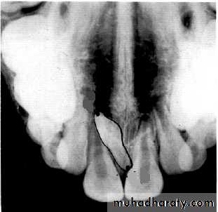
incisor film
canine filmIs the displaced incisor (arrows) located on the buccal or the lingual?
5
The lateral incisor is displaced to the lingual. The tube head moves distally from the incisor film to the canine film. The lateral incisor also moves distally, covering half the canine on the canine film.
0
canine film
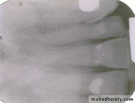
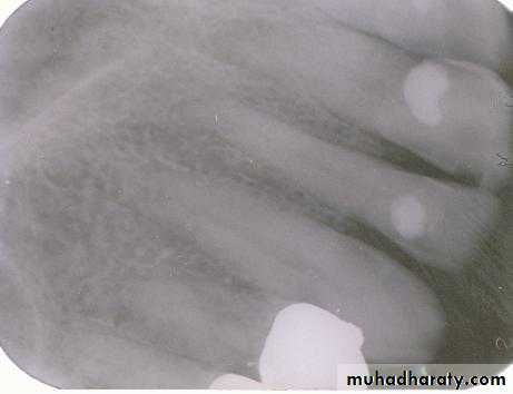
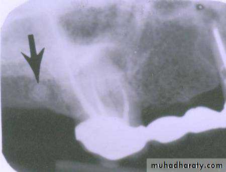
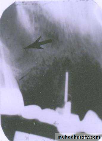
premolar filmIs the radiopaque object identified by the arrows located on the buccal or the lingual?
6
Lingual. The tube head moves mesially from the premolar film to the canine film. The object also moves mesially, starting out distal to the first molar on the premolar film and ending up mesial to the first molar on the canine film. This object represents the tip of the palatal root of the second molar and is located distal to the first molar and in a lingual relationship
0
film placement for canine film
film placement for premolar filmPID placement for premolar film
PID placement for canine film
root tip
0
A
B