The Respiratory System
د.رند عبداللطيف
Lecture 2
Trachea
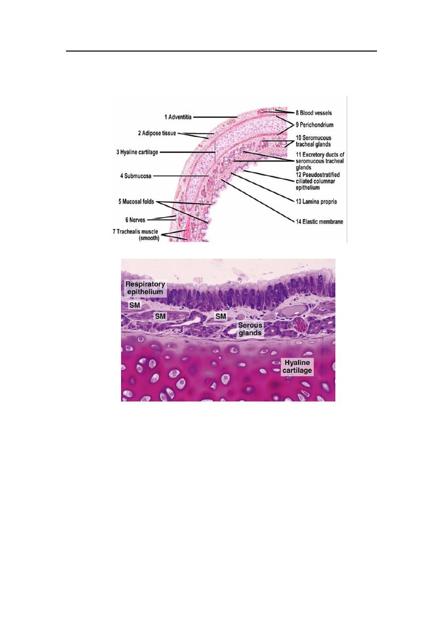
Is a thin walled tube about 10 cm long & 2-3 cm. in diameter its wall
contain about 15-20 incomplete circular rings of hyaline cartilage, a
small narrow area in the posterior wall of the trachea is devoid of
cartilage, this gab is bridged by dense fibrous ligament & bundles of
smooth muscle called trachealis muscle, contraction of this muscle will
produce narrowing of the tracheal lumen as a part of the cough reflex, this
narrowing leads to increased velocity of expired air which aids in the
cleaning of the air passges. The trachea give rise to the main bronchus
The Respiratory System
د.رند عبداللطيف
Lecture 2
which divided into Rt and Lt bronchus, their histological structure
corresponds largely to that of the trachea. The trachea is lined by:
1. typical
respiratory
epithelium
(pseudostratified
ciliated
columnar cells with goblet cells) the number of goblet cells is
variable depending on physical or chemical irritation of the
epithelium which will increase the goblet cell number.
2. lamina propria consists of loose connective tissue with many
elastic
fibers.subepithelial
sero-mucous
tracheal
glands
supplement the secretions of goblet cells in the epithelium, these
glands are particularly numerous in the posterior band of the
trachea devoid of cartilage.
3. hyaline cartilage surrounded by perichodrium present in the
submucosa.
4. adventitia containing blood vessels, lymphatics & adipose tissue.
Bronchial tree:
The Respiratory System
د.رند عبداللطيف
Lecture 2
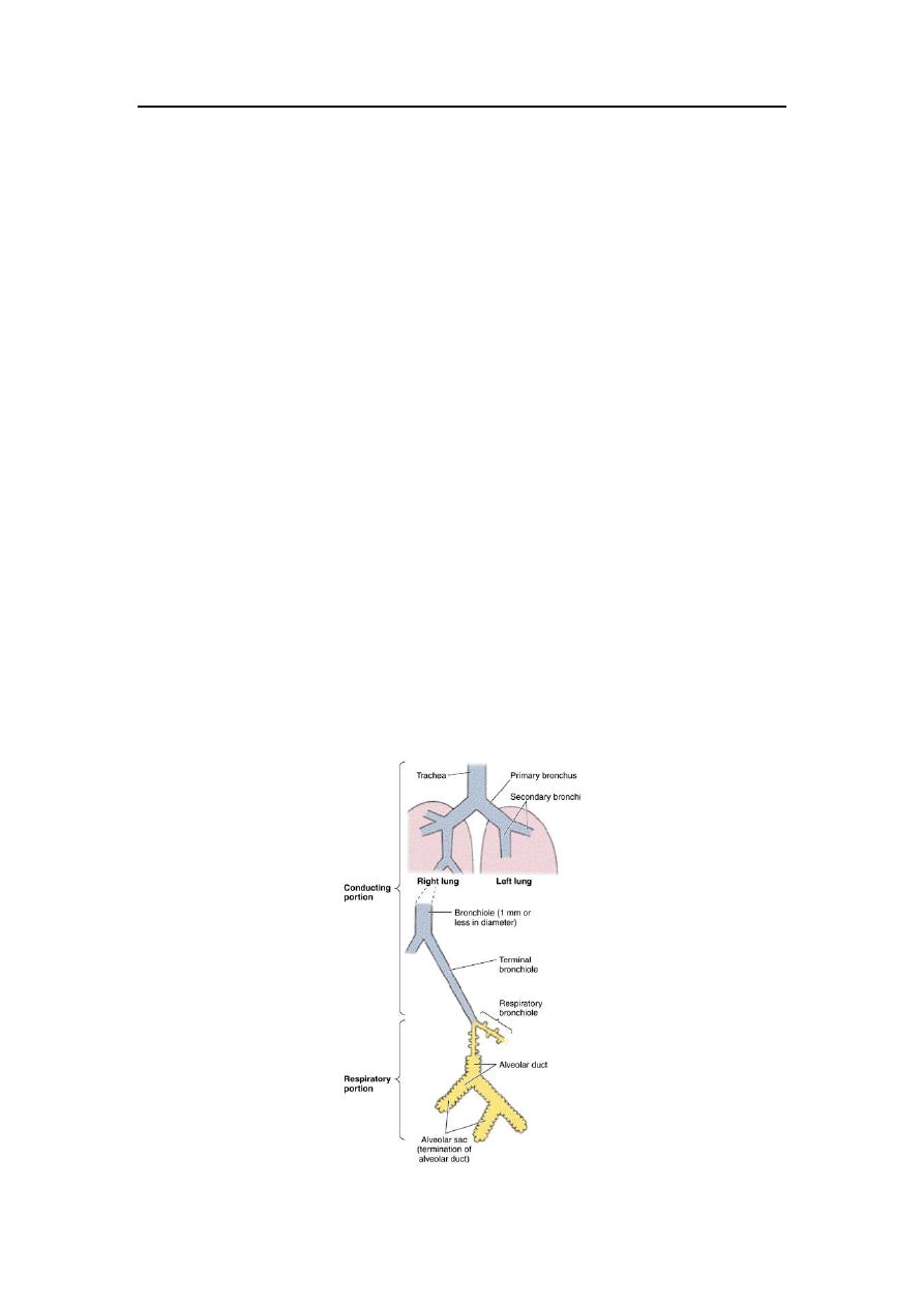
The trachea divide into 2 main bronchi (called right & left
primary bronchi) that enter the lung through the hilum and divide to
give secondary bronchi (lobar bronchi) one for each lobe of the lung.
These lobar bronchi divide repeatedly giving rise to smaller & smaller
bronchi whose terminal branches are called bronchioles & each
bronchiole then divide to form 5-7 terminal bronchioles. Bronchial
branches are accompanied by branches of the pulmonary artery, nerves
and lymph vessels. Throughout their coarse the bronchi have a similar
structure to that of the trachea but with few variations only.
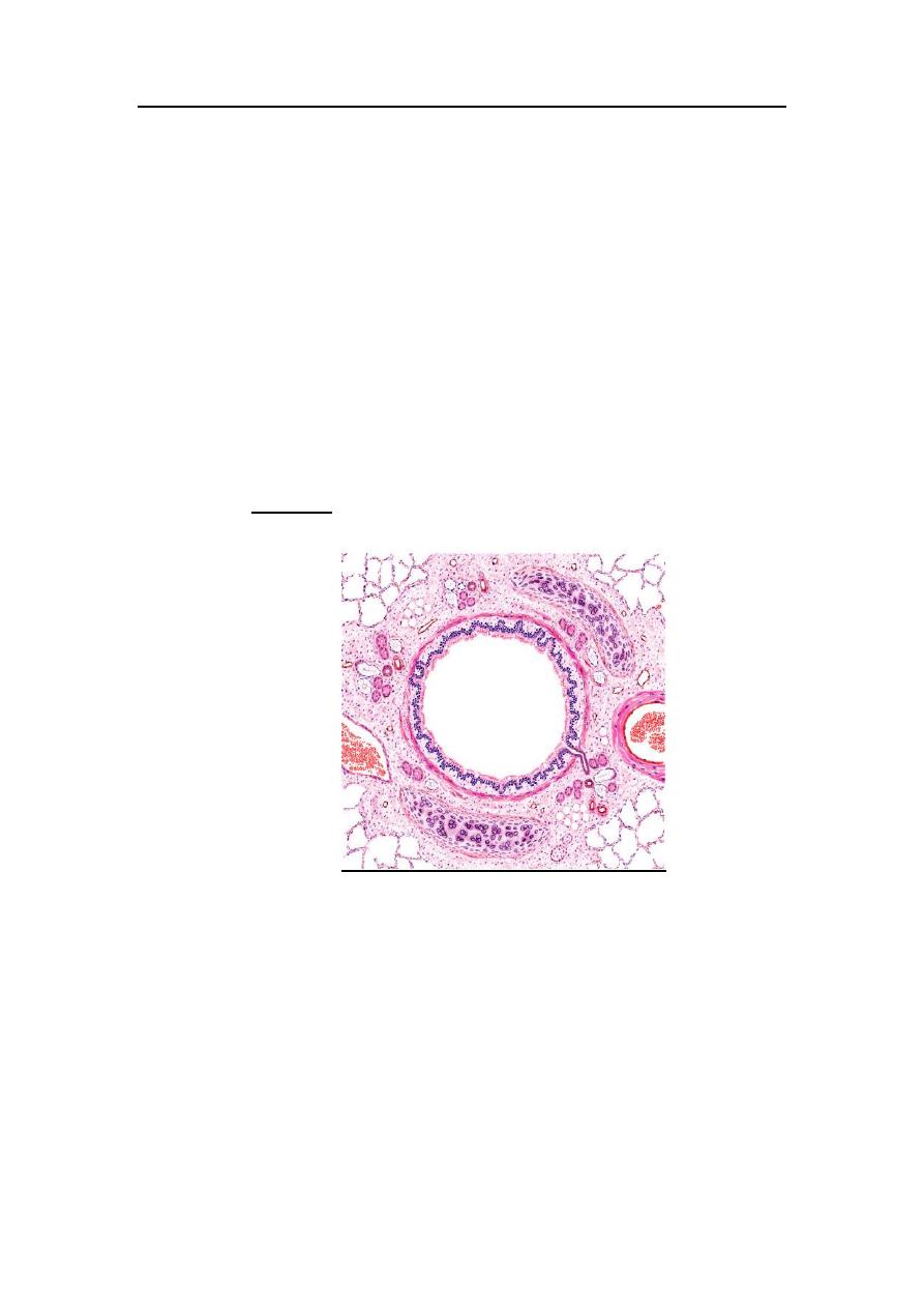
Bronchi:
The primary bronchi generally have the same histological appearance
as the trachea so the basic structure of the bronchi is:
a pseudostratified columnar ciliated epithelium with goblet cells, this
epithelium become less columnar in the smaller branches, the
epithelium contains columnar ciliated cells, goblet cells, basal cells,
brush cells & neuroendocrine cells.
The Respiratory System
د.رند عبداللطيف
Lecture 2
a subepithelial fibrocollagenous lamina propria containing variable
quantities of seromucous glands which empty their secretions into
the lumen by short ducts. Other deeper glands with long ducts are
located in between & beneath the cartilaginous plates of the bronchi.
the lamina propria contains variable amounts of smooth muscle
fibers and elastic fibers that are arranged spirally around the
bronchus.
neumerous lymphocytes are present in the lamina propria &
lymphatic nodules are found mainly at the branching points of the
bronchial tree.
the bronchial cartilages become more irregular than that found in the
trachea, in the smaller bronchi the cartilaginous rings will
incompletely encircle the lumen, as the bronchial diameter decrease
the cartilagenious rings are replaced by isolated plates of hyaline
cartilage.
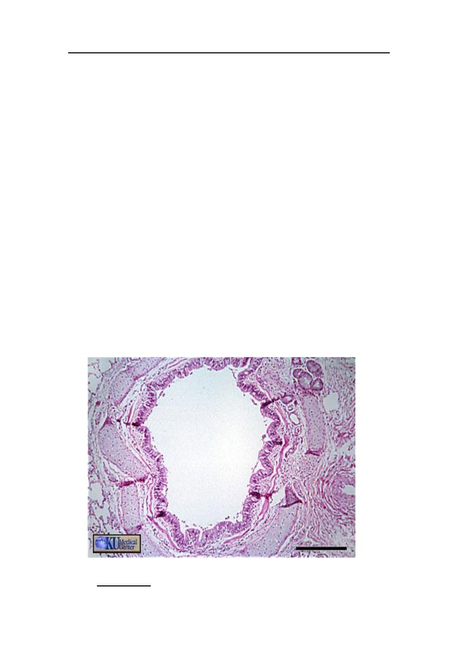
Bronchioles

The Respiratory System
د.رند عبداللطيف
Lecture 2
:
Are the smallest parts of the conducting portion of the
respiratory system, their diameter is 5 mm. or less. They have
no cartilage & no glands in their walls, and with absence of the
cartilage the smooth muscle fibers become the major
component of their wall.
The larger bronchioles are lined with ciliated columnar
epithelium, these cells become low columnar and even cuboidal
in the smaller terminal bronchioles.
Goblet cells are only found occasionally in the wall of the
bronchioles and there are no seromucous glands.
A special type of cells found in the bronchiolar wall called
Clara cell which are more numerous in the terminal
bronchioles. These cells are non-ciliated columnar to cuboidal
in shape , have many secretory granules in their cytoplasm with
many mitochondria and smooth endoplasmic reticulum near the
luminal surface of the cell that bulge above the level of adjacent
ciliated cells, they have many functions:
The Respiratory System
د.رند عبداللطيف
Lecture 2
protection against the effect of inhaled toxins & carcinogenous
substances.
play a role in surfactant production.
possibly act as stem cells capable of producing other types of
cells.
The lamina propria of the bronchioles composed mainly of
smooth muscle and elastic fibers which are under the effect of
both parasympathetic (by Vagus nerve) and sympathetic
innervations, thus stimulation of Vagus nerve produces
contraction of these muscle & leads to decrease in the
bronchiolar diameter, while the stimulation of the sympathetic
nervous system produces the opposite effect.
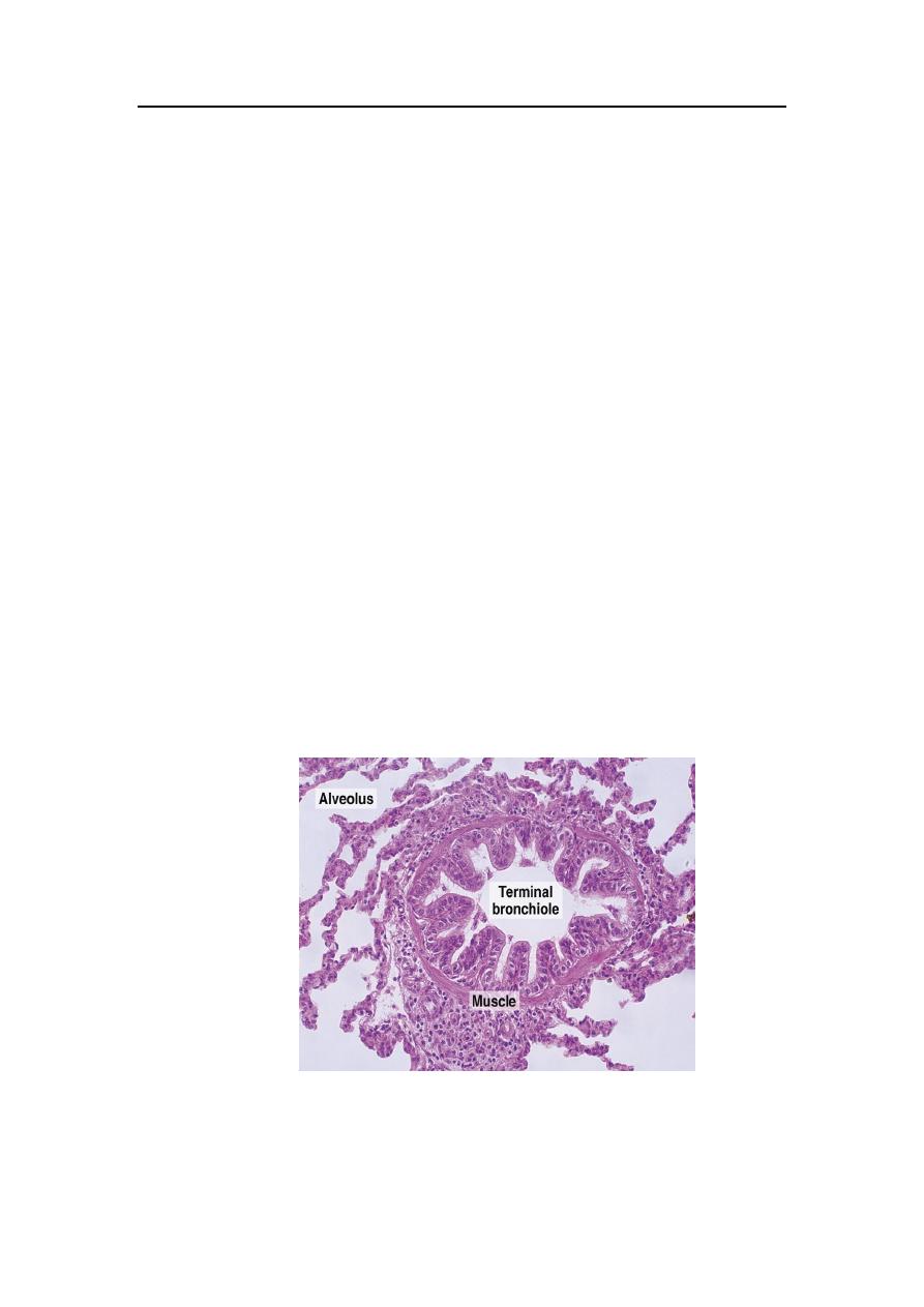
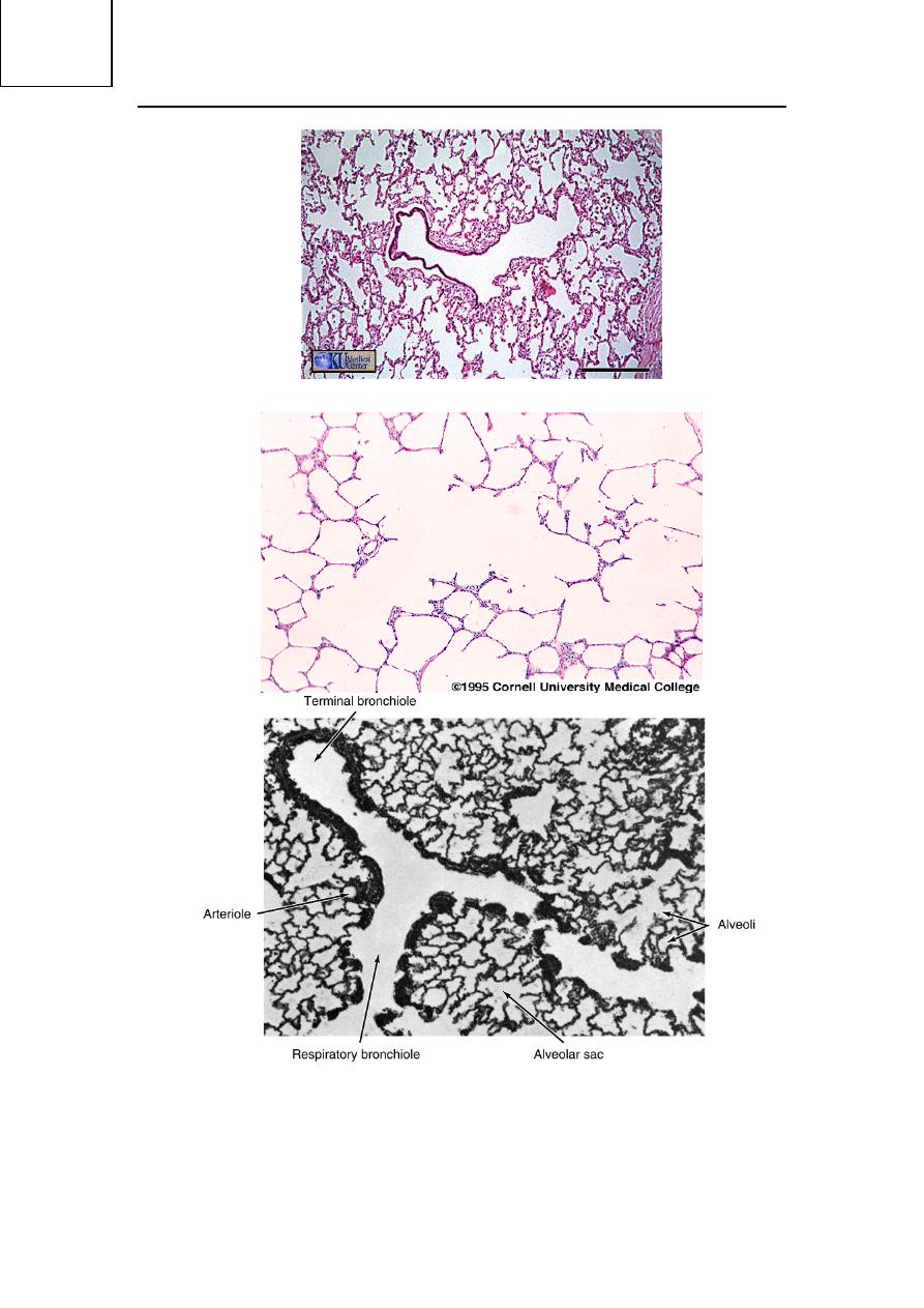
The final bifurcation of the bronchiole leads to the formation of
terminal bronchioles which are the smallest bronchioles
concerned only with air conduction.

The Respiratory System
د.رند عبداللطيف
Lecture 2
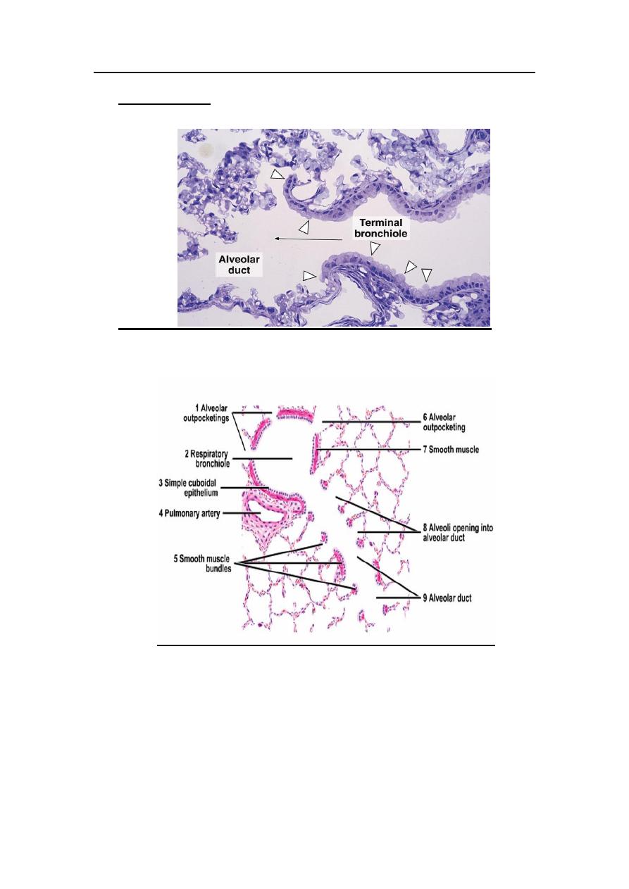
Distal respiratory tract :
Respiratory Bronchioles:
The first part of the distal respiratory tract which is concerned
mainly with the gaseous exchange, each terminal bronchiole divides
into 2 or more respiratory bronchioles which is considered as a
region of transition between the conducting part and the respiratory
part of the system.
o The mucosa of the respiratory bronchioles is structurally
identical to that of the terminal bronchioles (low cuboidal
cells plus Clara cells) except that their walls are interrupted
by numerous sac-like alveoli where gaseous exchange
occur.
o The wall of the respiratory bronchiole is lined with ciliated
cuboidal epithelial cells and Clara cells, but near the
opening of the alveoli this epithelium become continuous
with the cells that line the alveolar wall (i.e squamous
alveolar lining cells- type I alveolar cells)
o Beneath the epithelium the connective tissues of the
respiratory bronchiolar wall contain smooth muscles &
elastic fibers.
o Proceeding distally along the respiratory bronchioles, the
alveoli increase greatly in number & the distance between
them is markedly reduced.
The Respiratory System
د.رند عبداللطيف
Lecture 2
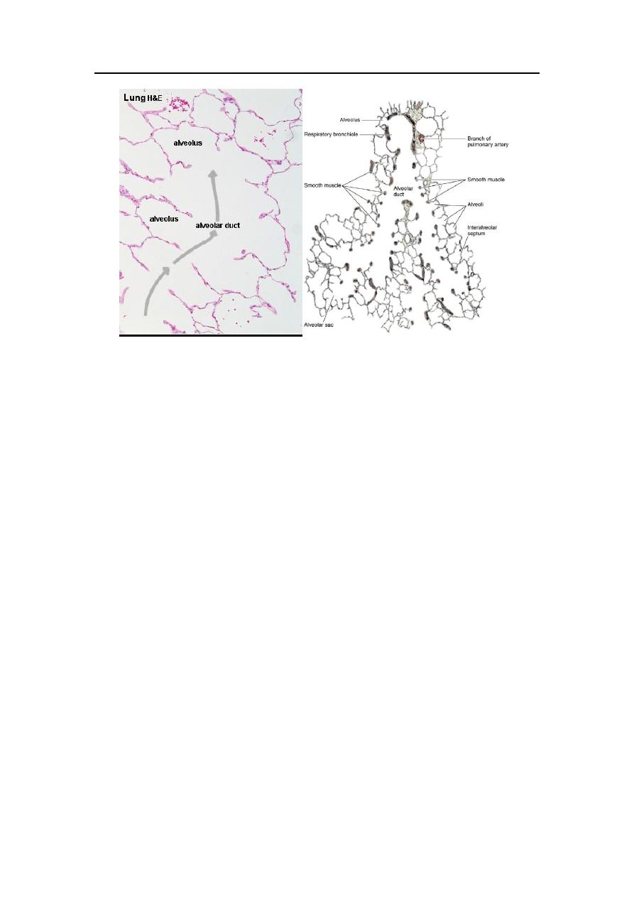
Alveolar ducts:
The Respiratory System
د.رند عبداللطيف
Lecture 2
Proceeding distally along the respiratory bronchioles, the number
of alveolar openings into the bronchiolar wall become more and
more until the wall consist only of the openings of the alveoli, and
this tube is now called alveolar duct.
Both the alveolar ducts and alveoli are lined with extremely thin
squamous alveolar cells. The lamina propria is only formed of a
thin network of smooth muscle cells and it will appear as knobs
between adjacent alveoli, these alveolar ducts will lead to the
alveolar sacs.
Elastic and reticular fibers form a complex network of fibers that
surround the alveolar sacs and alveoli. The elastic fibers enable the
alveoli to expand with inspiration & contract passively with
expiration, while the reticular fibers serve as a support that prevent
over distention & damage to the delicate capillaries & thin inter-
alveolar septa.
The Respiratory System
د.رند عبداللطيف
Lecture 2