Lec.2 Tooth development د0سحرغانم
4. Bell stage:
A.Early bell stage:
As the invagination of the epithelium deepens &it's margins continue to grow, the enamel organ assumes a bell shape. in this stage the crown shape is determined due to pressure exerted by the growing dental papilla cells on the inner enamel epithelium, the folding of enamel organ to cause different crown shapes is shown due to differential rates of mitosis & differences in cell differentiation time.
Cells begin to differentiate only when cells cease to divide, the inner enamel epithelial cells which lie in the future cusp tip or incisor region stop dividing earlier & begin to differentiate first.
4 types of cells can be distinguished under light microscope:
1.inner enamel epithelium2.the stratum intermedium
3.the stellate reticulum.
4.the outer enamel epithelium.
The junction between the inner & outer enamel epithelium is called
cervical loop which is area of intense mitotic activity.1.inner enamel epithelium
consist of single layer of cells that differentiate prior to amelogenesis in to tall columnar cells called ameloblasts. The cells of the inner enamel epithelium exert an organizing influence on the underlying ectomesenchymal cells in the dental papilla, which later differentiate in to odontoblasts.2.the stratum intermedium
a few layers of squamous cells form the stratum intermedium, between the inner enamel epithelium & the stellate reticulum,these layers seems to be essential to enamel formation. so it's absent in the part of the tooth germ that outlines the root portions of the tooth which does not form the enamel.it is rich in alkaline phosphatase enzymes essential for mineralization of enamel.
3.the stellate reticulum.
they expands further by an increase in the amount of intercellular fluid, the cells are
star shaped, with long processes that anastomose with those of adjacent cells.
Before enamel formation begins, the stellate reticulum collapses occur. reducing
The distance between inner enamel epithelium (ameloblast)and the nutrient capillaries near the the outer enamel epithelium.
4.the outer enamel epithelium.
cells are flat to low cuboidal cells in this stage.
dental papilla
Before the inner enamel epithelium begin to form the enamel, The peripheral cells of the ectomesenchymal dental papilla differentiate in to odontoblasts which are first assume a cuboidal form, later they assume a columnar form. The basement membrane that separates the enamel organ & the dental papilla just prior to dentin formation is called the membrane preformativa.dental sac
Before formation of the dental tissues begins, the dental sac shows a circulararrangement of it's fibers & resembles acapsular structure, with development of
the root the fibers of the dental sac differentiate in to periodontal fibers that
become embedded in the developing cementum & alveolar bone.
B.Advanced bell stage
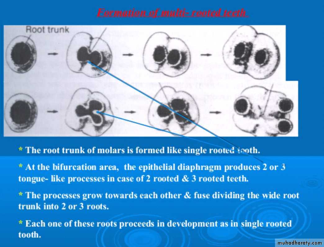
at this stage the boundary between the inner enamel epithelium & the odontoblasts outlines the future dentinoenamel junction, first layer of dentin formed along the dentino enamel junction in the region of future cusps & continuous apically. This stimulate the ameloblasts to lay down enamel over the dentin , the cervical portion of the enamel organ gives rise to the epithelial root sheath of Hertwig,s. The Hertwig,s epithelial root sheath (HERS) outlines the future root so it responsible for the shape,length, size & the number of roots.
Hertwig's epithelial root sheath & root formation:
After the crown of the tooth is completely formed , a structure called the epithelial sheath of Hertwig,s is derived from both the outer and inner dental epithelia at
where these layers are continuous with each other at the cervical loop region, they are responsible to induce differentiation of adjacent dental papilla cells into
odontoblasts. The sheath also determines the shape and number of roots .Once
sheath disintegrates at that sits allow sac cells to come in contact with root dentin. these cells differentiate into cementoblasts. Remnants of the epithelial root sheath
persist in the ligament throughout life. These rests are called the epithelial cell rests of Malassez.
Tooth Development
A. Bud StageB. Cap Stage
C. Bell Stage
D and E. Dentinogenesis and amelogenesis
F. Crown formation
G. Root Formation and eruption H. Function