
Bone Tumors
Clues and Cues
Bone Tumors
Clues and Cues
William Herring, M.D. © 2002
In Slide Show mode, advance the slides by pressing the spacebar
All Photos Retain the Copyright of their Authors

Clues by Appearance
of Lesion

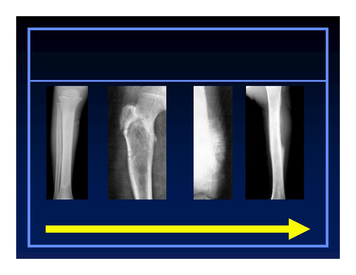
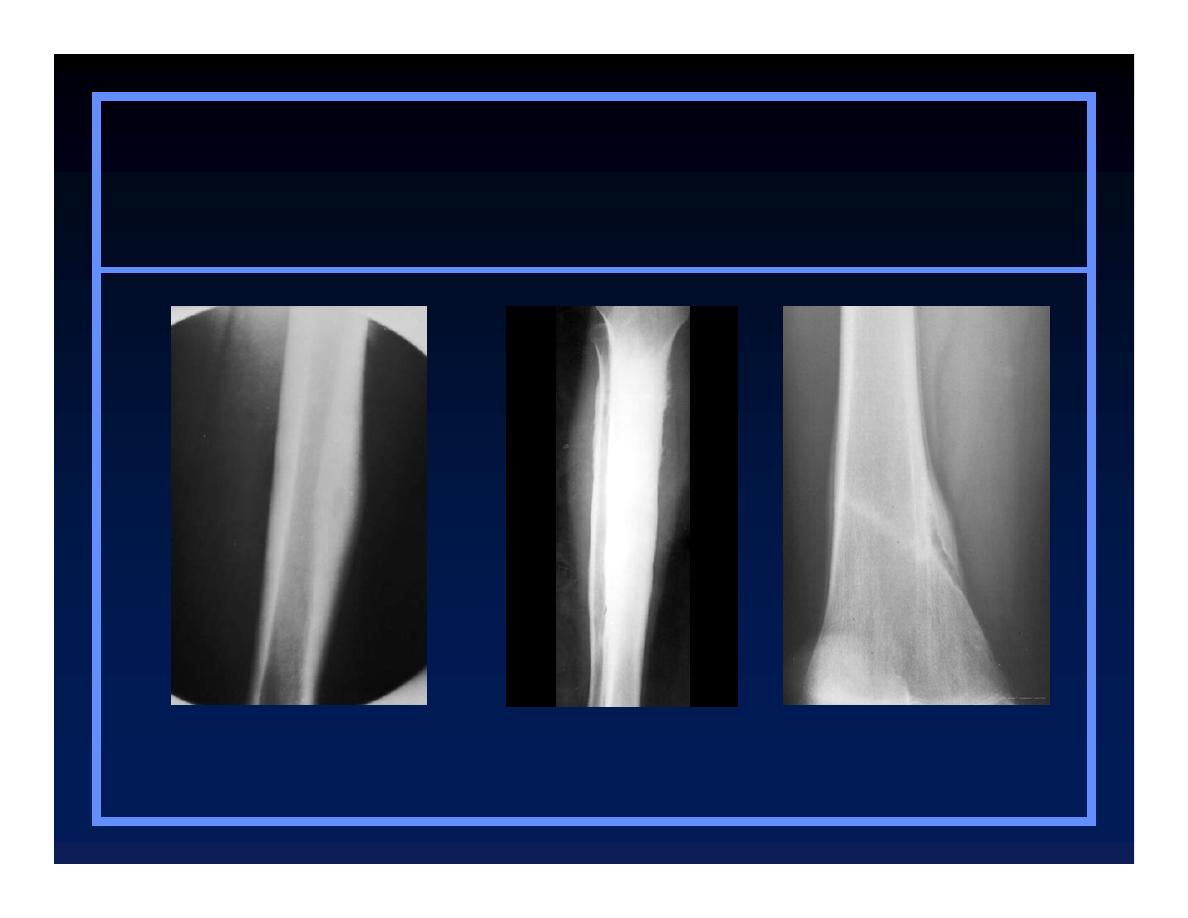
Patterns of Bone Destruction
O
Geographic
O
Moth-eaten
O
Permeative

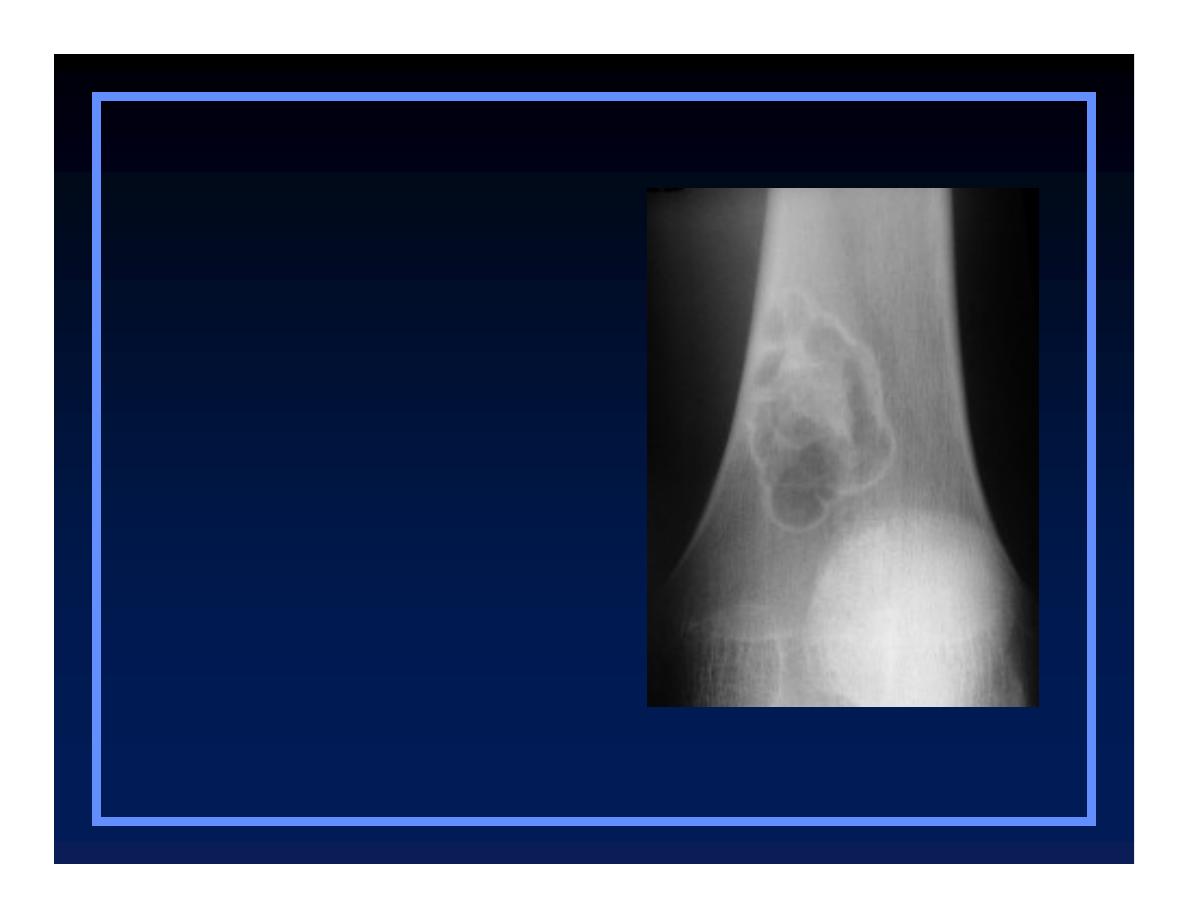
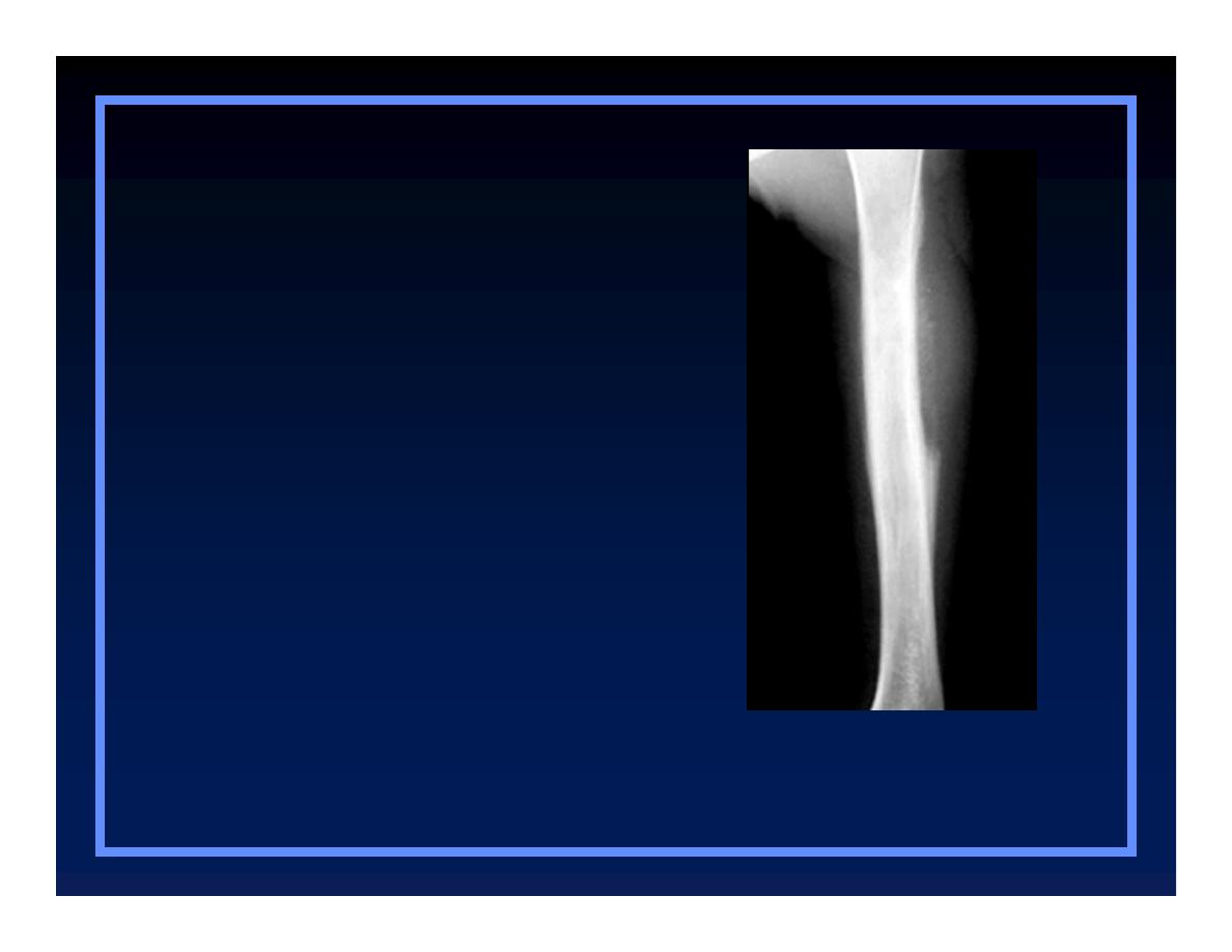
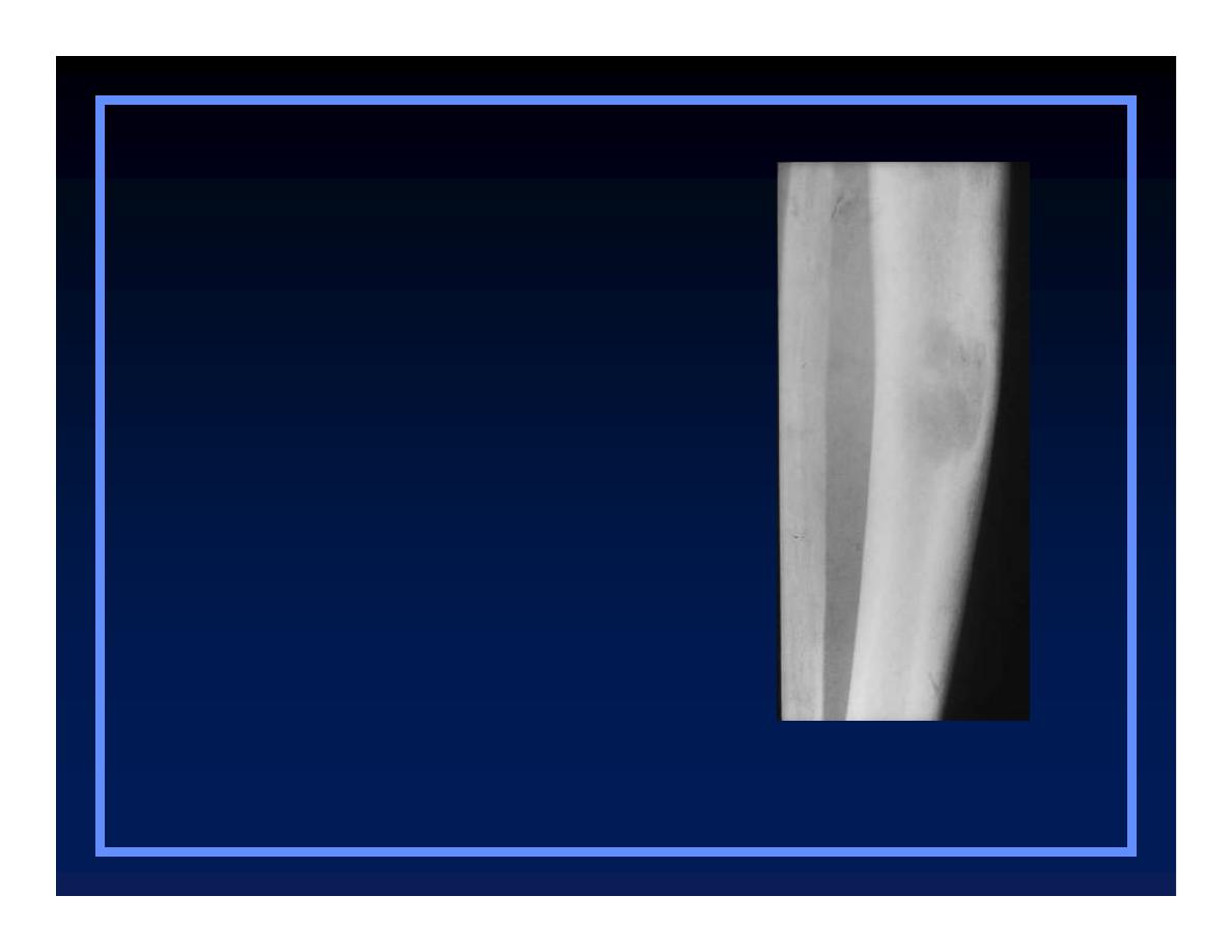
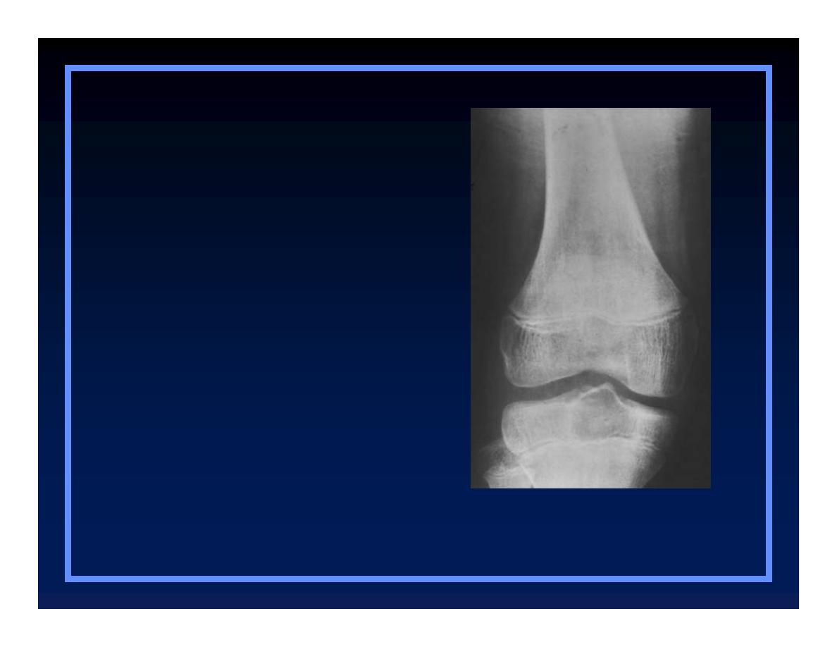
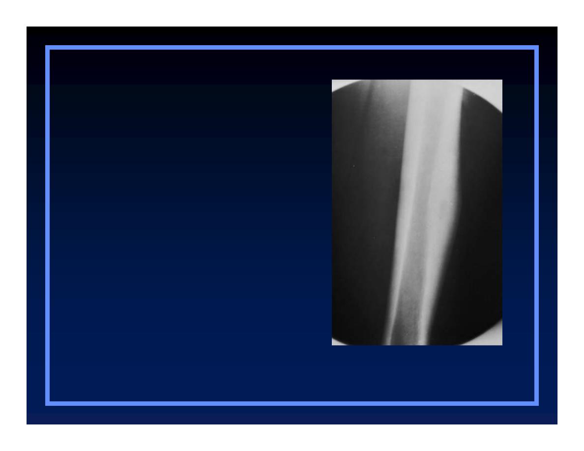
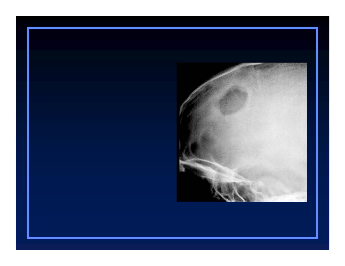
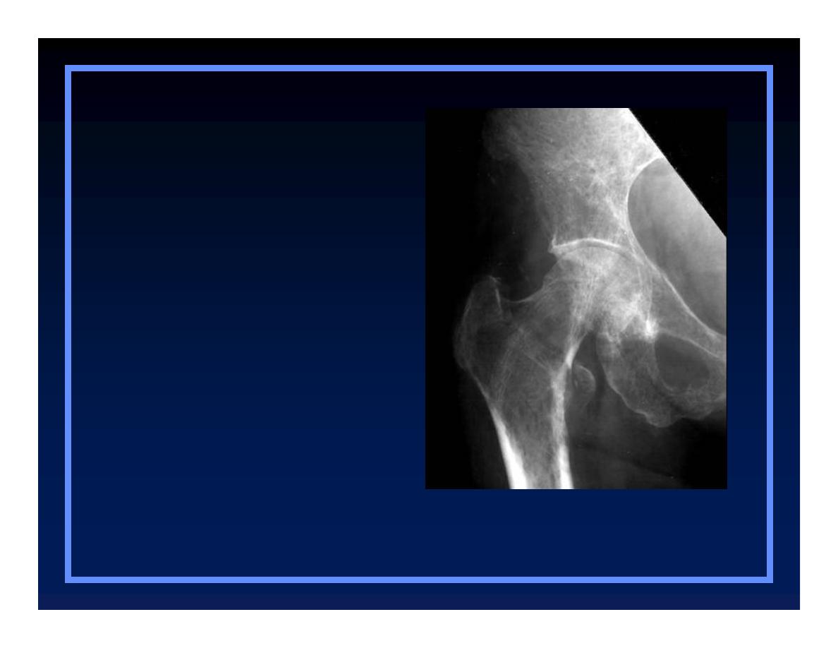
Geographic Bone Destruction
O
Destructive lesion with sharply
defined border
O
Implies a less-aggressive, more
slow-growing, benign process
O
Narrow transition zone
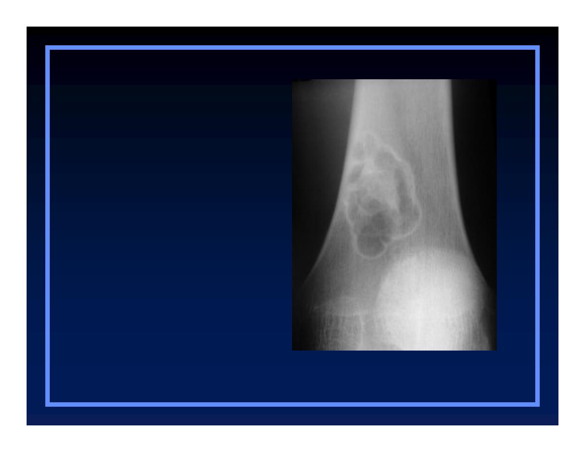
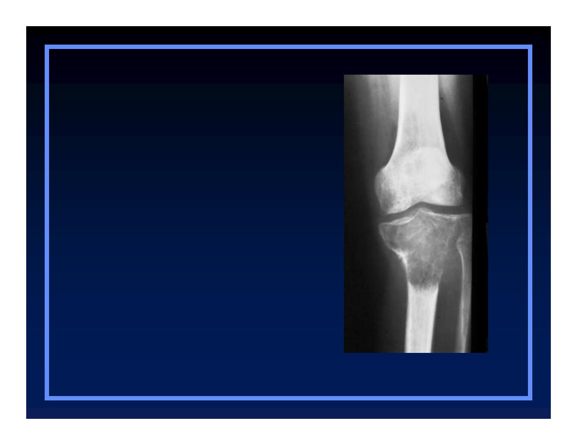
Non-ossifying fibroma
O
Geographic
O
Moth-eaten
O
Permeative
Patterns of Bone Destruction
Patterns of Bone Destruction
© R
3
, 2000

Geographic Lesions
Examples
O
Non-ossifying fibroma
O
Chondromyxoid fibroma
O
Eosinophilic granuloma

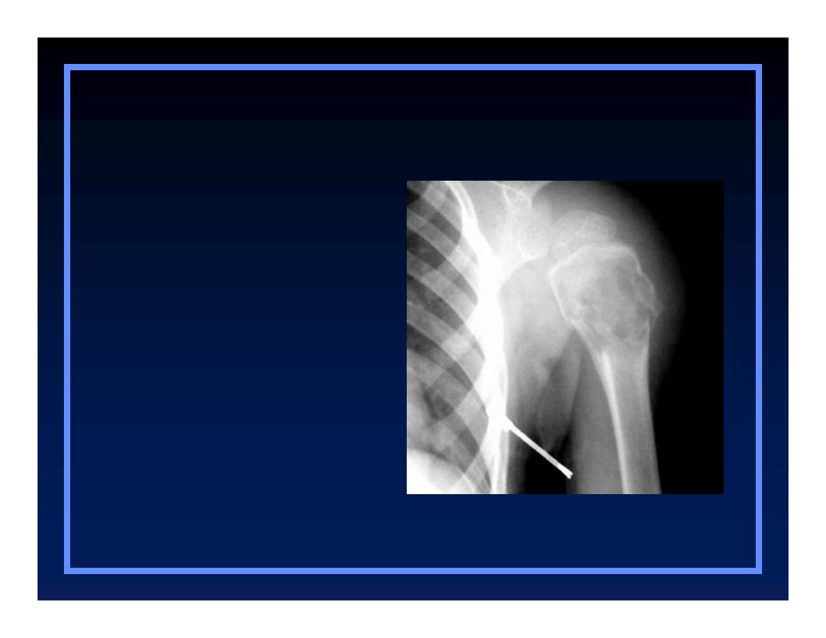
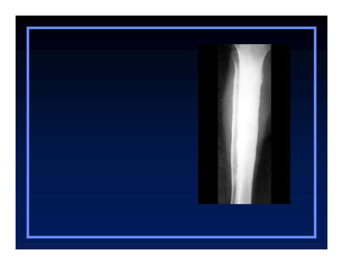
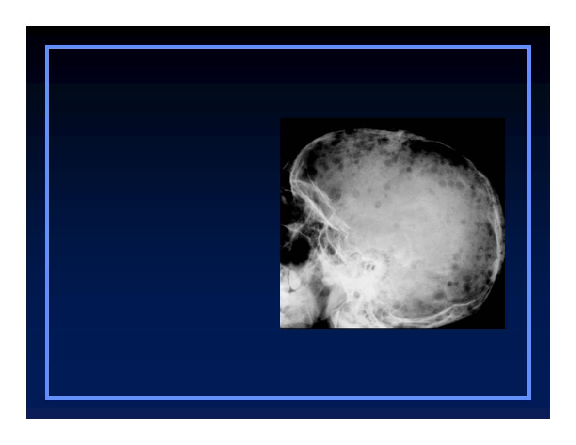
Moth-eaten Appearance
O
Areas of destruction with ragged
borders
O
Implies more rapid growth
Q
Probably a malignancy
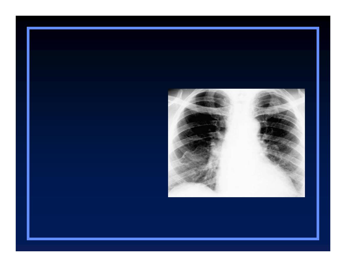
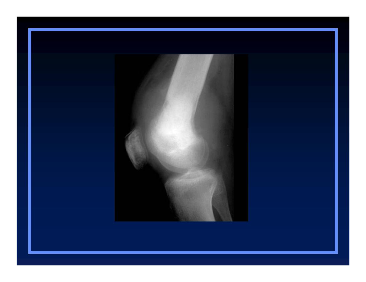
Multiple Myeloma
Patterns of Bone Destruction
Patterns of Bone Destruction
O
Geographic
O
Moth-eaten
O
Permeative
© R
3
, 2000

Moth-eaten Appearance
Examples
O
Myeloma
O
Metastases
O
Lymphoma
O
Ewing’s sarcoma

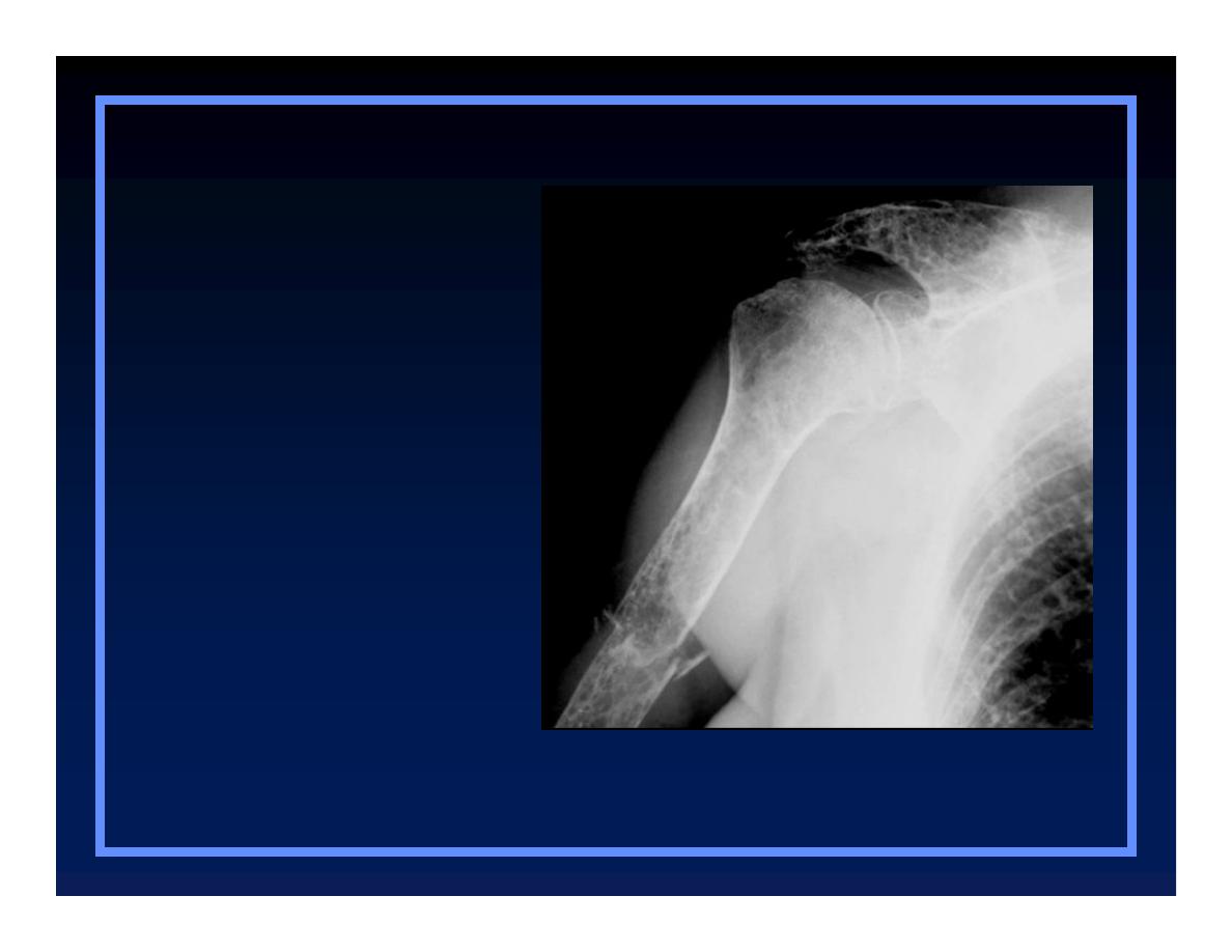
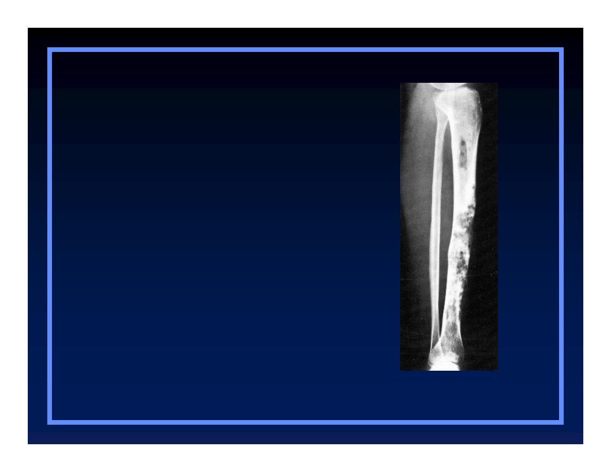
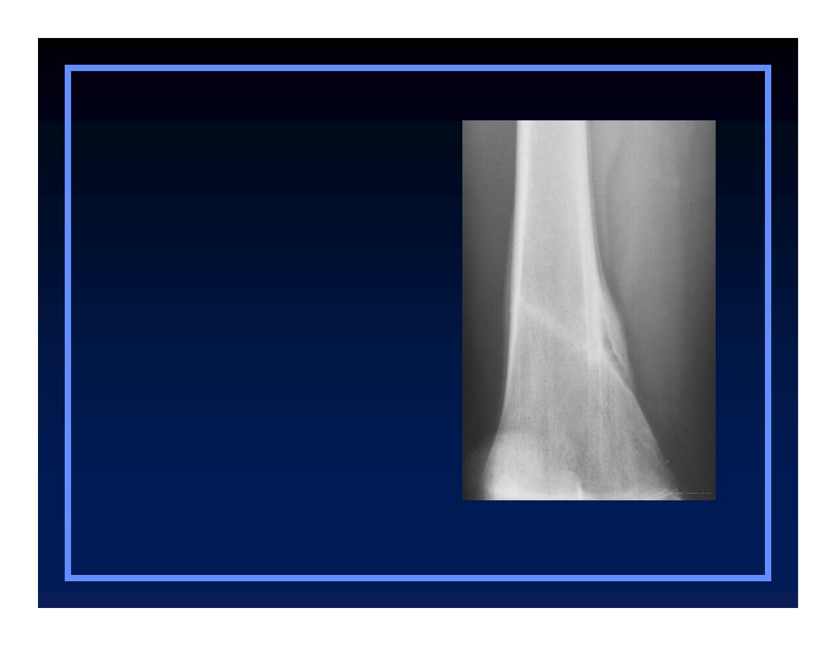
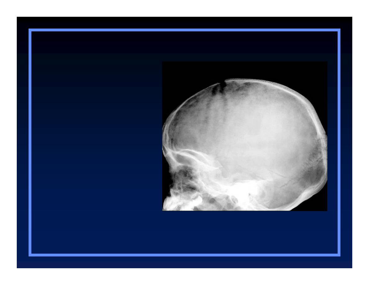
Permeative Pattern
O
Ill-defined lesion with multiple “worm-
holes”
O
Spreads through marrow space
O
Wide transition zone
O
Implies an aggressive malignancy
Q
Round-cell lesions
Leukemia
Patterns of Bone Destruction
Patterns of Bone Destruction
O
Geographic
O
Moth-eaten
O
Permeative

Permeative Pattern
Round cell lesions
O
Lymphoma, leukemia
O
Ewing’s Sarcoma
O
Myeloma
O
Osteomyelitis
O
Neuroblastoma
Patterns of Destruction
Geographic
Moth-eaten
Permeative
Less malignant
More malignant

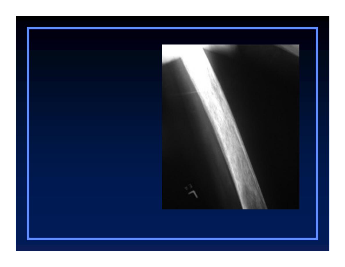
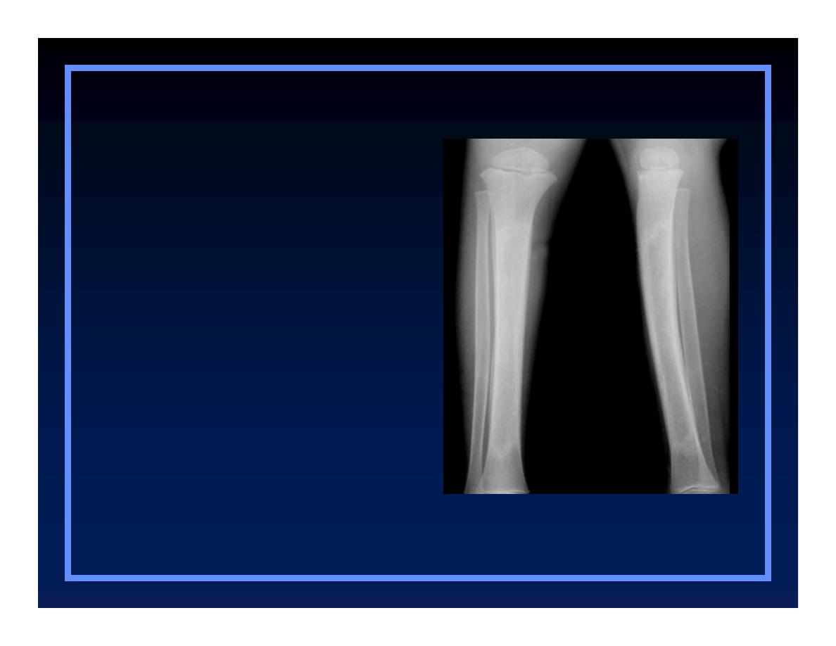
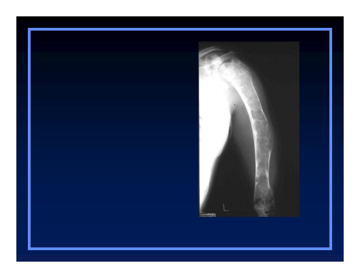
Periosteal Reactions
O
Benign
Q
None
Q
Solid
O
More aggressive or malignant
Q
Lamellated or onion-skinning
Q
Sunburst
Q
Codman’s triangle
Non-ossifying fibroma
O
Benign
Q
None
Q
Solid
O
Aggressive/malignant
Q
Onion-skinning
Q
Sunburst
Q
Codman’s triangle
Periosteal Reactions
© R
3
, 2000
Chronic osteomyelitis
O
Benign
Q
None
Q
Solid
O
Aggressive/malignant
Q
Onion-skinning
Q
Sunburst
Q
Codman’s triangle
Periosteal Reactions
© R
3
, 2000
O
Benign
Q
None
Q
Solid
O
Aggressive/malignant
Q
Onion-skinning
Q
Sunburst
Q
Codman’s triangle
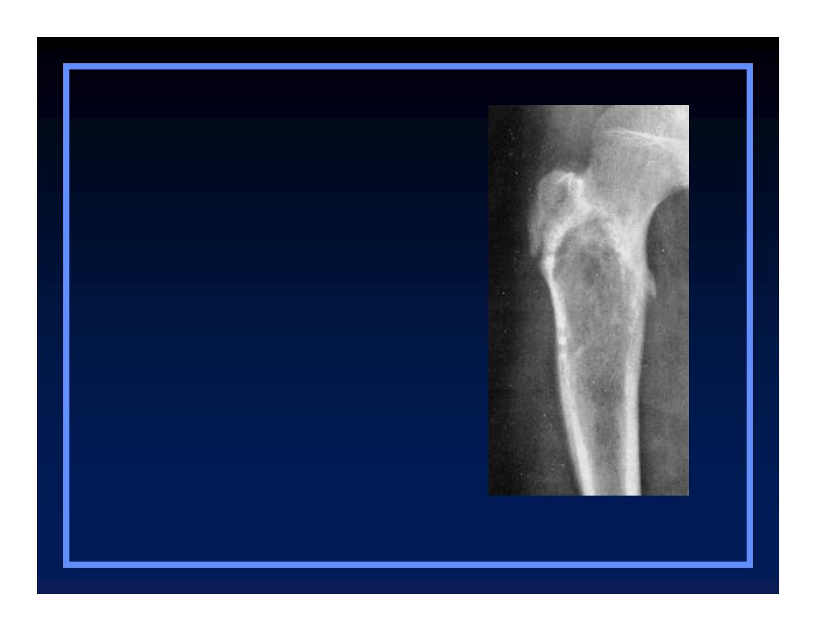
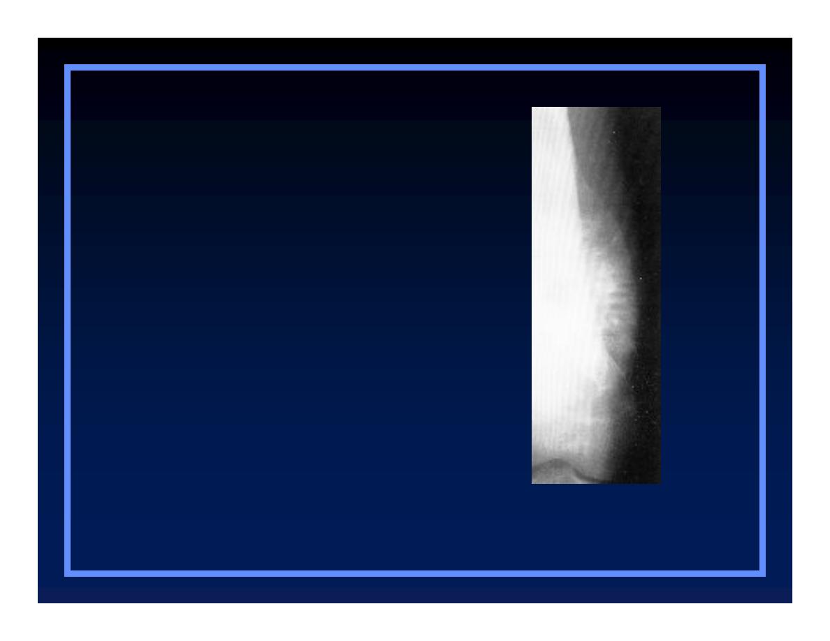
Periosteal Reactions
Ewing’s sarcoma
© Greenspan, 2000
Osteosarcoma
O
Benign
Q
None
Q
Solid
O
Aggressive/malignant
Q
Onion-skinning
Q
Sunburst
Q
Codman’s triangle
Periosteal Reactions
© Greenspan, 2000
Ewing’s-Codman’s
triangle
O
Benign
Q
None
Q
Solid
O
Aggressive/malignant
Q
Onion-skinning
Q
Sunburst
Q
Codman’s triangle
Periosteal Reactions
© R
3
, 2000
Periosteal Reactions
Solid
Lamellated
Sunburst
Codman’s
Less malignant
More malignant

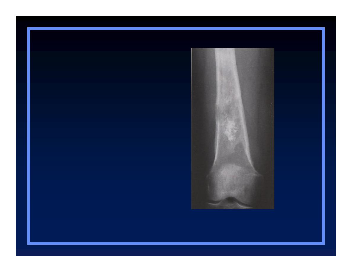
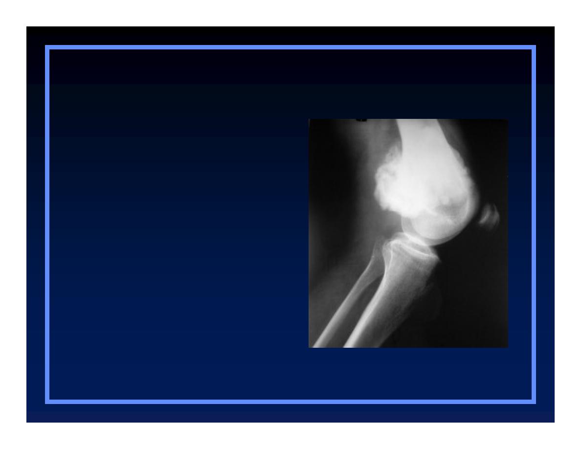
Tumor Matrix
O
Osteoblastic
Q
Fluffy, cotton-like or cloud-like densities
V
Osteosarcoma
O
Cartilaginous
Q
Comma-shaped, punctate, annular, popcorn-
like
V
Enchondroma, chondrosarcoma, chondromyxoid
fibroma
O
Osteoblastic
O
Cartilaginous
Tumor matrix
Osteosarcoma
© R
3
, 2000
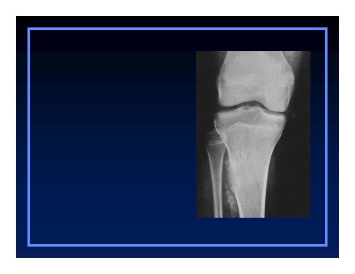
O
Osteoblastic
O
Cartilaginous
Tumor matrix
Chondrosarcoma
© R
3
, 2000

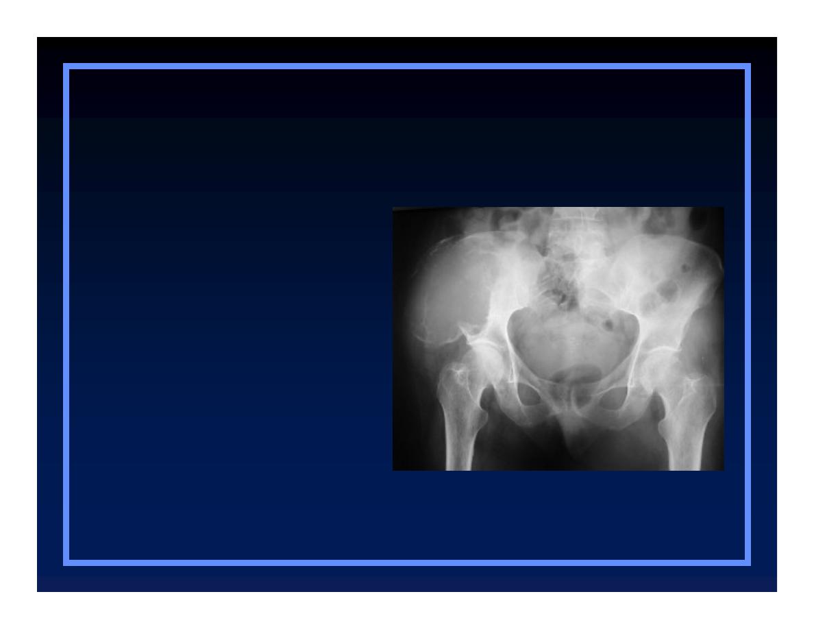
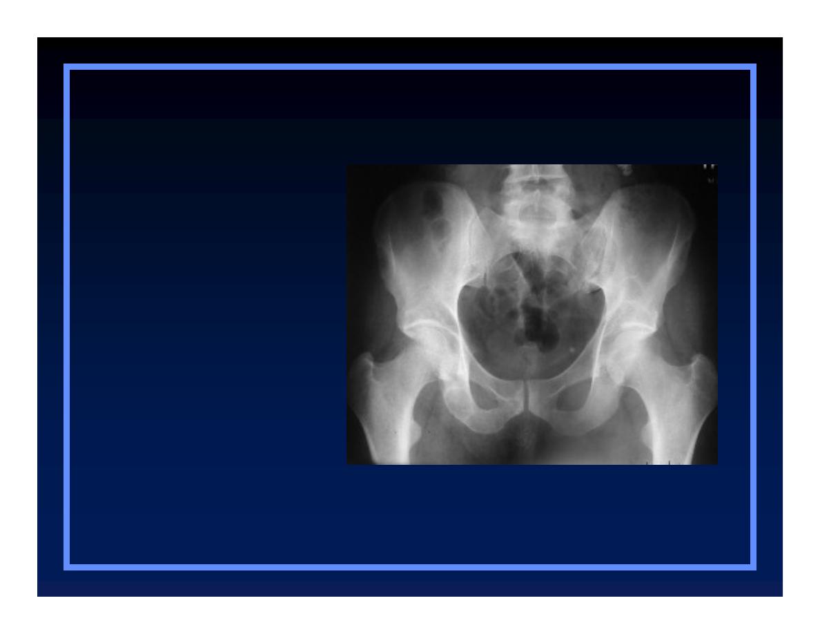
Expansile Lesions of Bone
O
Multiple myeloma
O
Brown tumor
O
Mets
O
Enchondroma
O
Aneurysmal bone cyst
O
Lymphoma
O
Fibrous dysplasia
Multiple Myeloma
Expansile lesions
O
Multiple myeloma
O
Mets
O
Aneurysmal bone cyst
O
Fibrous dysplasia
O
Brown tumor
O
Enchondroma
O
Lymphoma
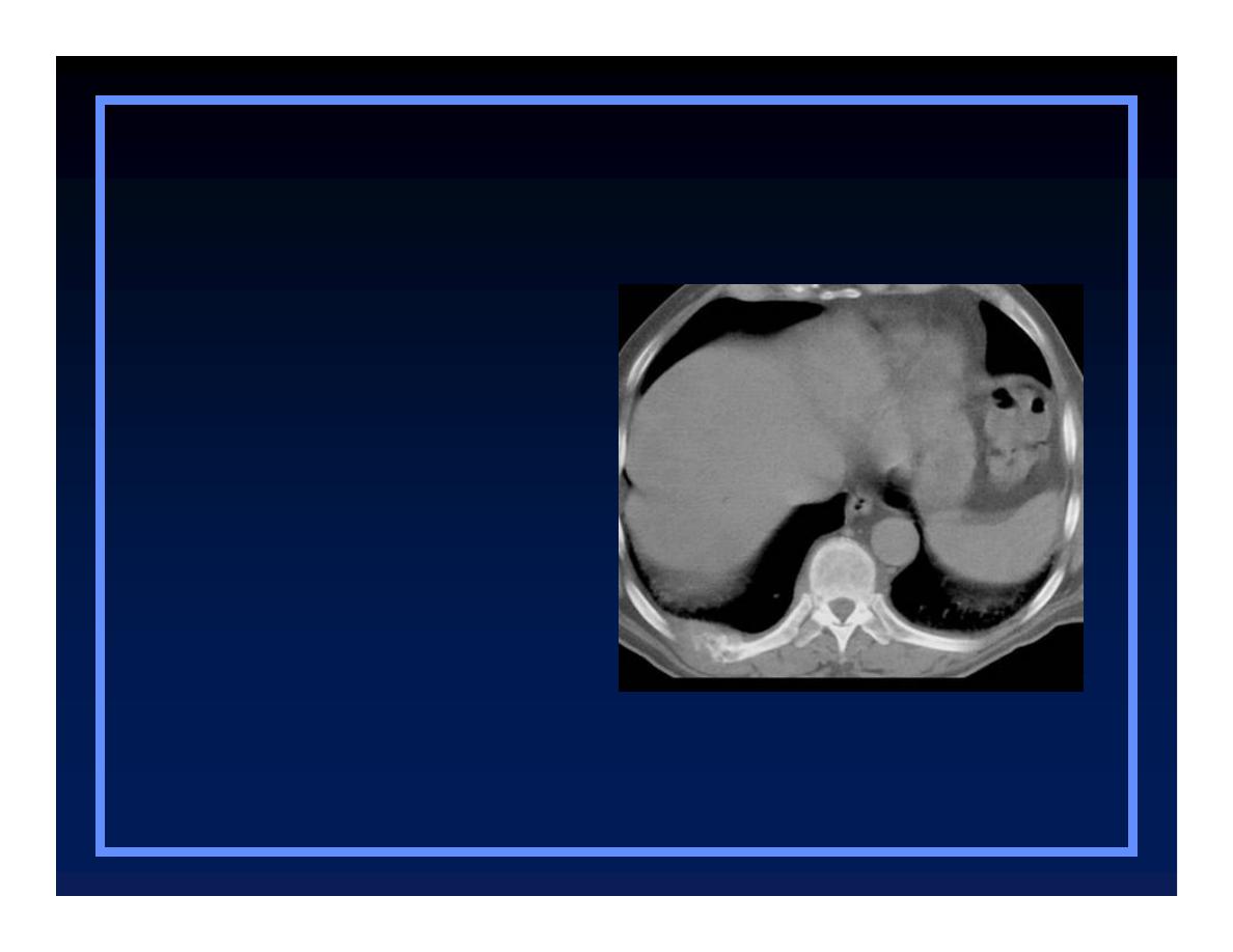
Renal Cell Carcinoma
Expansile lesions
O
Multiple myeloma
O
Mets
O
Aneurysmal bone cyst
O
Fibrous dysplasia
O
Brown tumor
O
Enchondroma
O
Lymphoma
Aneurysmal Bone Cyst
Expansile lesions
O
Multiple myeloma
O
Mets
O
Aneurysmal bone cyst
O
Fibrous dysplasia
O
Brown tumor
O
Enchondroma
O
Lymphoma
Fibrous Dysplasia
Expansile lesions
O
Multiple myeloma
O
Mets
O
Aneurysmal bone cyst
O
Fibrous dysplasia
O
Brown tumor
O
Enchondroma
O
Lymphoma
Brown Tumor
Expansile lesions
O
Multiple myeloma
O
Mets
O
Aneurysmal bone cyst
O
Fibrous dysplasia
O
Brown tumor
O
Enchondroma
O
Lymphoma
© R
3
, 2000
Enchondroma
Expansile lesions
O
Multiple myeloma
O
Mets
O
Aneurysmal bone cyst
O
Fibrous dysplasia
O
Brown tumor
O
Enchondroma
O
Lymphoma
© R
3
, 2000
Lymphoma
Expansile lesions
O
Multiple myeloma
O
Mets
O
Aneurysmal bone cyst
O
Fibrous dysplasia
O
Brown tumor
O
Enchondroma
O
Lymphoma
© R
3
, 2000

Clues by Location
of Lesion

In the Transverse Plane
O
Central
Q
Enchondroma
O
Eccentric
Q
GCT, osteosarcoma, chondromyxoid fibroma
O
Cortical
Q
Non-ossifying fibroma, osteoid osteoma
O
Parosteal
Q
Parosteal osteosarcoma, osteochondroma
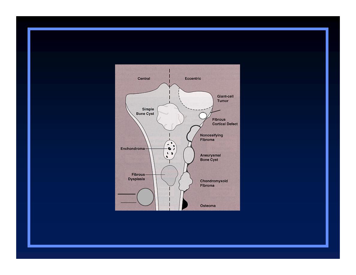
Parosteal sarcoma
Osteochondroma
Osteosarcoma
In The Transverse Plane
© Greenspan, Lippincott, 2000

In the Longitudinal Plane
O
Epiphyseal
Q
GCT, chondroblastoma
O
Metaphyseal
Q
Osteomyelitis, osteo- and chondrosarcoma
O
Diaphyseal
Q
Round cell lesions, ABC, enchondroma

Tumor Types
Characteristic Locations
O
Simple bone cyst
Q
Proximal humerus
O
Chondroblastoma
Q
Epiphyses
O
Giant Cell tumor
Q
Epiphyses
O
Simple bone cyst
Q
Proximal humerus
O
Chondroblastoma
Q
Epiphyses
O
Giant Cell tumor
Q
Epiphyses
Characteristic locations
© R
3
, 2000
Chondroblastoma
O
Simple bone cyst
Q
Proximal humerus
O
Chondroblastoma
Q
Epiphyses
O
Giant Cell tumor
Q
Epiphyses
Characteristic locations
© R
3
, 2000
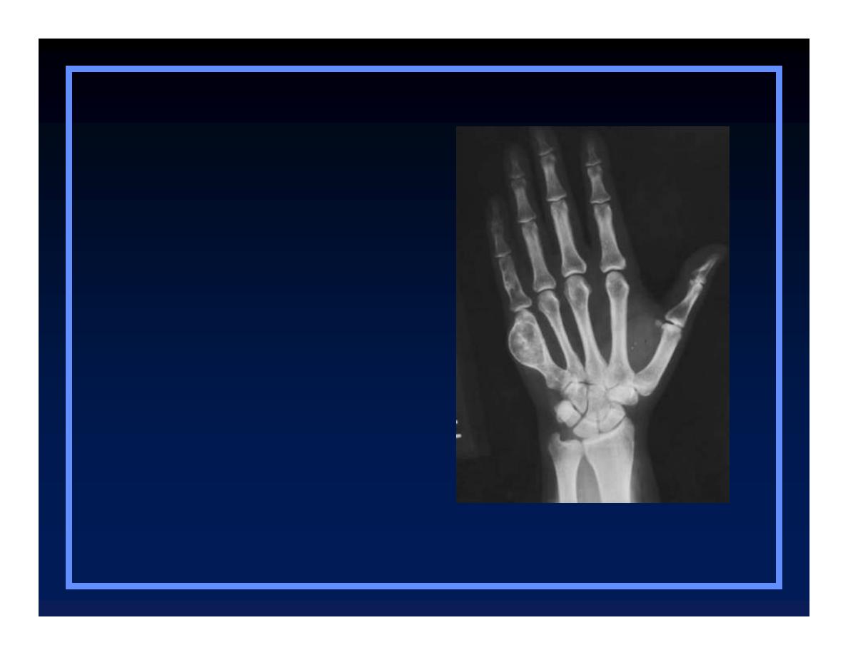
Giant Cell Tumor
O
Simple bone cyst
Q
Proximal humerus
O
Chondroblastoma
Q
Epiphyses
O
Giant Cell tumor
Q
Epiphyses
Characteristic locations
© R
3
, 2000

Tumor Types
Characteristic Locations
O
Adamantinoma
Q
Tibia
O
Chordoma
Q
Sacrum, clivus
O
Osteoblastoma
Q
Spine, posterior
O
Adamantinoma
Q
Tibia
O
Chordoma
Q
Sacrum, clivus
O
Osteoblastoma
Q
Spine, posterior
Characteristic locations
Adamantinoma
© R
3
, 2000
O
Adamantinoma
Q
Tibia
O
Chordoma
Q
Sacrum, clivus
O
Osteoblastoma
Q
Spine, posterior
Chordoma
Characteristic locations
© R
3
, 2000
Osteoblastoma
O
Adamantinoma
Q
Tibia
O
Chordoma
Q
Sacrum, clivus
O
Osteoblastoma
Q
Spine, posterior
Characteristic locations
Characteristic locations
© R
3
, 2000

Tumor Types
Characteristic Locations
O
Parosteal sarcoma
Q
Distal femur
O
Periosteal sarcoma
Q
Tibia
Characteristic locations
O
Parosteal sarcoma
Q
Distal femur
O
Periosteal sarcoma
Q
Tibia
Parosteal sarcoma
Characteristic locations
O
Parosteal sarcoma
Q
Distal femur
O
Periosteal sarcoma
Q
Tibia
© R
3
, 2000

Characteristic Tumors
By Body Site

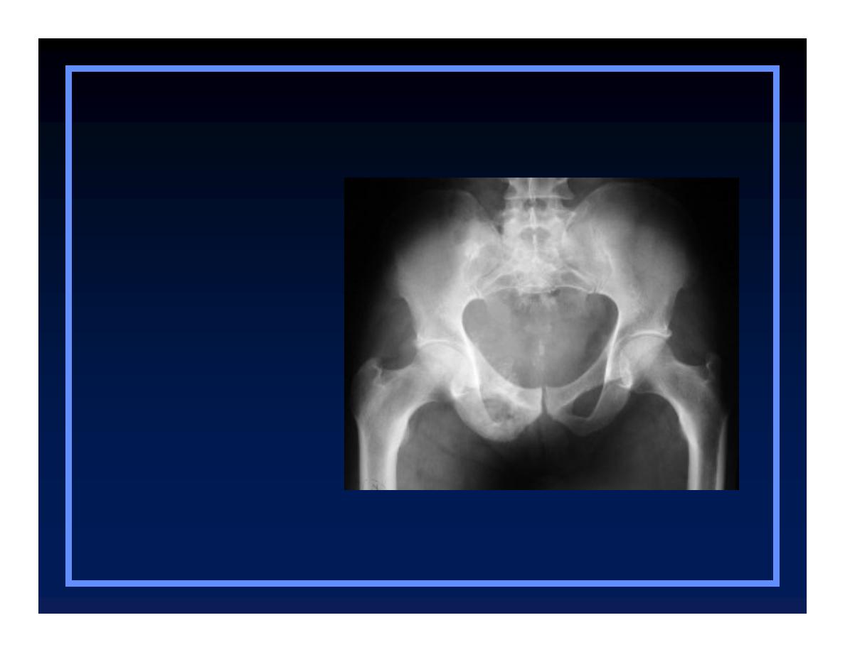
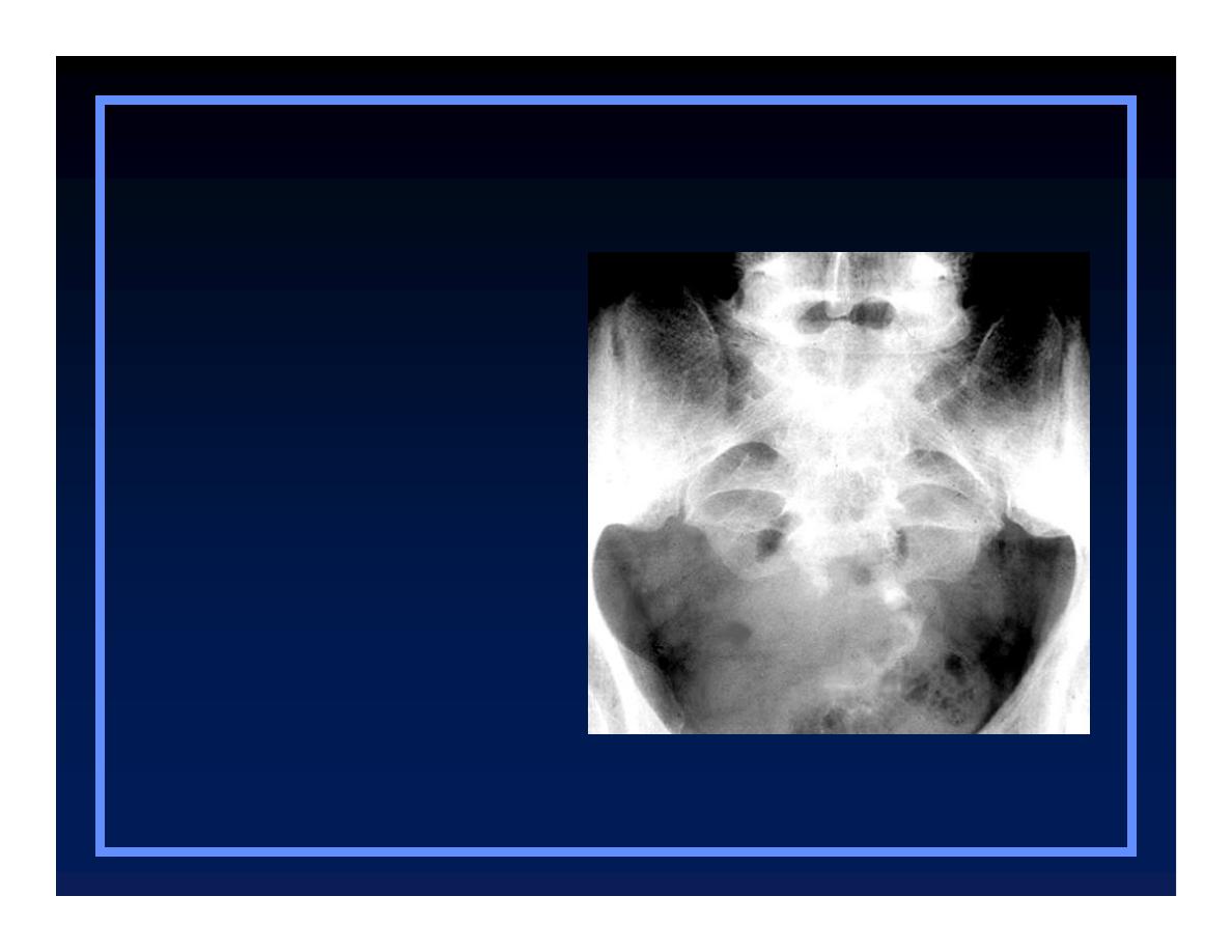
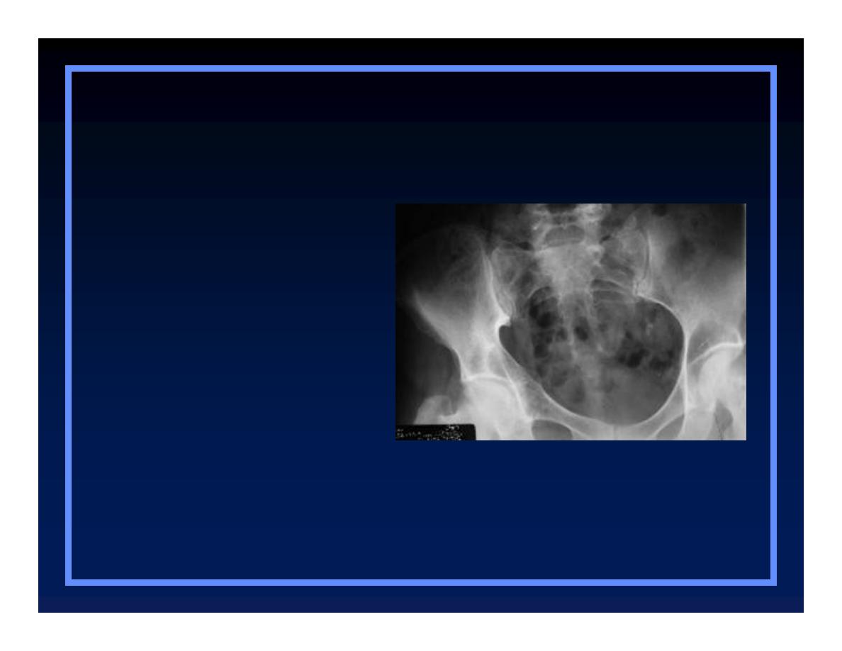
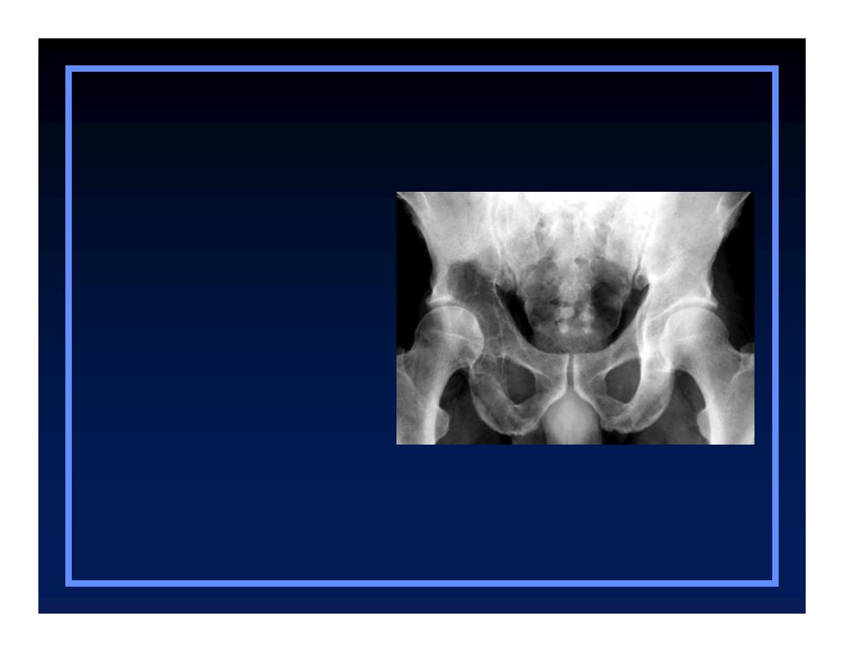
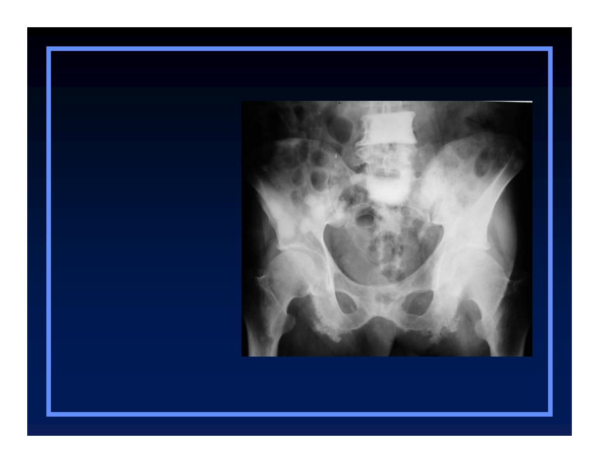
Pelvic Lesions
O
Chondrosarcoma
O
Solitary plasmacytoma
O
Chordoma
O
Chondrosarcoma
O
Solitary
plasmacytoma
O
Chordoma
Pelvic lesions
Plasmacytoma
O
Chondrosarcoma
O
Solitary
plasmacytoma
O
Chordoma
Pelvic lesions

Chordoma
O
Chondrosarcoma
O
Solitary
plasmacytoma
O
Chordoma
Pelvic lesions
© R
3
, 2000

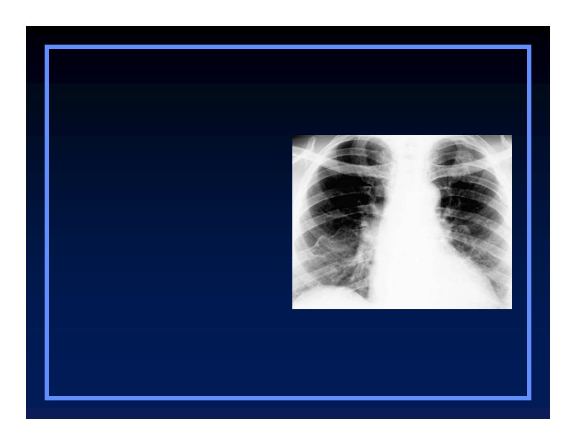
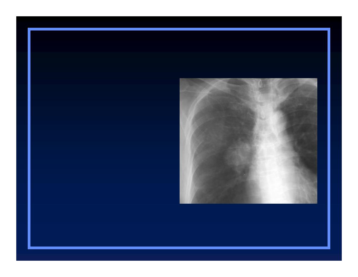
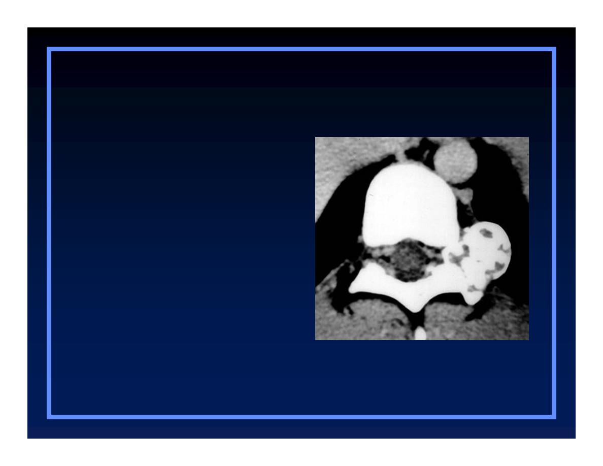
Expansile Rib Lesions
O
Plasmacytoma
O
Metastases
O
Chondrosarcoma
O
Eosinophilic granuloma
O
Neurofibromatosis
O
Fibrous dysplasia
O
Plasmacytoma
O
Metastases
O
Chondrosarcoma
O
Eosinophilic
granuloma
O
Neurofibromatosis
O
Fibrous dysplasia
Expansile rib lesions
O
Plasmacytoma
O
Metastases
O
Chondrosarcoma
O
Eosinophilic
granuloma
O
Neurofibromatosis
O
Fibrous dysplasia
Expansile rib lesions
Thyroid Carcinoma
O
Plasmacytoma
O
Metastases
O
Chondrosarcoma
O
Eosinophilic
granuloma
O
Neurofibromatosis
O
Fibrous dysplasia
Expansile rib lesions
© R
3
, 2000
O
Plasmacytoma
O
Metastases
O
Chondrosarcoma
O
Eosinophilic
granuloma
O
Neurofibromatosis
O
Fibrous dysplasia
Expansile rib lesions
© R
3
, 2000
O
Plasmacytoma
O
Metastases
O
Chondrosarcoma
O
Eosinophilic
granuloma
O
Neurofibromatosis
O
Fibrous dysplasia
Expansile rib lesions
© R
3
, 2000
O
Plasmacytoma
O
Metastases
O
Chondrosarcoma
O
Eosinophilic
granuloma
O
Neurofibromatosis
O
Fibrous dysplasia
Expansile rib lesions
© R
3
, 2000

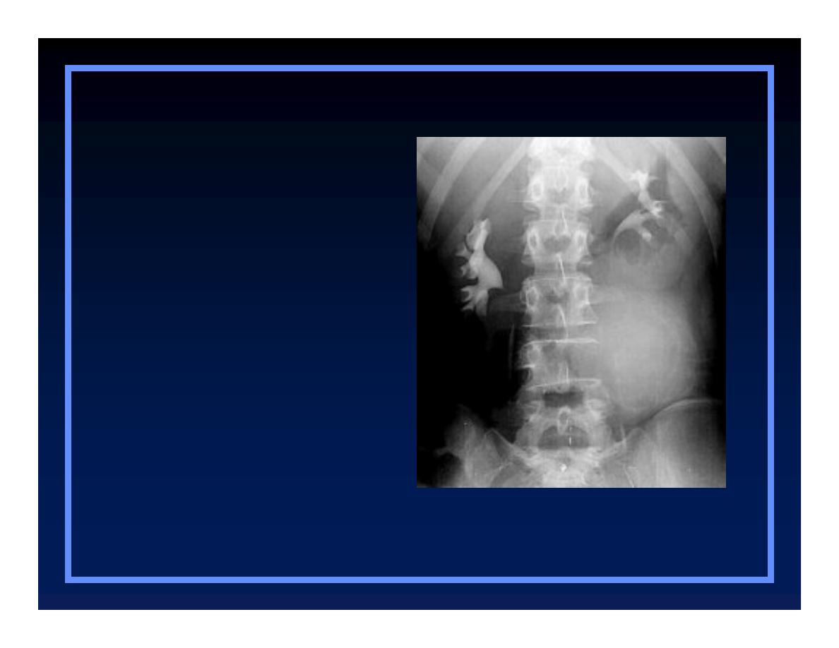
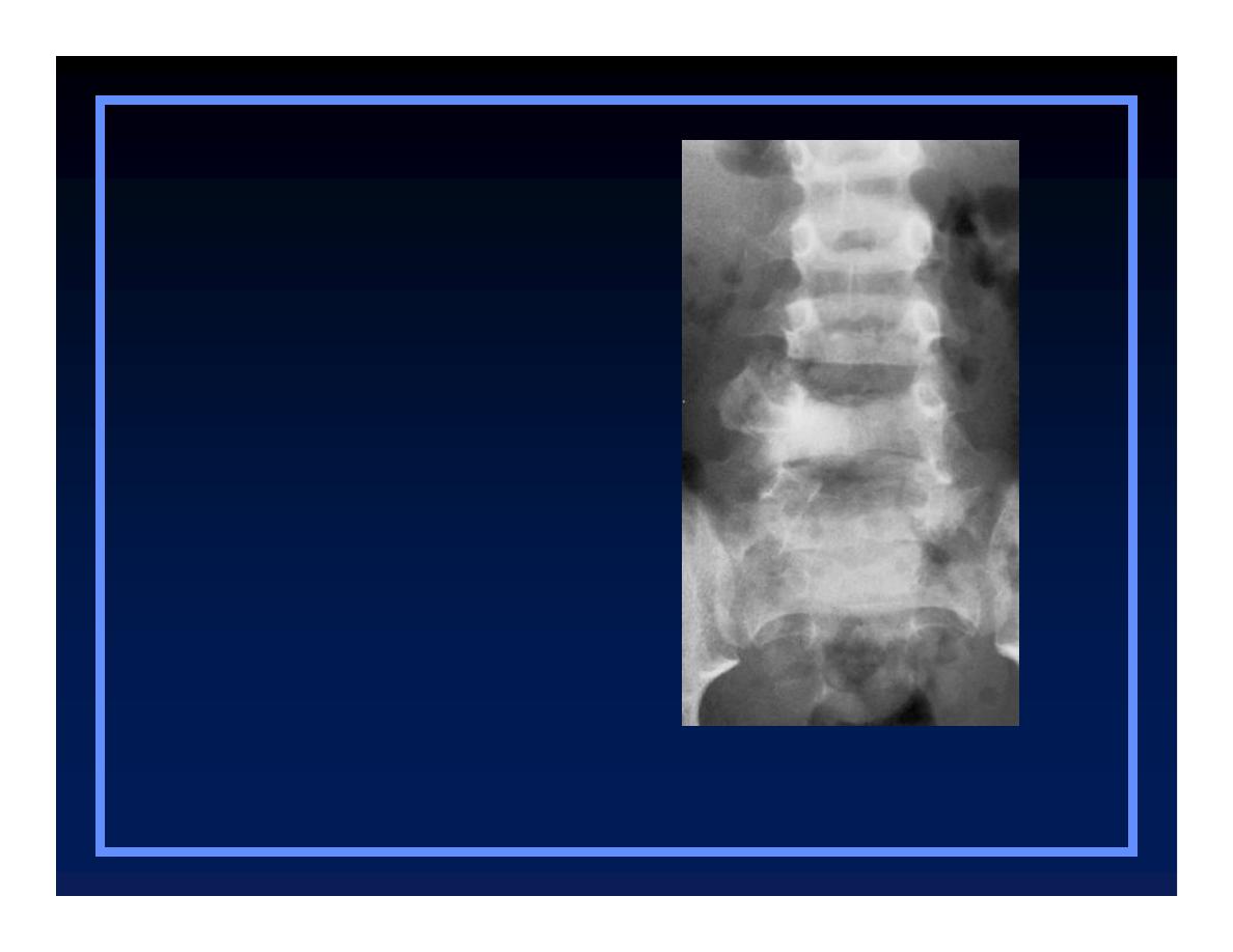
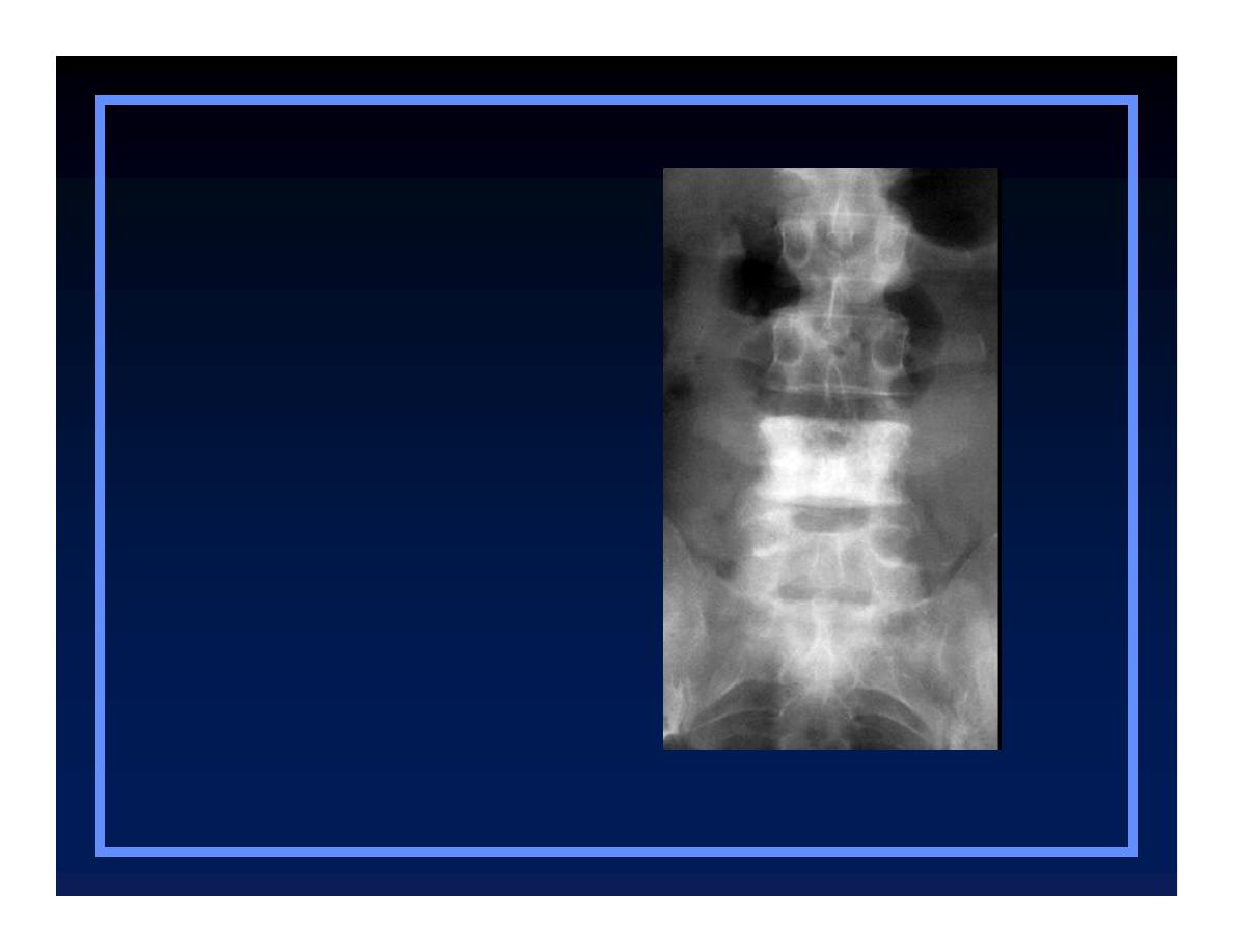
Lesions of the Spine
O
Osteoblastoma
Q
Expansile, with punctate densities
within
O
Chordoma
O
ABC
O
Metastatic disease
Osteoblastoma
O
Osteoblastoma
O
Chordoma
O
ABC
O
Metastatic
disease
Spine lesions
© R
3
, 2000
Chordoma
O
Osteoblastoma
O
Chordoma
O
ABC
O
Metastatic
disease
Spine lesions
© R
3
, 2000
Aneurysmal bone cyst
O
Osteoblastoma
O
Chordoma
O
ABC
O
Metastatic
disease
Spine lesions
Metastatic Breast Carcinoma
O
Osteoblastoma
O
Chordoma
O
ABC
O
Metastatic
disease
Spine lesions
© R
3
, 2000

Clues by Density
Of Lesion

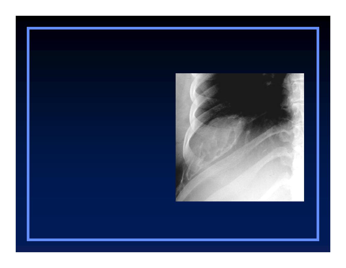
Sclerotic Cortical Lesions
O
Osteoid osteoma
O
Brodie’s abscess
O
Stress fracture
Osteoid Osteoma
O
Osteoid osteoma
O
Brodie’s abscess
O
Stress fracture
Sclerotic cortical lesions
Brodie’s abscess
O
Osteoid osteoma
O
Brodie’s abscess
O
Stress fracture
Sclerotic cortical lesions
© R
3
, 2000
Healing Stress Fracture
O
Osteoid osteoma
O
Brodie’s abscess
O
Stress fracture
Sclerotic cortical lesions
Sclerotic cortical lesions
Sclerotic Cortical Lesions
Osteoid Osteoma
Brodie’s abscess
Healing Stress
Fracture

Lytic Lesions in Children
O
Eosinophilic granuloma
O
Neuroblastoma
O
Leukemia
O
Eosinophilic
granuloma
O
Neuroblastoma
O
Leukemia
Lytic Lesions in Children
Eosinophilic granuloma
© R
3
, 2000
Neuroblastoma
O
Eosinophilic
granuloma
O
Neuroblastoma
O
Leukemia
Lytic Lesions in Children
© R
3
, 2000
Leukemia
O
Eosinophilic
granuloma
O
Neuroblastoma
O
Leukemia
Lytic Lesions in Children
© R
3
, 2000

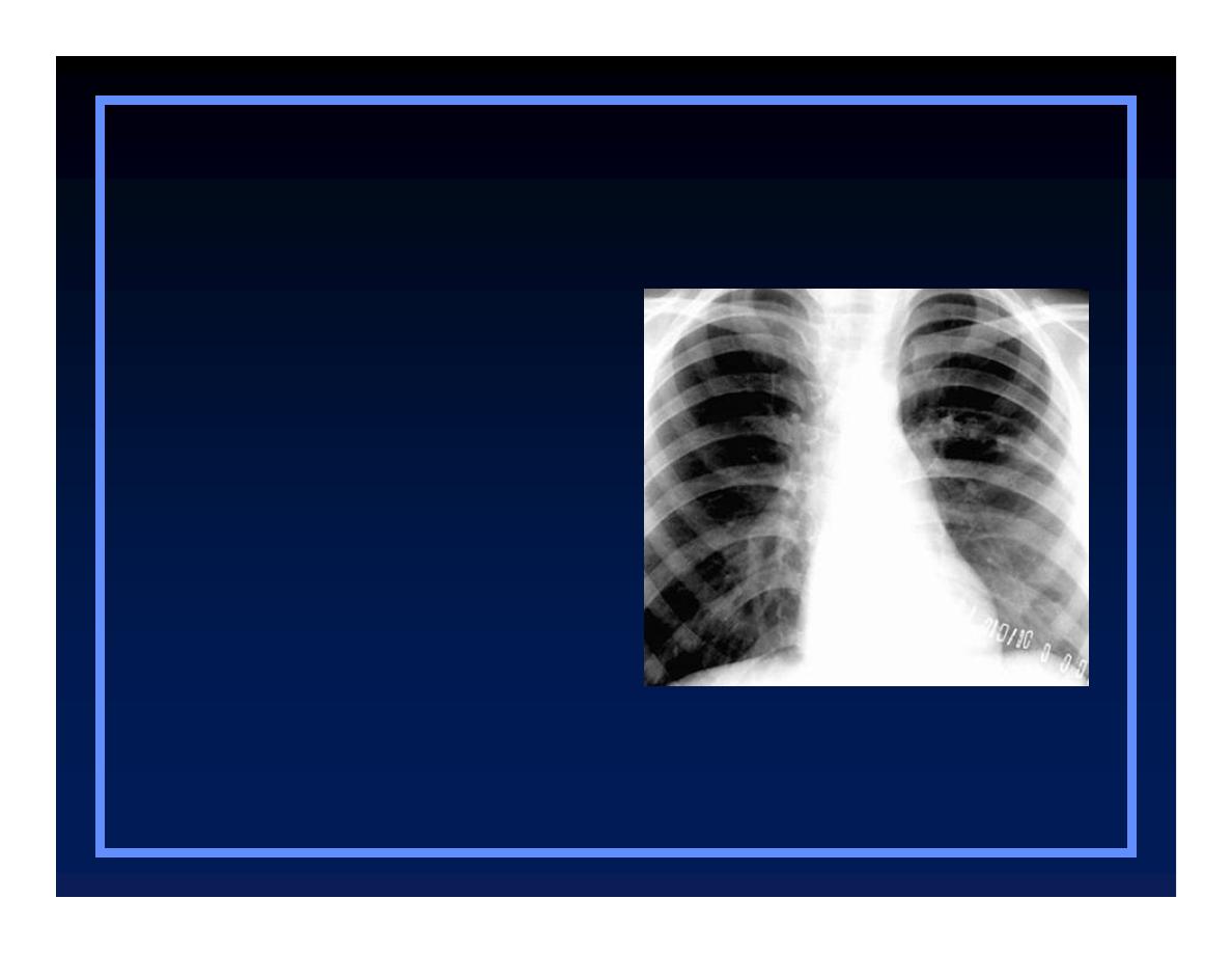
Lytic Lesions in Adults
O
Metastatic lesions
Q
Lung
Q
Renal
Q
Thyroid
O
Multiple myeloma
O
Primary bone tumor
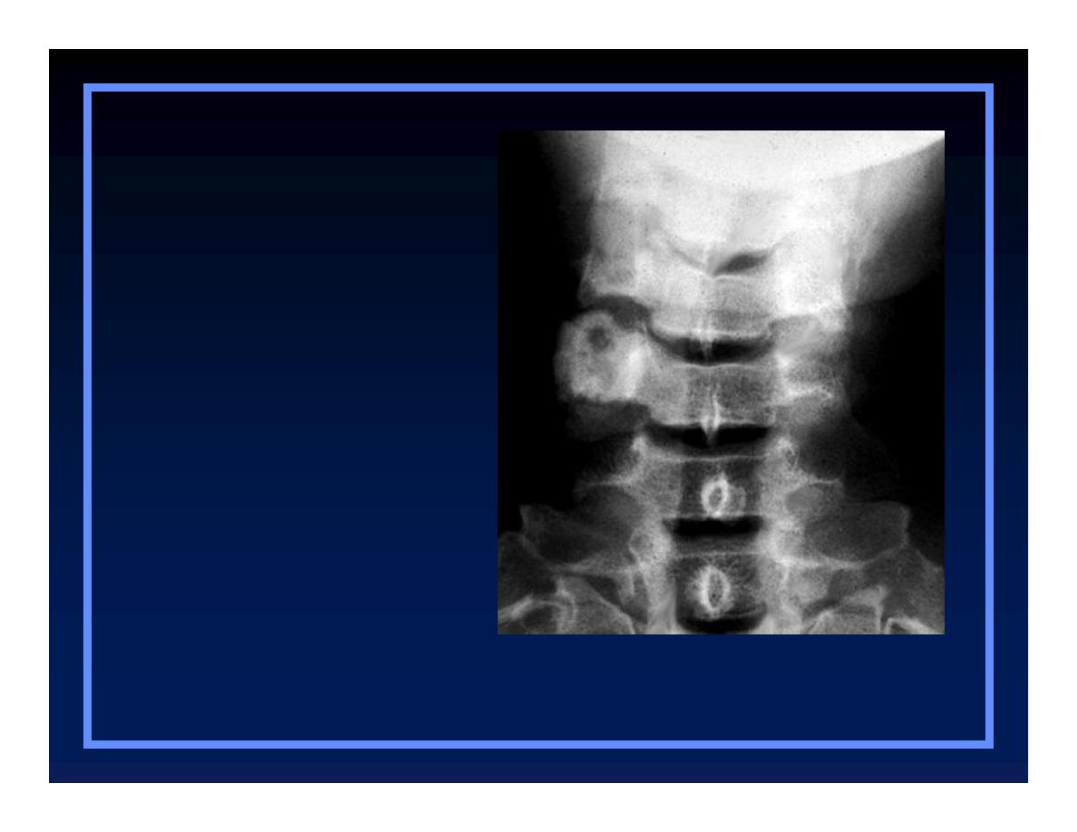
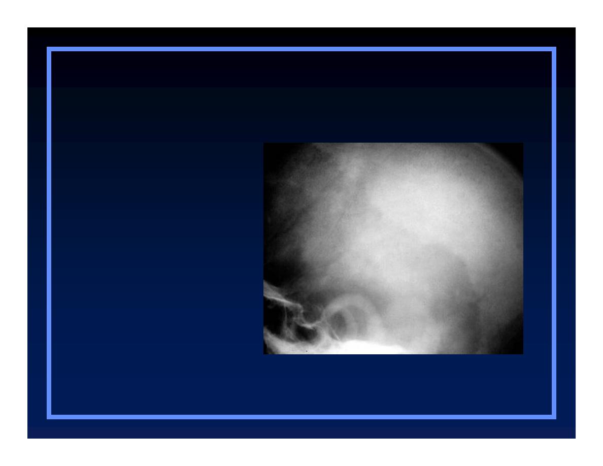
Met from Thyroid Carcinoma
O
Mets
O
Myeloma
O
Primary bone
tumor
Lytic Lesions in Adults
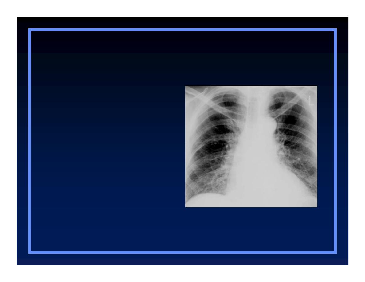
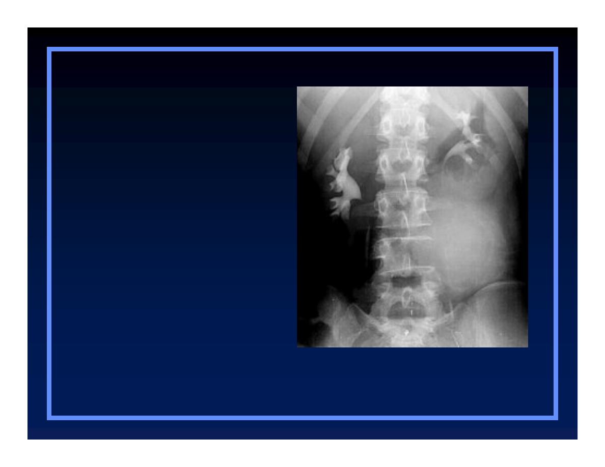
Multiple myeloma
O
Mets
O
Myeloma
O
Primary bone
tumor
Lytic Lesions in Adults
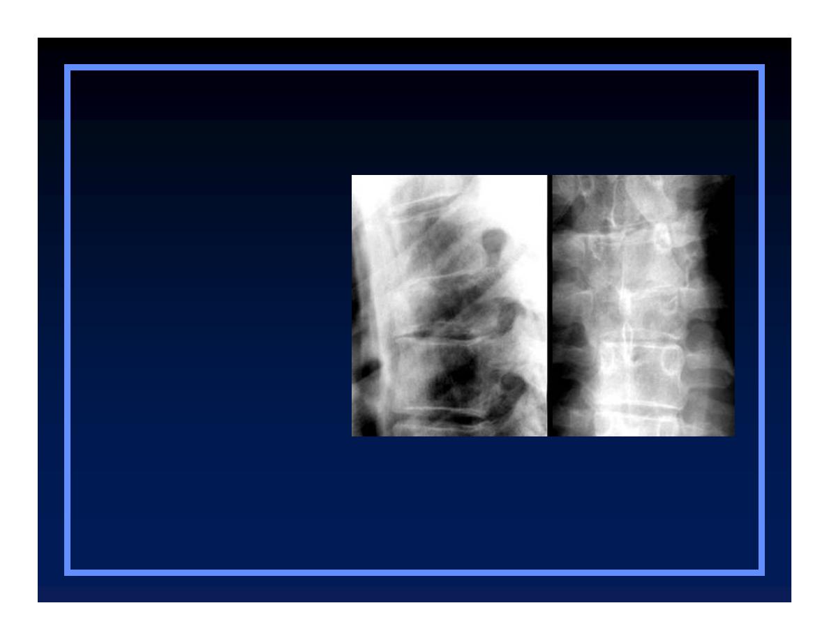
Chondrosarcoma
O
Mets
O
Myeloma
O
Primary bone
tumor
Lytic Lesions in Adults

Blastic Lesions in Children
O
Medulloblastoma
O
Lymphoma
Medulloblastoma
O
Medulloblastoma
O
Lymphoma
Blastic Lesions in Children
Blastic Lesions in Children
© R
3
, 2000
Lymphoma
O
Medulloblastoma
O
Lymphoma
Blastic Lesions in Children
Blastic Lesions in Children

Blastic Lesions in Adults
O
Metastatic disease
Q
Breast – female
Q
Prostate – male
O
Lymphoma
O
Paget’s disease
O
Etcetera-mastocytosis, fluorosis
Prostate Mets
O
Mets
O
Lymphoma
O
Paget’s
Blastic Lesions in Adults
Breast Mets
O
Mets
O
Lymphoma
O
Paget’s
Blastic Lesions in Adults
Lymphoma
O
Mets
O
Lymphoma
O
Paget’s
Blastic Lesions in Adults
Blastic Lesions in Adults
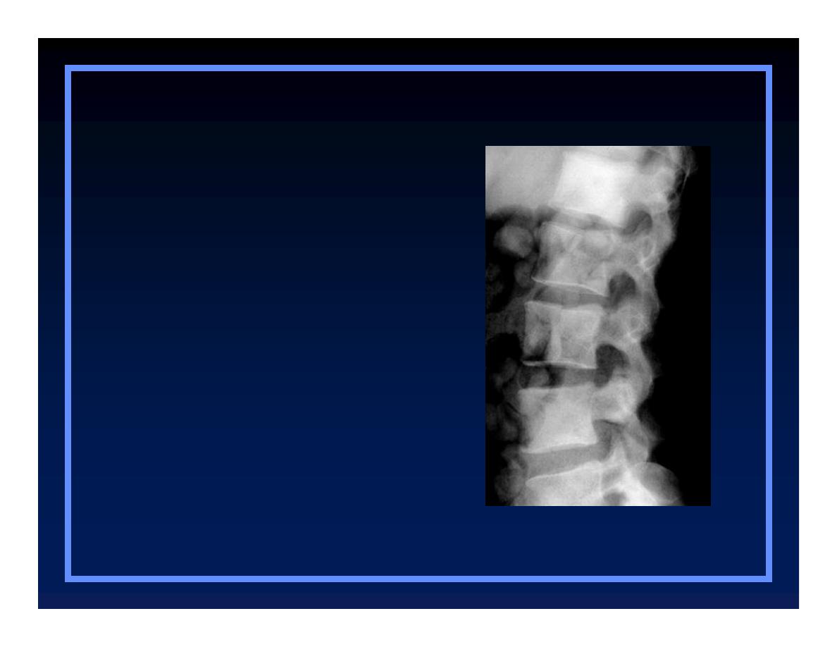
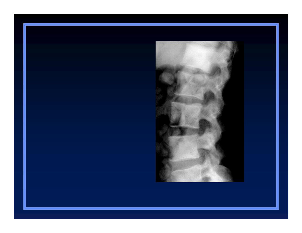
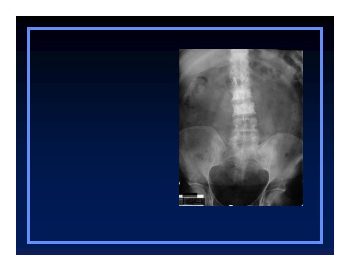
Paget’s of Spine
O
Mets
O
Lymphoma
O
Paget’s
Blastic Lesions in Adults
Blastic Lesions in Adults

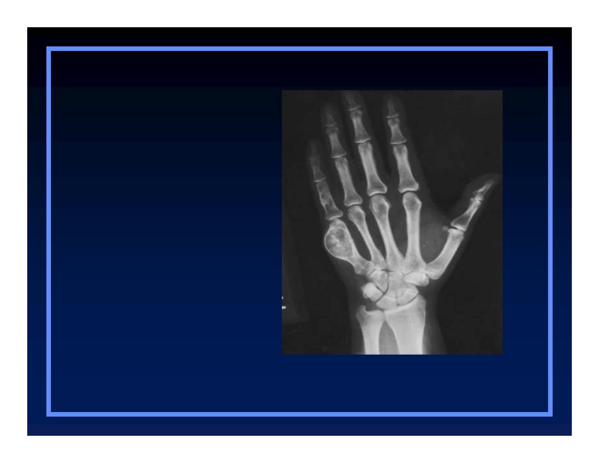
Other Clues

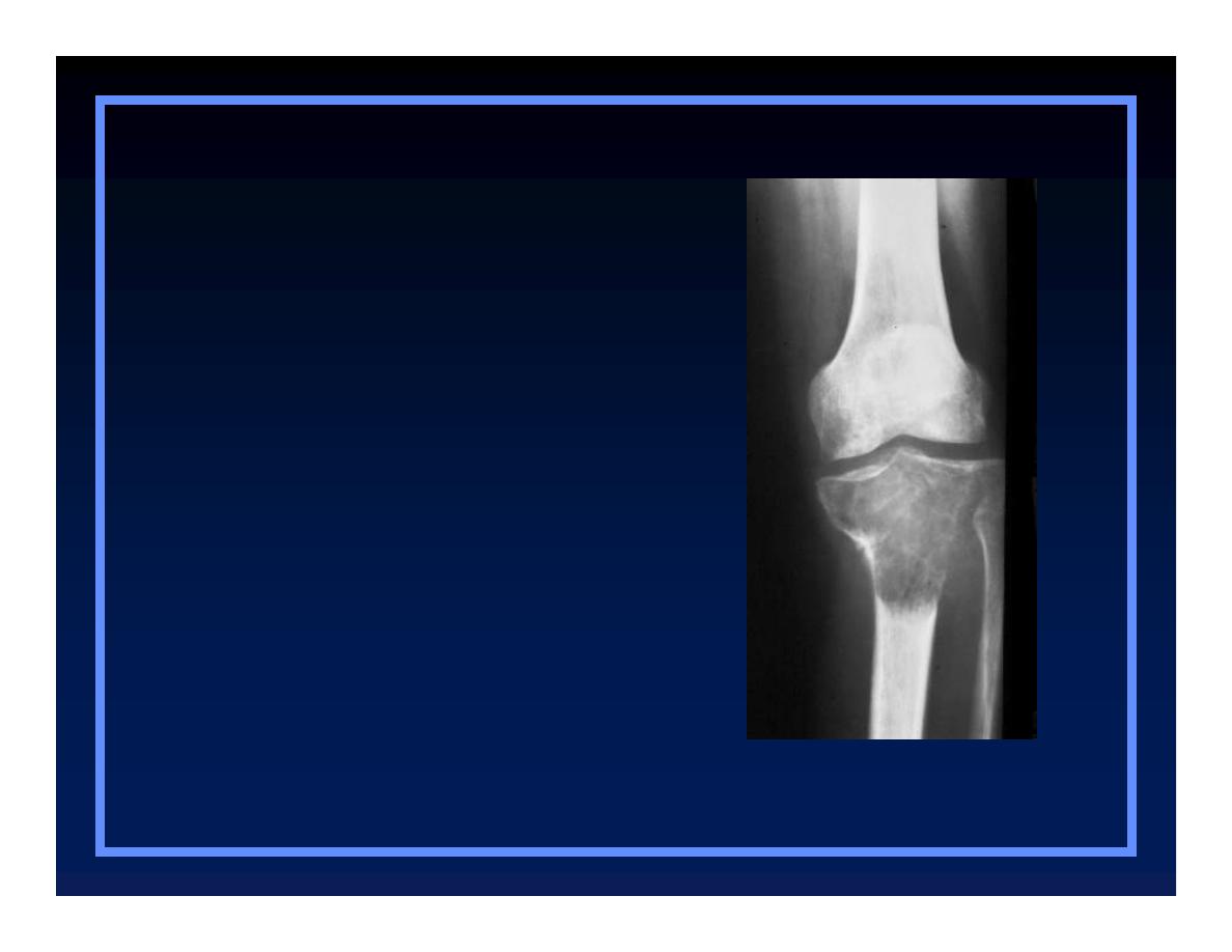
Benign Lesions
Without Sclerotic Boarders
O
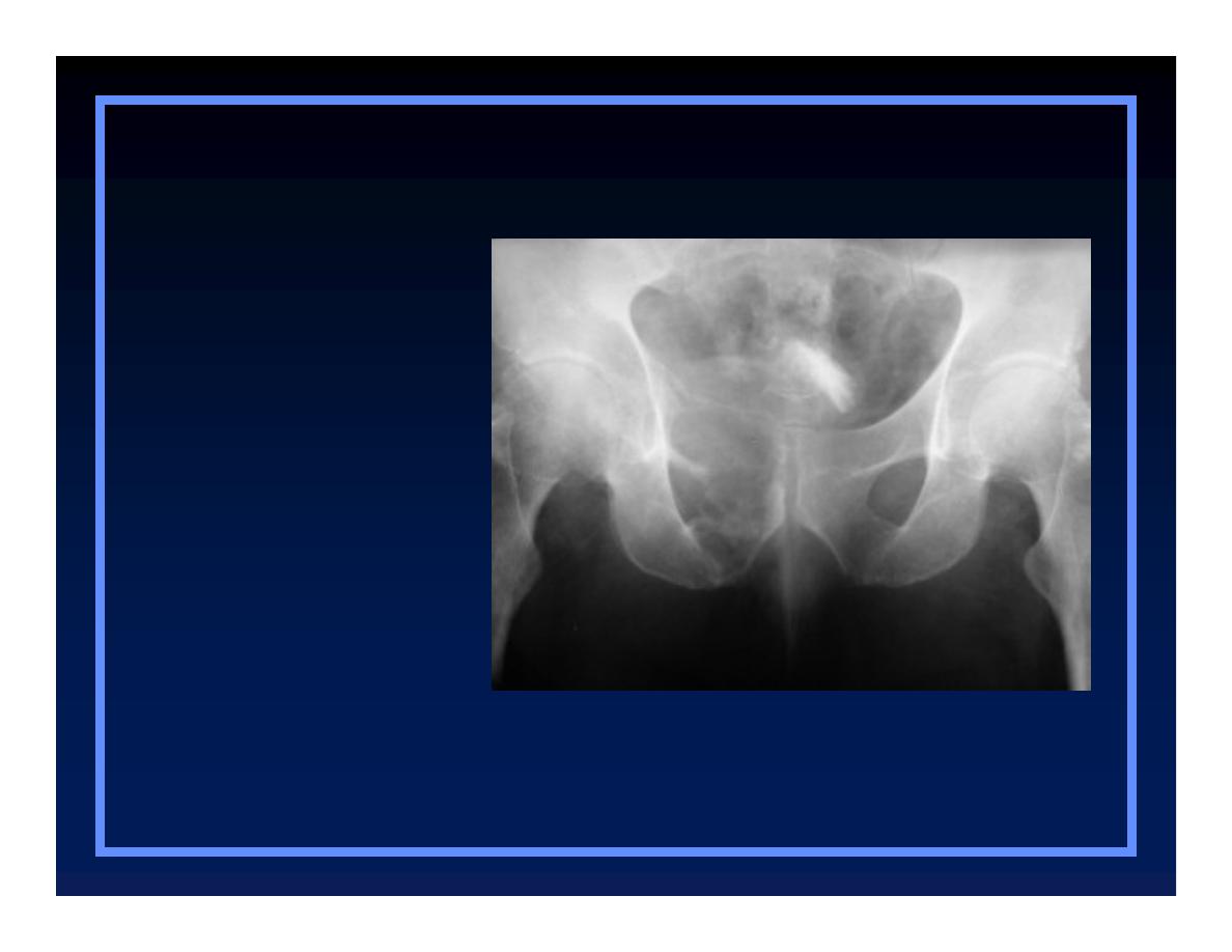
Giant Cell tumor
O
Brown tumor
O
Osteolytic phase of Paget’s Disease
Benign Lesions without Sclerotic
Benign Lesions without Sclerotic
Borders
Borders
O
Giant cell tumor
O
Brown tumor
O
Osteolytic Paget’s
Giant Cell Tumor
© R
3
, 2000

Brown Tumor
Benign Lesions without Sclerotic
Benign Lesions without Sclerotic
Borders
Borders
O
Giant cell tumor
O
Brown tumor
O
Osteolytic Paget’s
© R
3
, 2000
Osteolytic Paget’s
Benign Lesions without Sclerotic
Benign Lesions without Sclerotic
Borders
Borders
O
Giant cell tumor
O
Brown tumor
O
Osteolytic Paget’s
© R
3
, 2000

Soft Tissue Extension
O
Usually implies malignancy
Q
More likely to form discrete soft tissue mass
O
Benign conditions with soft tissue
extension
Q
Osteomyelitis
V
Usually infiltration of fat
Osteosarcoma
© R
3
, 2000

Multiple Lesions
O
More often benign
O
Malignancies with multiple lesions
Q
Metastatic disease
Q
Multiple myeloma
Q
Lymphoma
Q
Ewing’s sarcoma (rarely)
Q
Osteosarcoma (rarely)
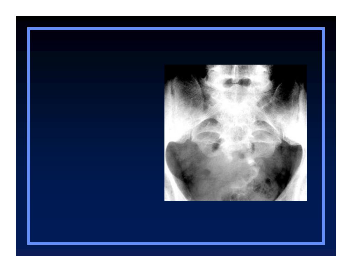
Mets from Ca of Prostate
Multiple lesions
Multiple lesions
O
Metastatic
O
Multiple myeloma
O
Lymphoma
© R
3
, 2000
Multiple Myeloma
Multiple lesions
Multiple lesions
O
Metastatic
O
Multiple myeloma
O
Lymphoma
© R
3
, 2000
Lymphoma
Multiple lesions
Multiple lesions
O
Metastatic
O
Multiple myeloma
O
Lymphoma
© R
3
, 2000
Osteosarcomatosis
Multiple lesions
Multiple lesions
O
Metastatic
O
Multiple myeloma
O
Lymphoma
O
Osteosarcomatosis
© R
3
, 2000
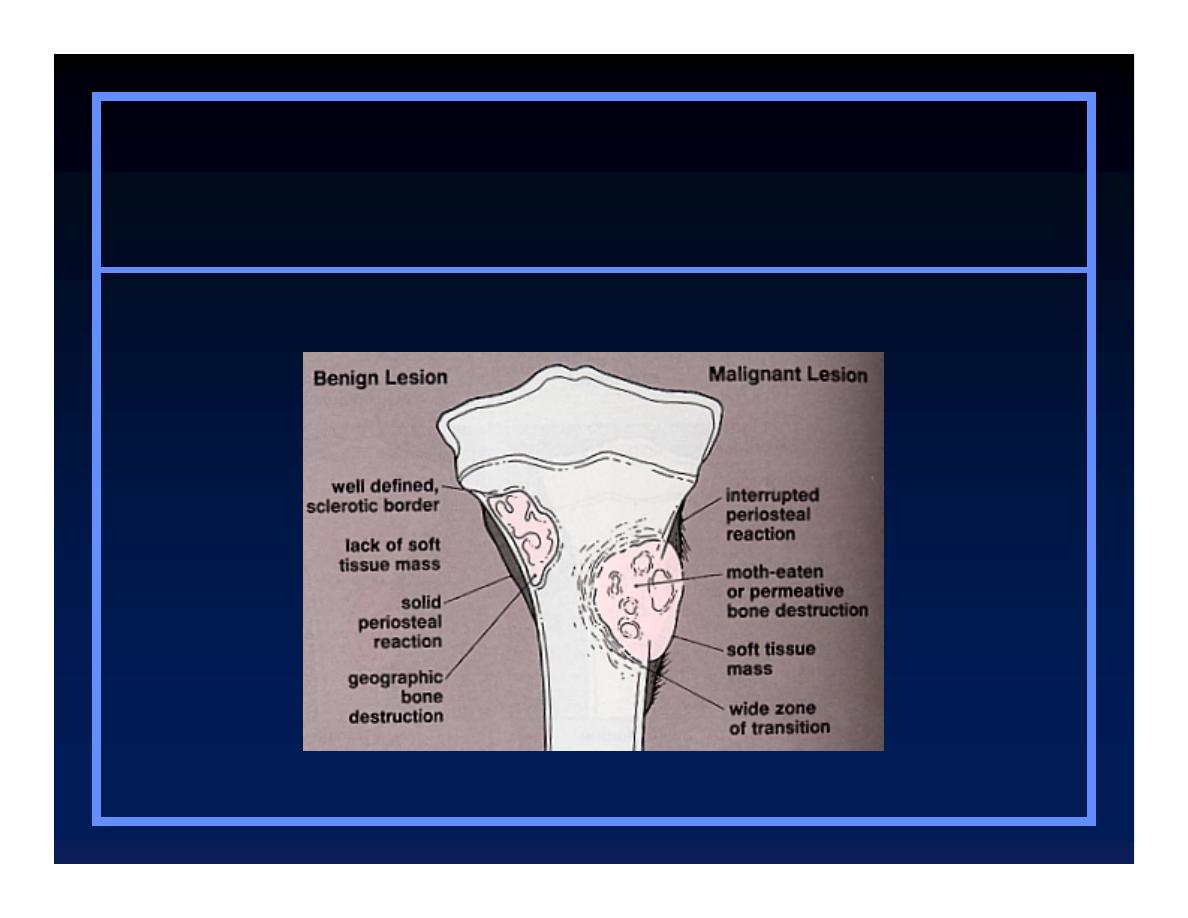
Benign vs. Malignant
© Greenspan, Lippincott, 2000

The End