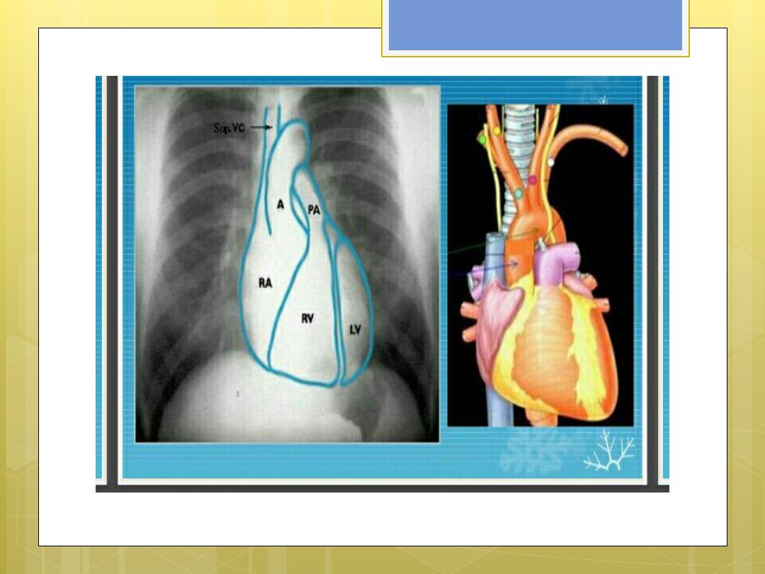
Cardiovascular radiology
Dr.Muma AG.Z


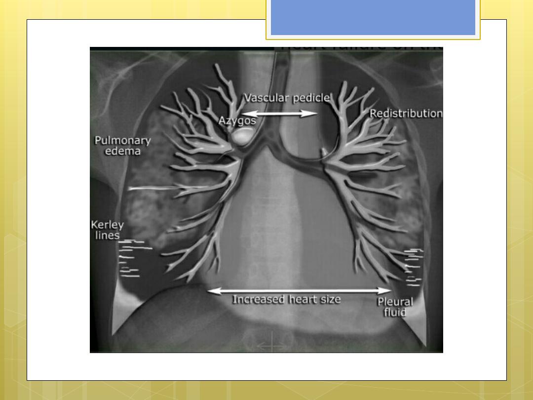
Heart failure
Congestive HF is the result of
insufficient output ..associated with
high resistance to circulation or
fluid over load .
Left ventricular failure is the most
common leading to decrease
cardiac output and increase
pulmonary venous pressure

This will lead to dilatation of
pulmonary vessels , leak of
fluid into the interstetium ,
pleural space and into the
alveoli resulting in
pulmonary odema
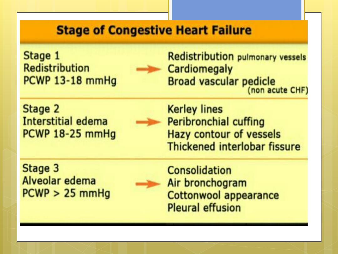

Stages of CHF
Stage I redistribution
…
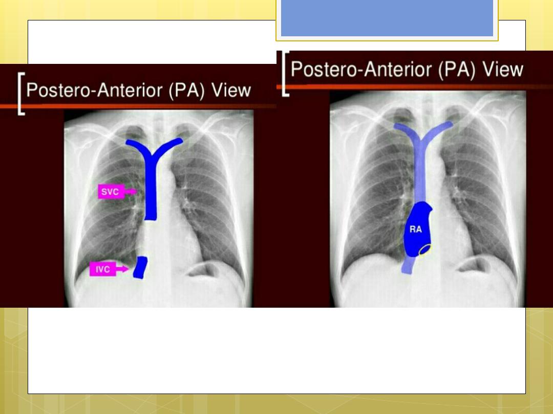
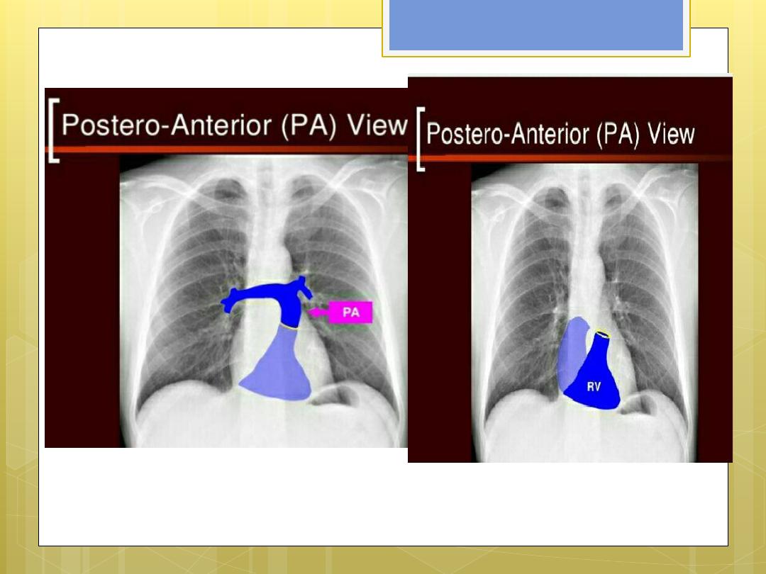
in normal erect plain chest film the
pulmonary vessels supplying the
upper lung fields are smaller and
fewer in no. than those supplying the
lung bases.
With CHF there will be redistribution of
pulmonary blood flow equal in upper
and lower lobes and subsequently
flow from the lower to upper lobe
vessels
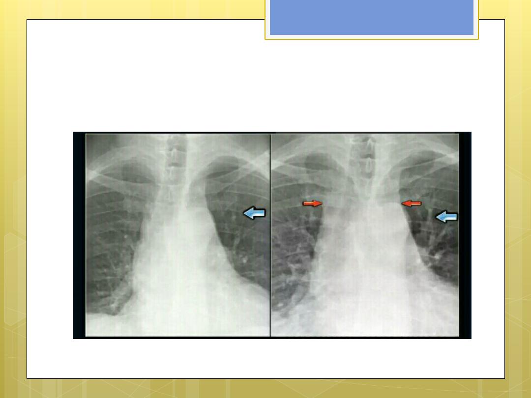
Redistribution ( upper lobe
blood diversion

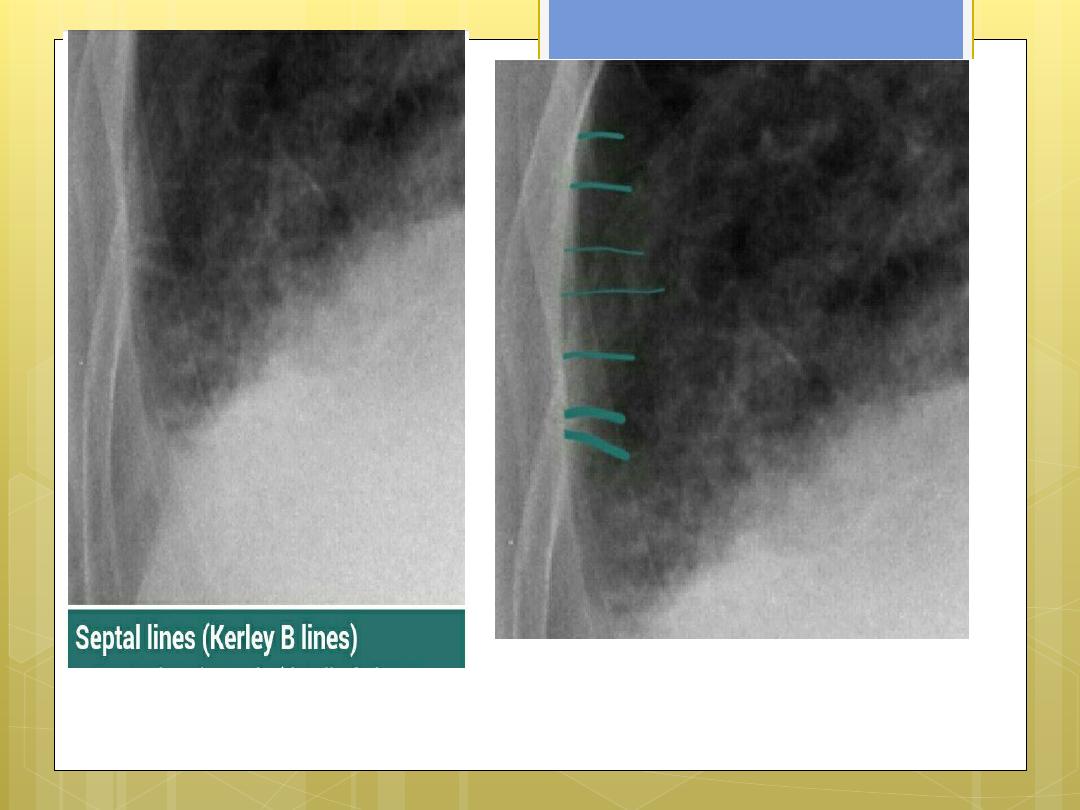
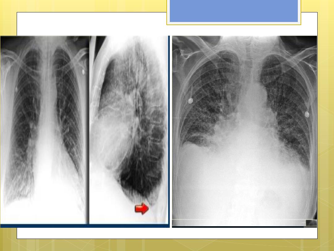
Stage II interstitial odema
fluid leak from the loaded
vessels in to the adjacent
interstetium …forming septal
lines ..kerley lines ….horizontal
lines seen near the
costophrenic angles

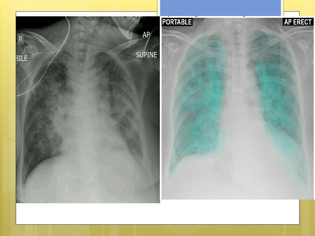
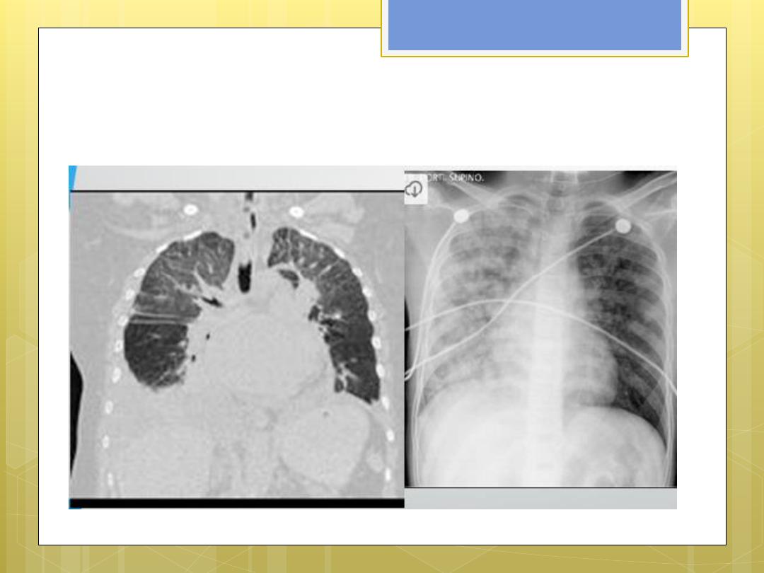
Alveolar odema
Fluid leak from the interstetium in to the
adjacent alveoli.
It is affected by
gravity
…
and
obstructive lung disease
( fluid leak in
the less obstructed lung region )
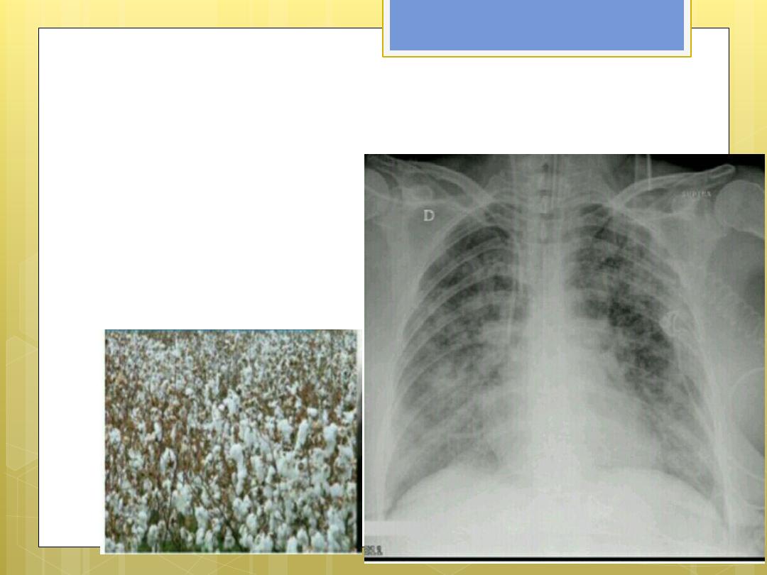
Cotton wool appearance of
alveolar odema

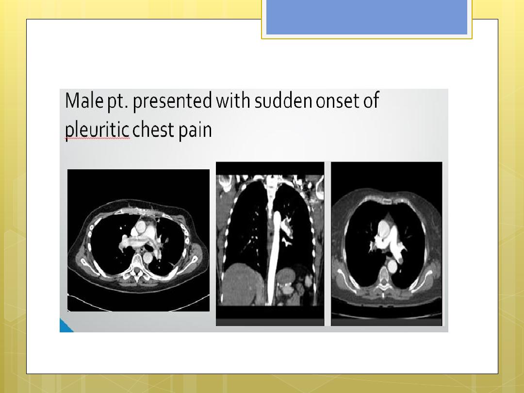
Pulmonary embolism
Hx. Post op., trauma , fracture
,DVT , birth control pills ,
previous PE , .
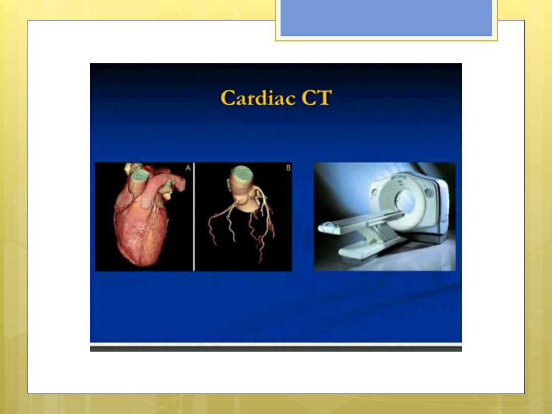
Imaging evaluation ,
need CT
scan with high dose i.v
contrast
…normal CXR do not
exclude occurrence of early
PE.
CT scan showing
filling defect

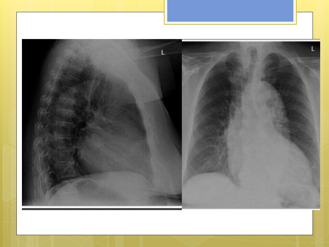
Aortic aneurysm
Causes ..
1.
Atherosclerosis
( most common)
2.
Vasculitis
3.
Trauma
4.
Chronic aortic dissection
5.
Connective tissue disorder ex. Marfan
syndrome

Role of imaging
1.
Detection
2.
Monitor growth rate
3.
Pre op. surgical
planning
4.
Post . Op. follow up

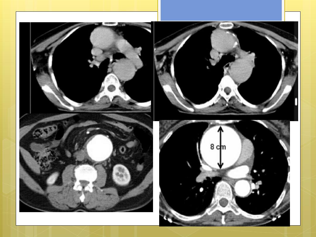
Imaging evaluation
CXR …
showing mediastinal mass,
enlarged affected segment , curvilinear
calcification .
Thoracic U/S
..
no role
Trans esophageal echo
…
but due to
invasive nature …it is not used routinely.

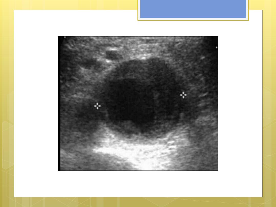
Abdominal U/S
…simple ,,, safe ,,,
available,,,highly sensitive and specific
detection rate .
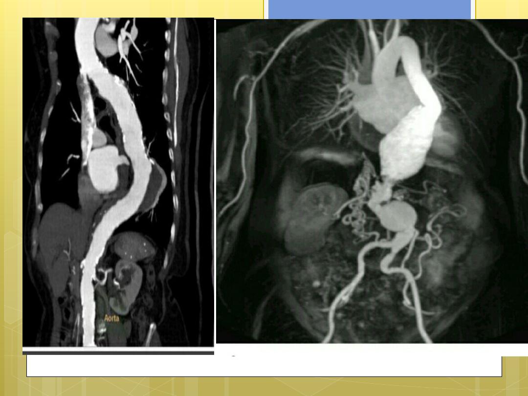
computed tomography
…
May detect rupture …
calcification …
Size of aneurysm …intra luminal thrombi ,
relation to adjacent structure ex.. Bone
erosion