Neuro imaging
Dr. Muna AG.Z

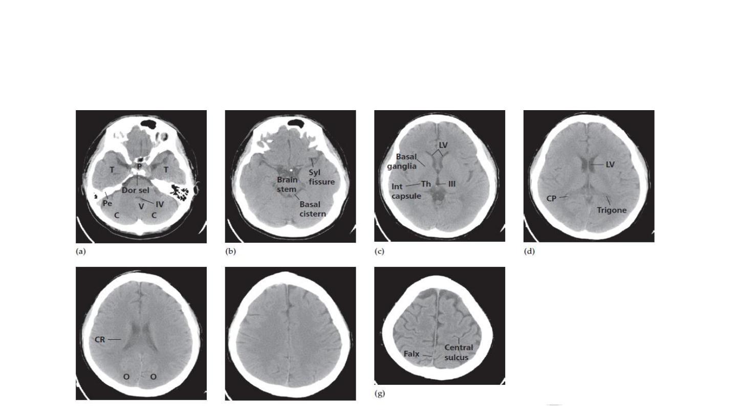
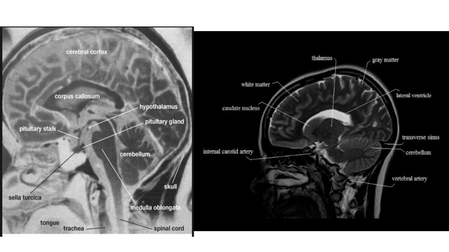
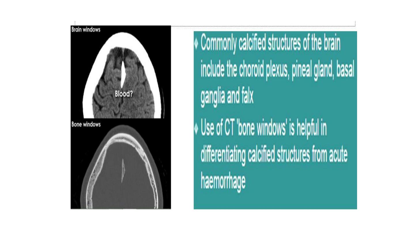
Normal CT anatomy
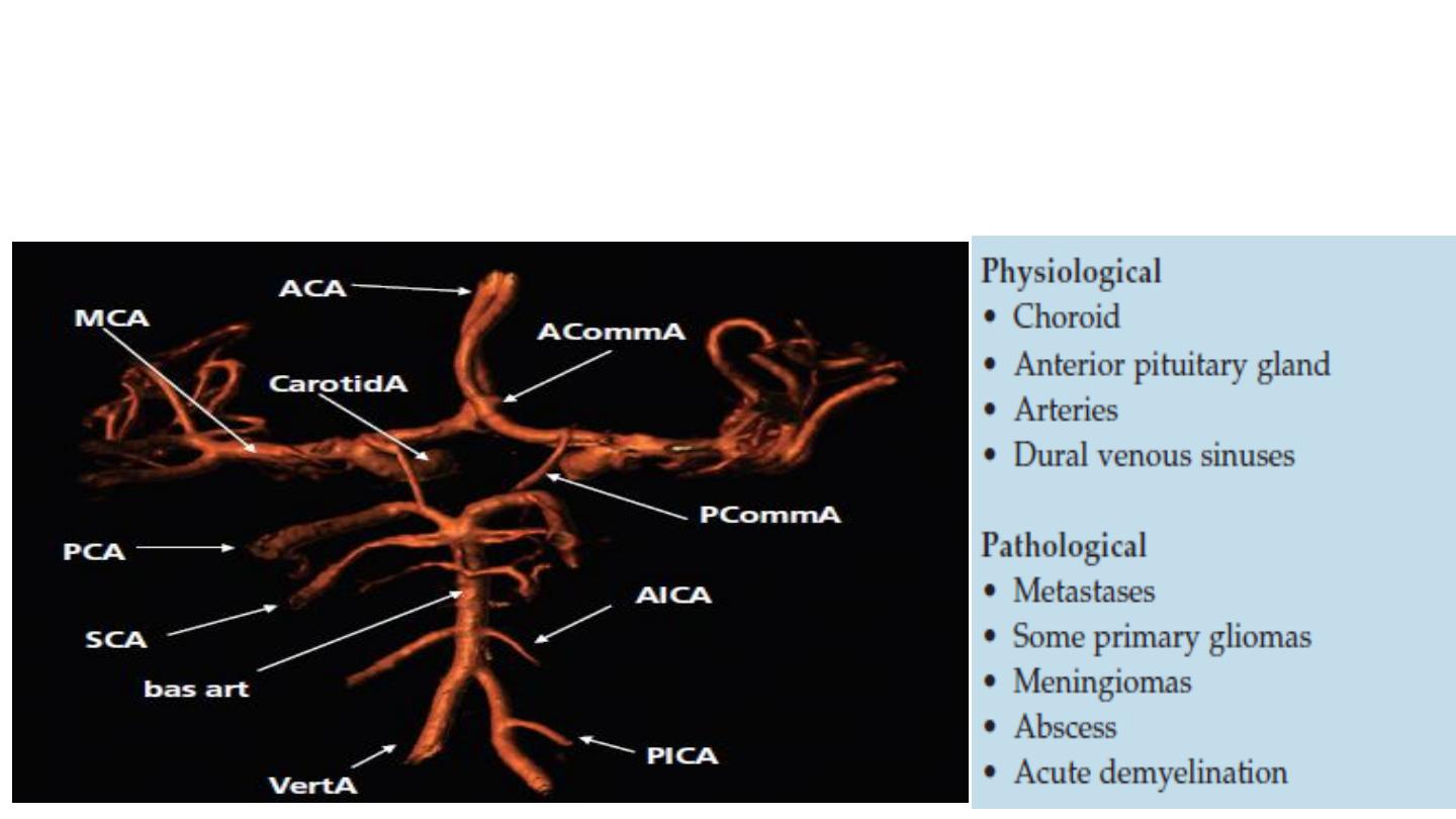
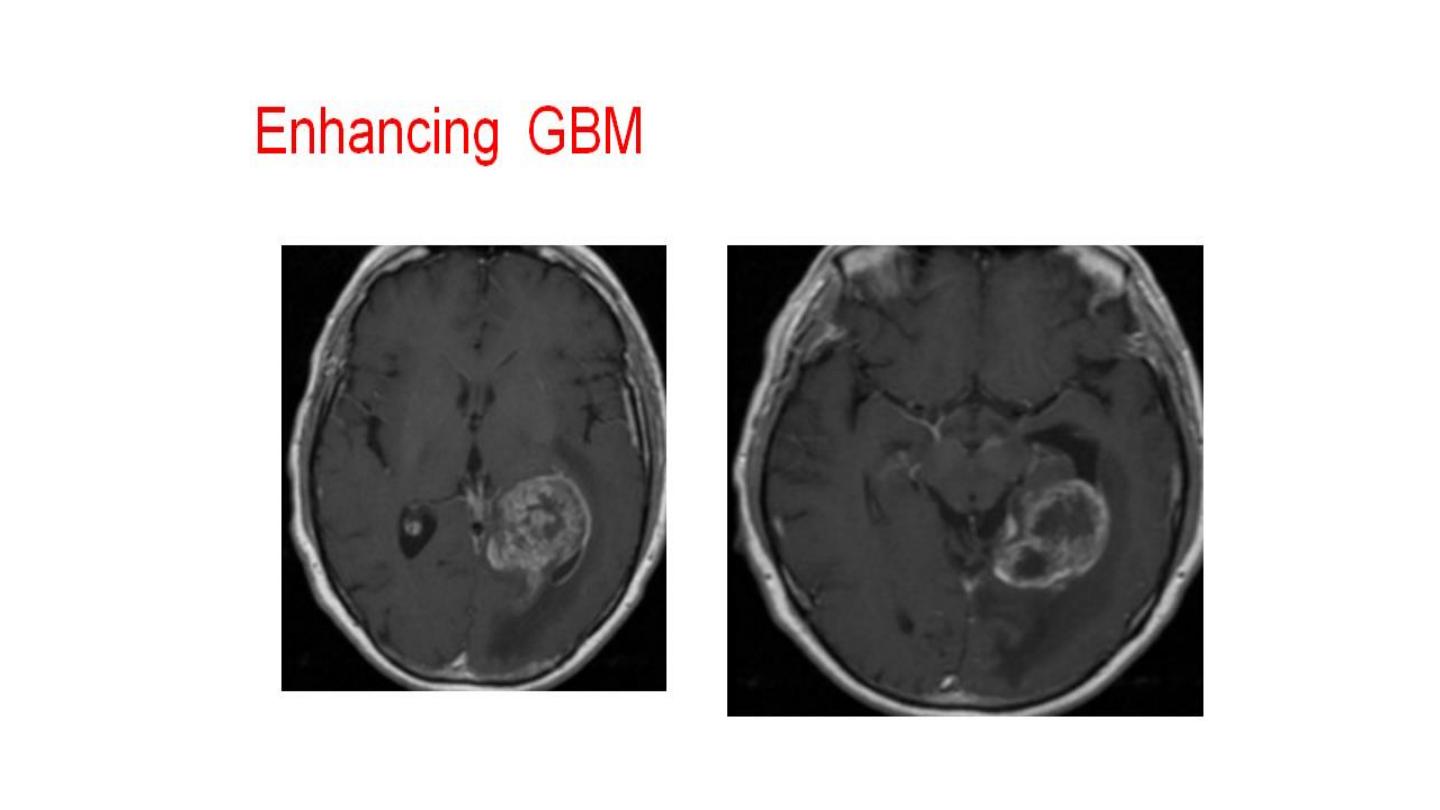
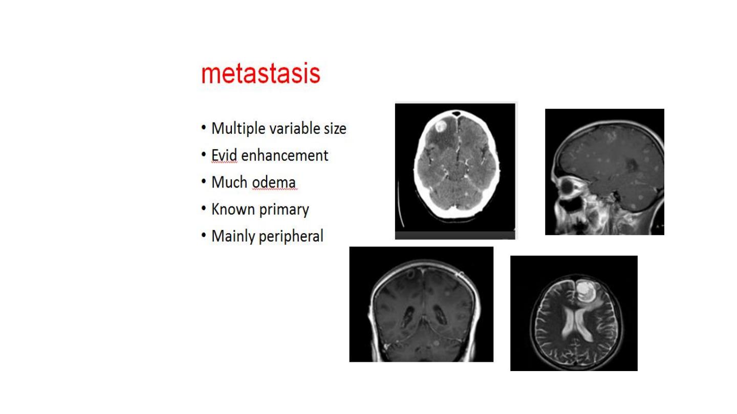
Enhancement with I.V contrast
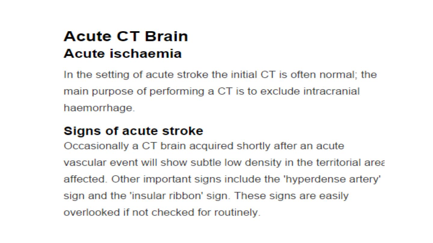

The goal of imaging in acute stroke
•Exclude hemorrhage
•Differentiate between reversible and
irreversible damaged brain tissue.
•CT scan is the initial imaging investigation
,
it is quick , 24 hrs. available , gold slandered
in showing hemorrhage when present .

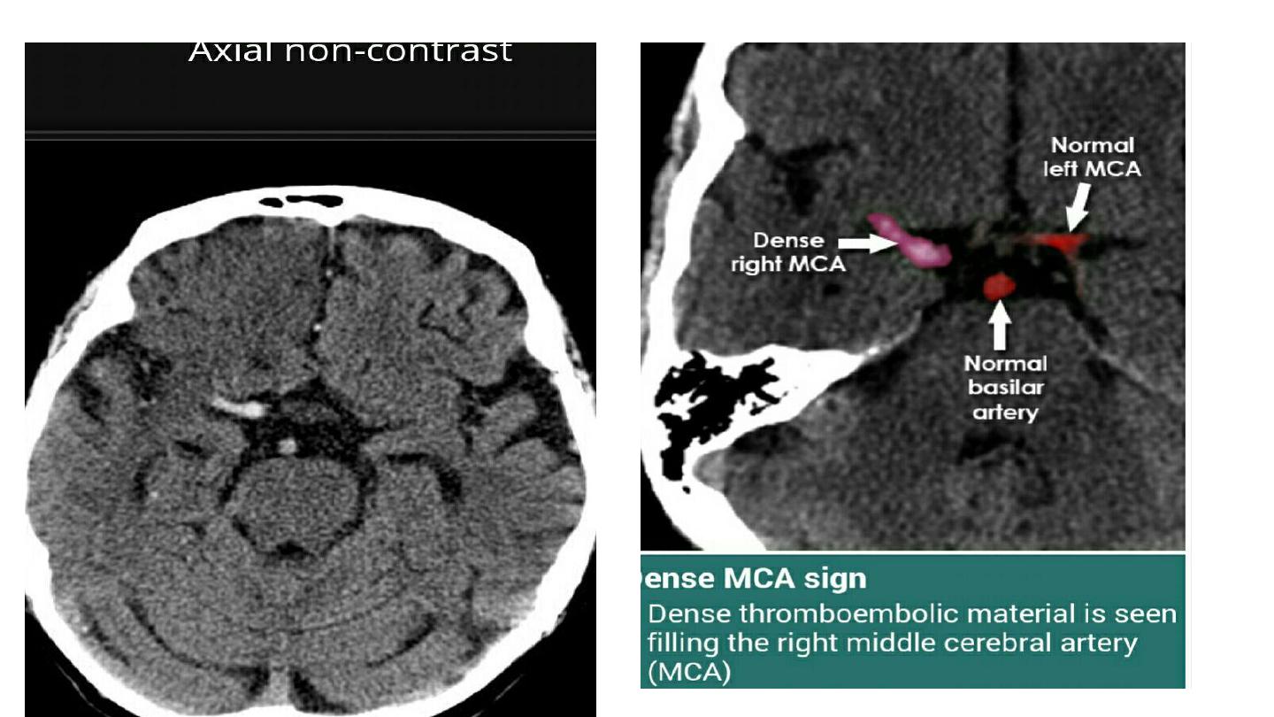
Early singe of MCA infarction (acute brain
ischemia )
•Focal increase of density of the MCA due to
the presence of thrombotic materials within
the lumen of the artery (
dense MCA singe
).
•Loss of definition of the grey- white matter
interface in the lateral margin of the insular
cortex (
insular ribbon singe
)
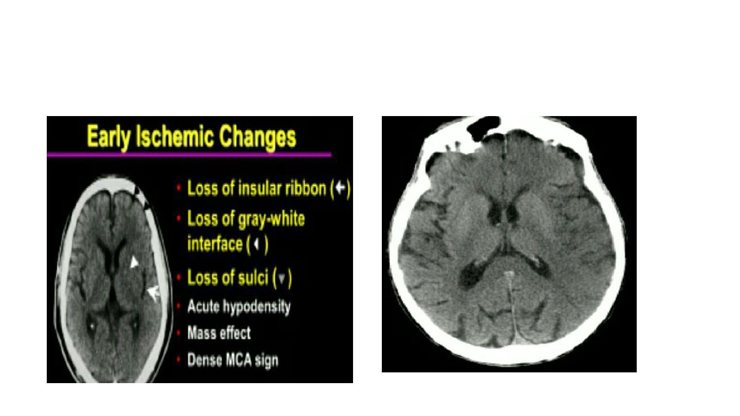
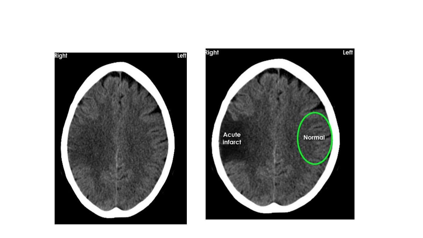
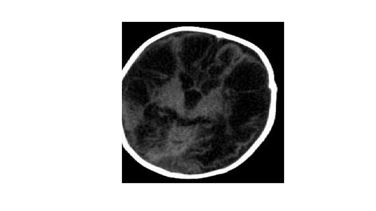
Loss of insula ribbon singe

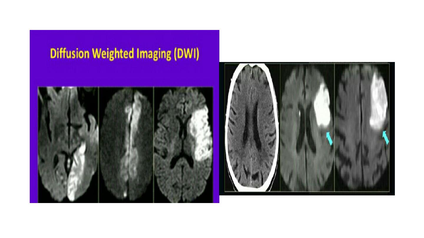
Diffusion weighted imaging DWI the
most
sensitive sequence in acute stork
evaluation
but not the initial, because of limited
availability , time consuming, not emergency
friendly
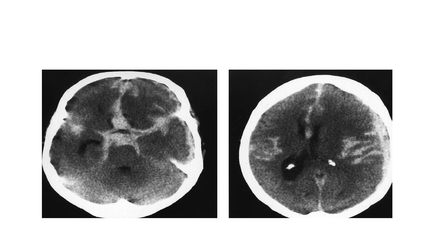
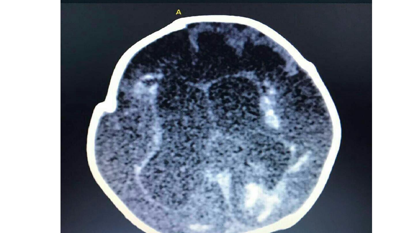
SAH
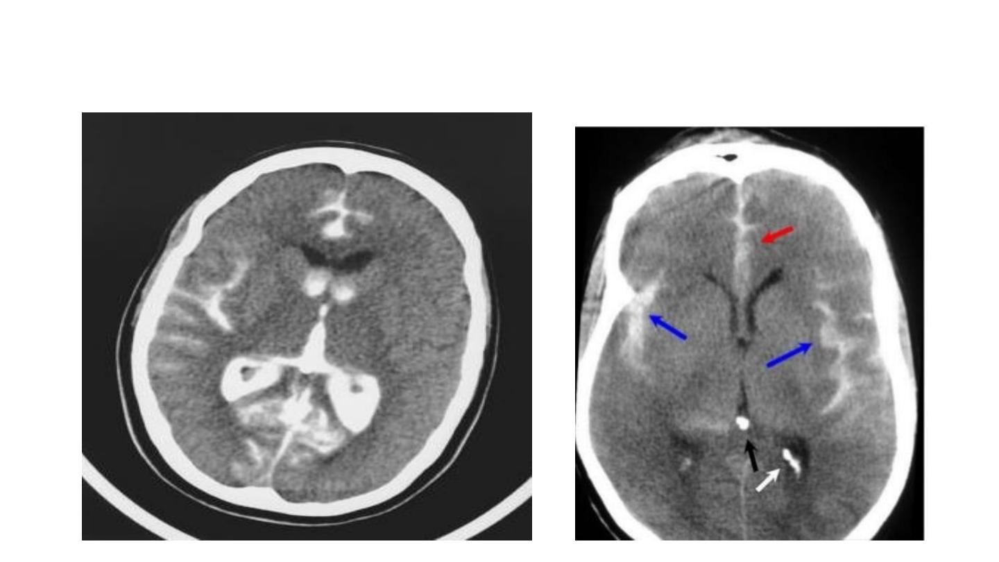
SAH + IVH

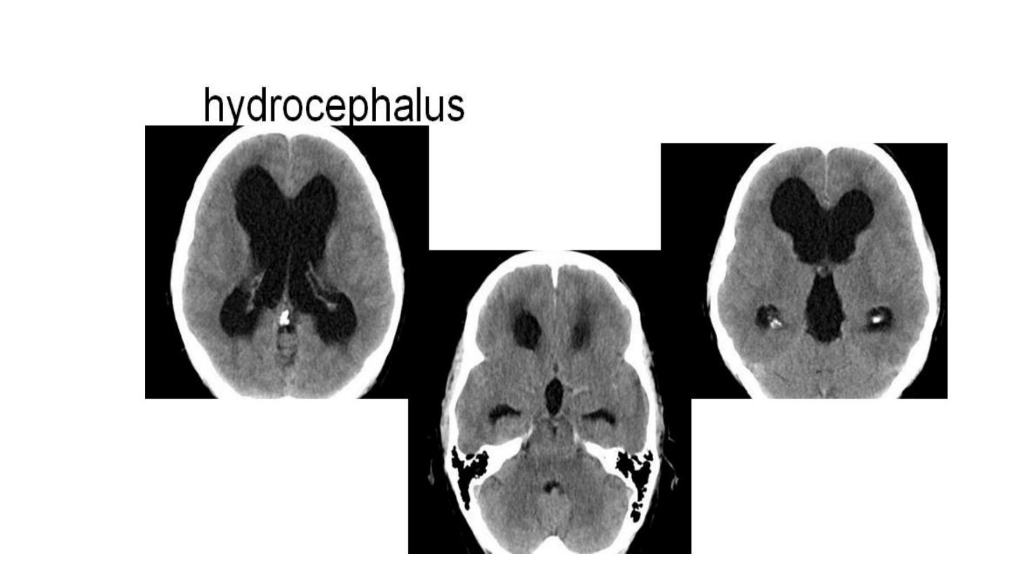

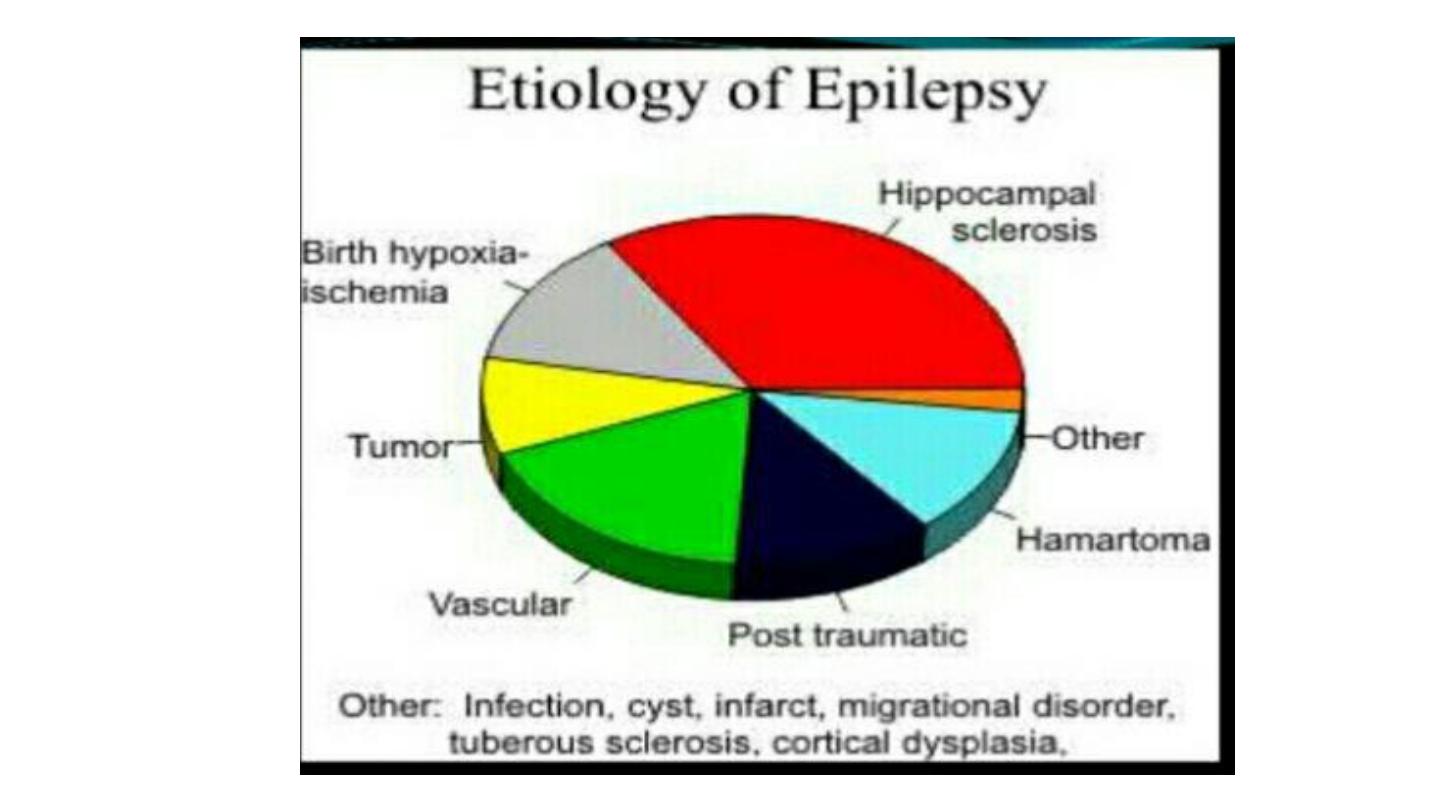
Neuro- imaging In epilepsy
• To identify structural abnormalities ..
•
Mesial (medial ) temporal sclerosis ( hippocampal sclerosis ).
• Vascular anomalies
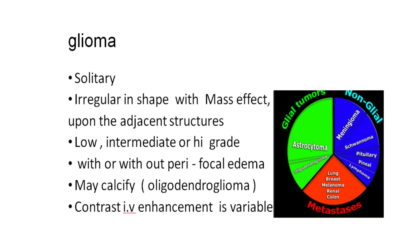
• Low grade glial tumors
• Malformations of cortical development
( heterotopia )
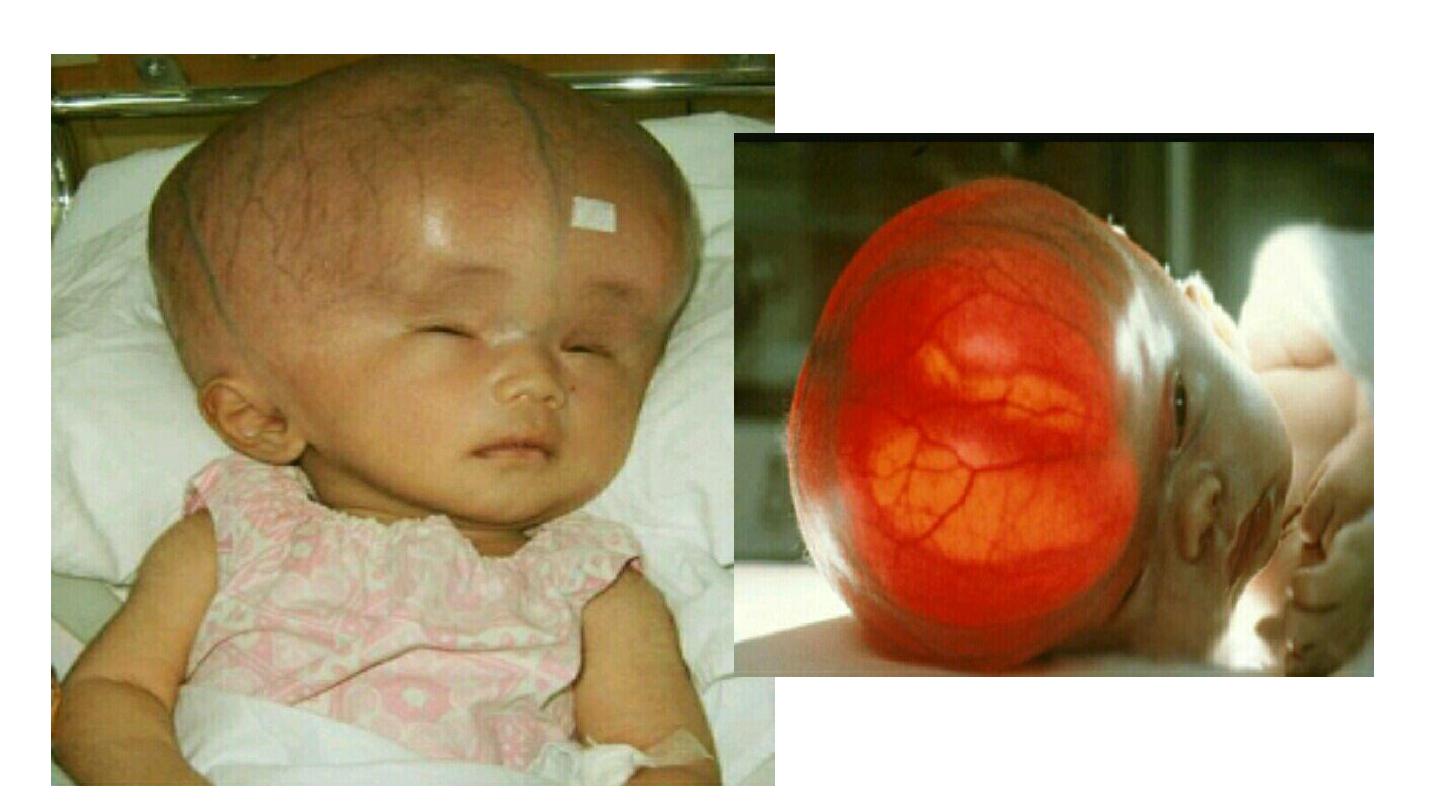
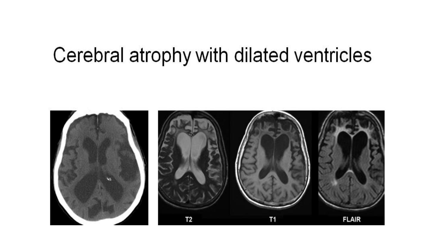
• In pediatrics …congenital anomalies ,,infection, hypoxic ischemic
encephalopathy .
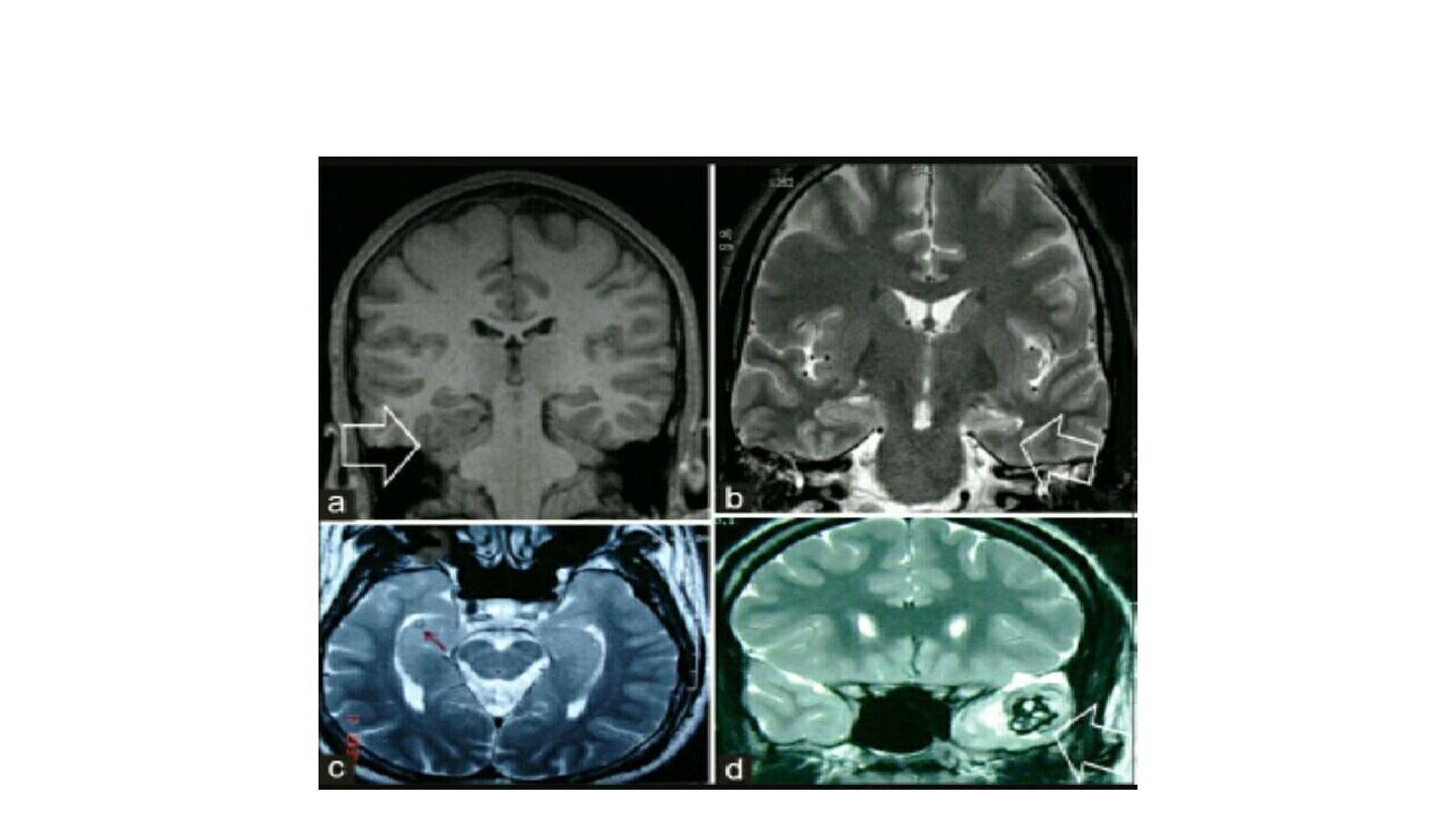
MTS
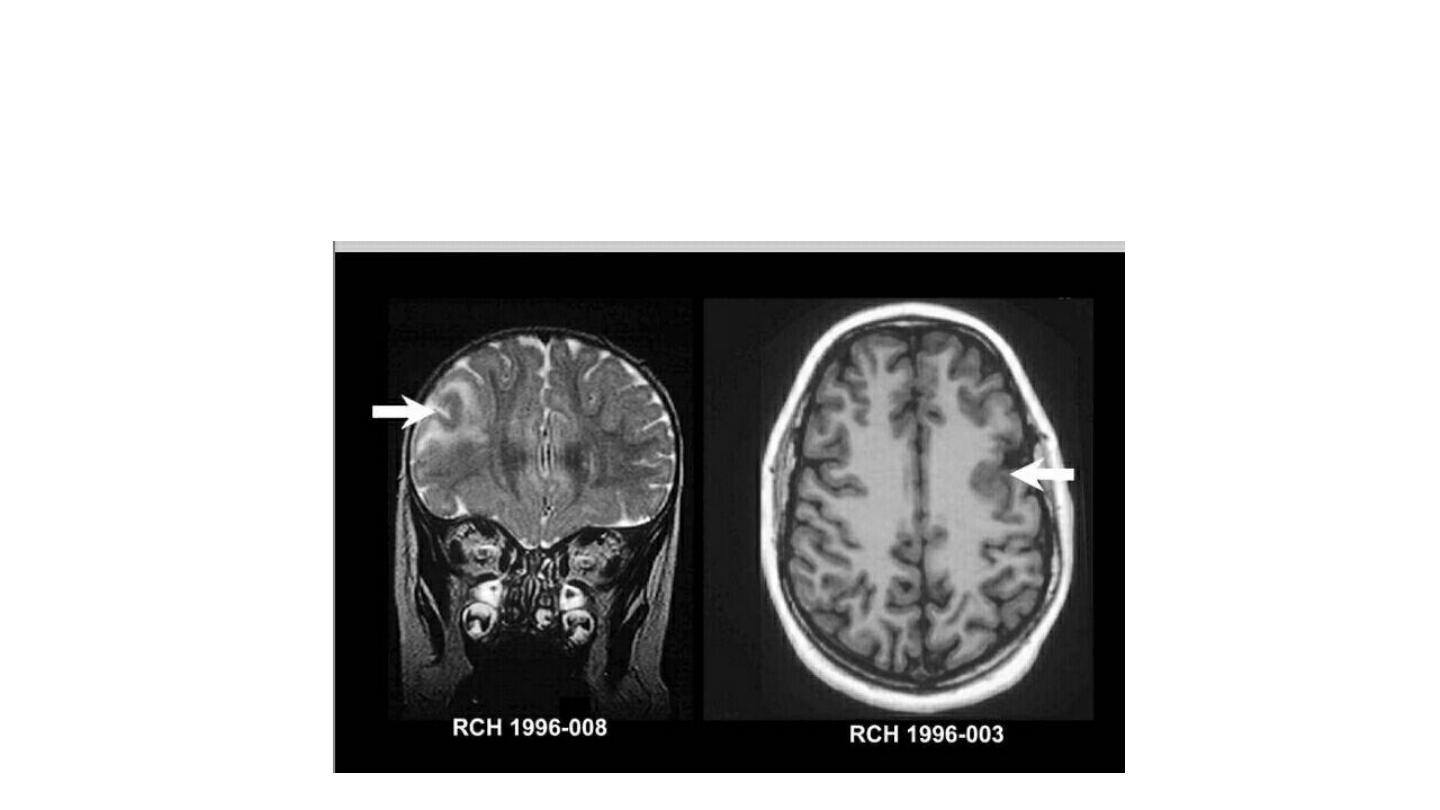
Focal cortical development
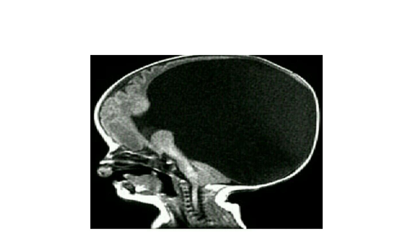
Congenital anomalies (haloprocencephaly)
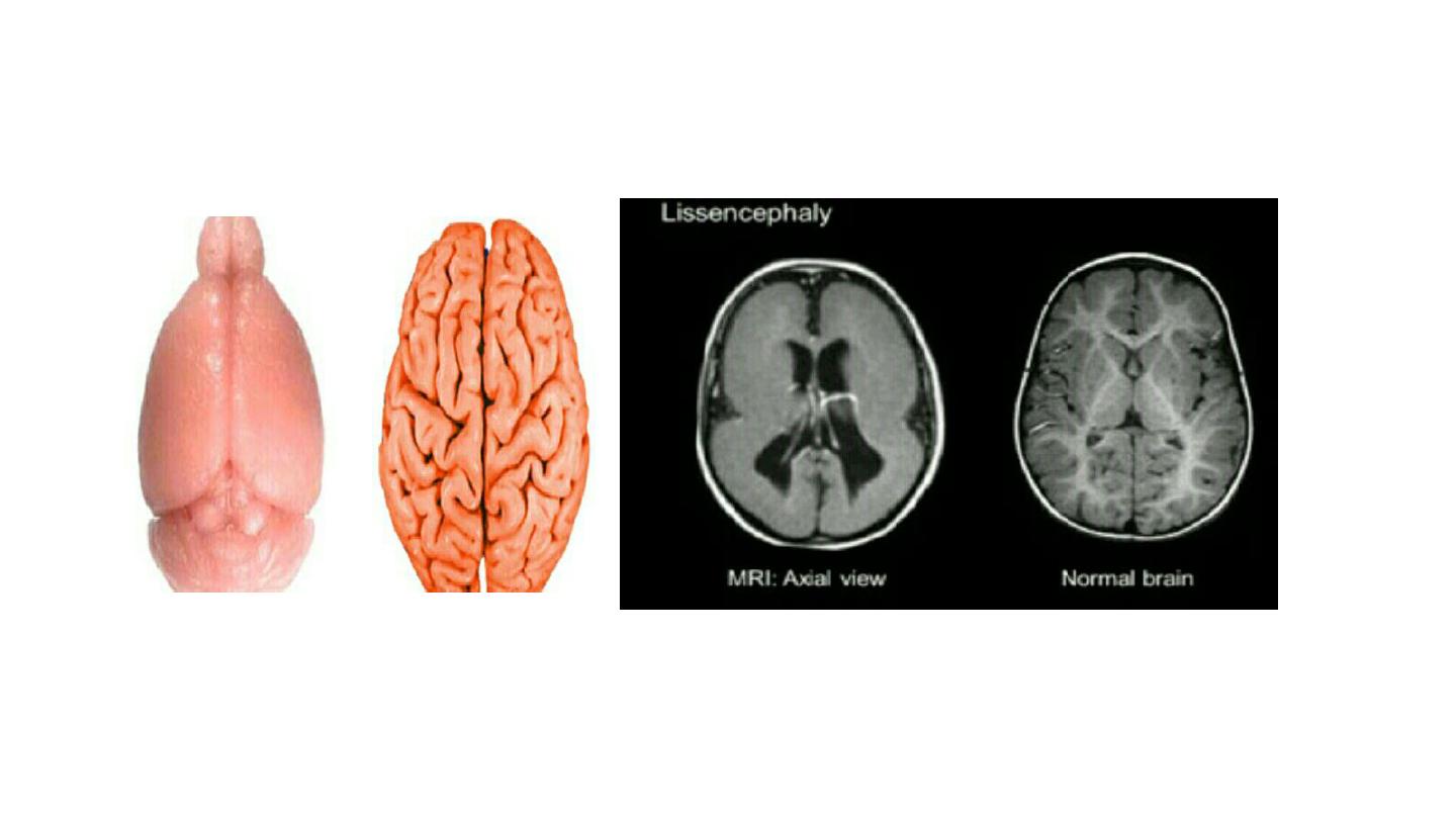
Migration anomaly
HIE