Development of the primary teeth
DR. Bushra Rashid NoamanMSc. pedodontics
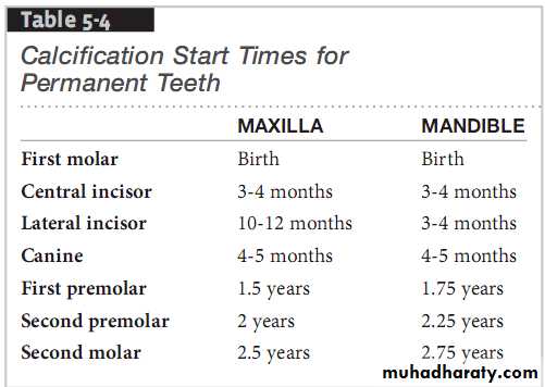
An accurate chronology of primary tooth calcification is of clinical significance to the dentist.
It is often necessary to explain to parents the time sequence of calcification in utero and during infancy.
Reasons to know the calcification times
The common observation of tetracycline pigmentation, developmental enamel defects, and generalized hereditary anomaliescan be explained if the calcification schedule is known.
Life cycle of the tooth
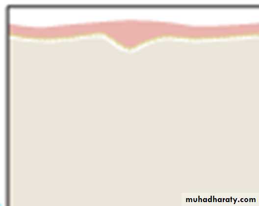
1. Initiation (bud stage)Begins as early as six weeks in the embryonic life.
Cells in the basic layer of oral epithelium proliferate a more rapid rate than do the adjacent cells.
The result is an epithelial thickening in the region of the future dental arch that extends along the entire free margin of the jaw.
result is called the dental lamina.

10 round or ovoid swellings
Formed from the dental laminaThose 10 swelling will form the 10 primary teeth

The permanent molars like the primary teeth rise from:
the dental lamina.The permanent incisors, canines and premolars develop from:
the buds of their primary predecessors.Proliferation (cap stage)
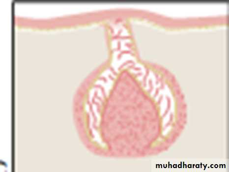
2. Proliferation (CAP STAGE)As a result of unequal growth in the different parts of the bud a cap is formed.

A shallow
invagination appears on the deep surface of the bud. The peripheral cells of the cap later form the outer and inner enamelepithelium.
Histodifferentiation and morphodifferentiation(bell stage)
Histodifferentiation• During this stage there is: differentiation of the cells of the dental papilla into odontoblasts and
2. the cells of the inner enamel epithelium into ameloblasts.
Ameloblast and odontoblast are the formative cells
morphodifferentiation
In morphodifferentiation stage the formative cells are arranged to outline the form and size of the tooth.
The process occur before matrix deposition. The morphologic pattern of the tooth established
the formative cells: ameloblasts and odontoblasts
which line up along the future dentino-enamel and the dentino-cemental junction at the stage of morphodifferentiation.APPOSITION
Appositional growth is the result of;a layer-like deposition of a nonvital extracellular secretion in a form of a tissue matrix.

This matrix is deposited by ;
the formative cells: ameloblasts and odontoblastsCALCIFICATION
It takes place after matrix depositionand involves the ;
precipitation of the inorganic calcium salts within the deposited matrix.
The process begins with the precipitation of a small nidus about which further precipitations occurs.
The original nidus increases in size ;
by addition of concentric laminations.There is an eventual approximation and fusion of these individual calco-spherites into a homogenously mineralized layer of tissue matrix.
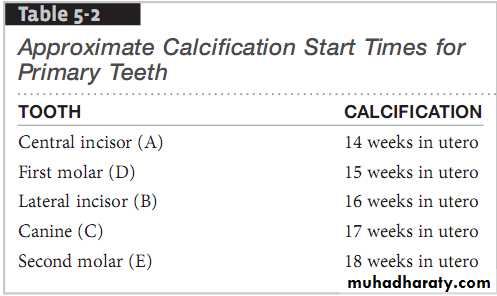
Table 1