The Back
To describe the skeleton of the back
To identify parts of the vertebrae
To define atypical cervical vertebrae
To demonstrate different articulations in the cervical
series
To relate to some vertebral fractures & disk prolapse
To list postvertebral muscles
To locate the suboccipital triangle
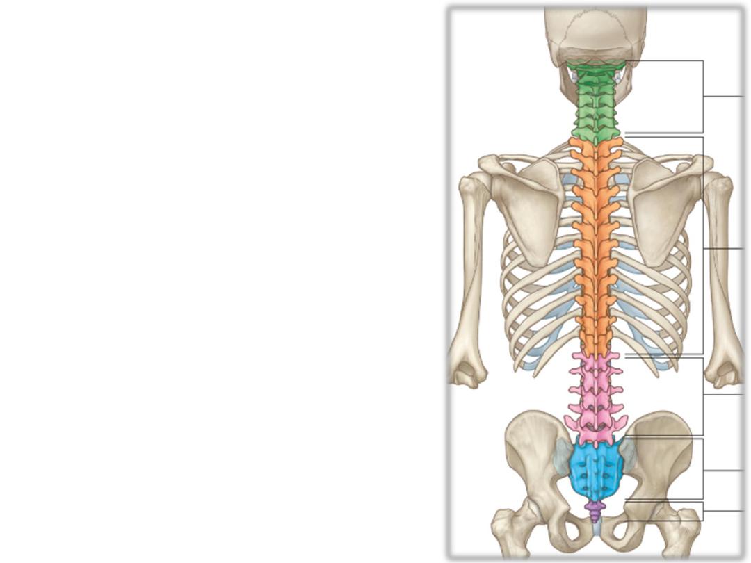
The vertebral column:
-Average length in the male is about 71
cm& in female about 61 cm
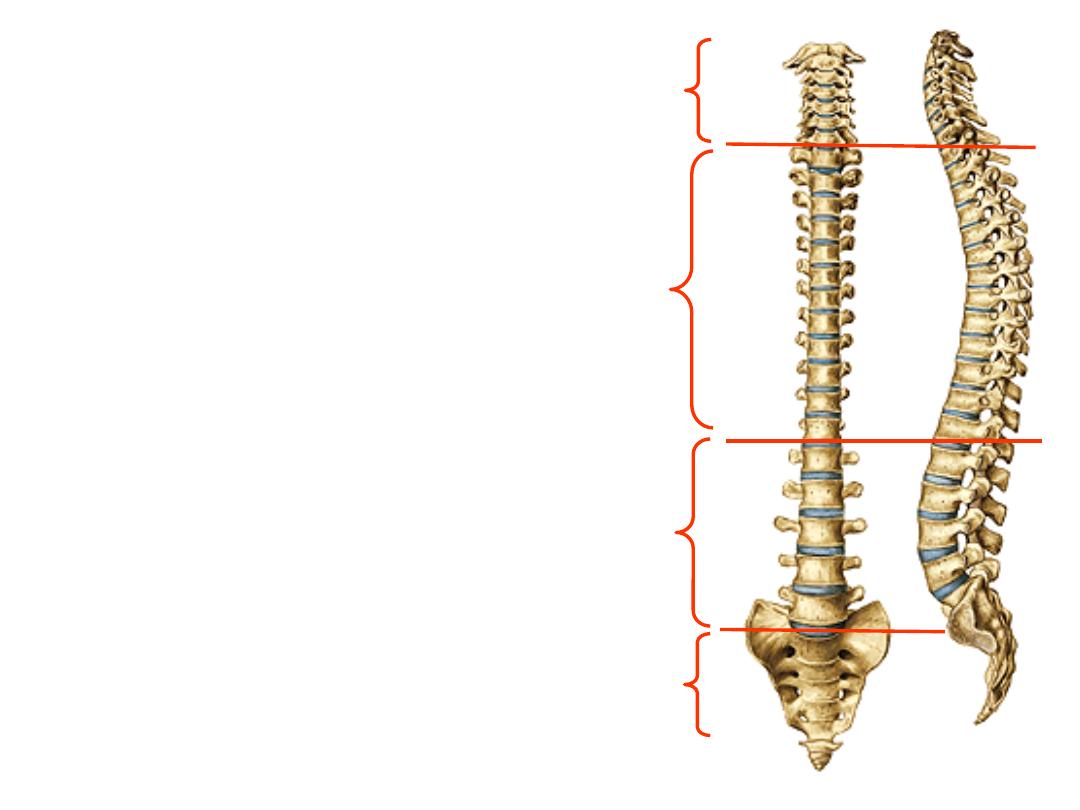
-The column constitutes the five known
regions:
Cervical; 7
Thoracic; 12
Lumbar; 5
Sacral; 5
Coccyx; 1
Measurements (male values):
-Cervical part: 12.5 cm.
(17.5%)
-Thoracic: 28 cm.
(40%)
-Lumbar: 18 cm.
(25%)
-Sacrum and coccyx: 12.5 cm.
(17.5%)
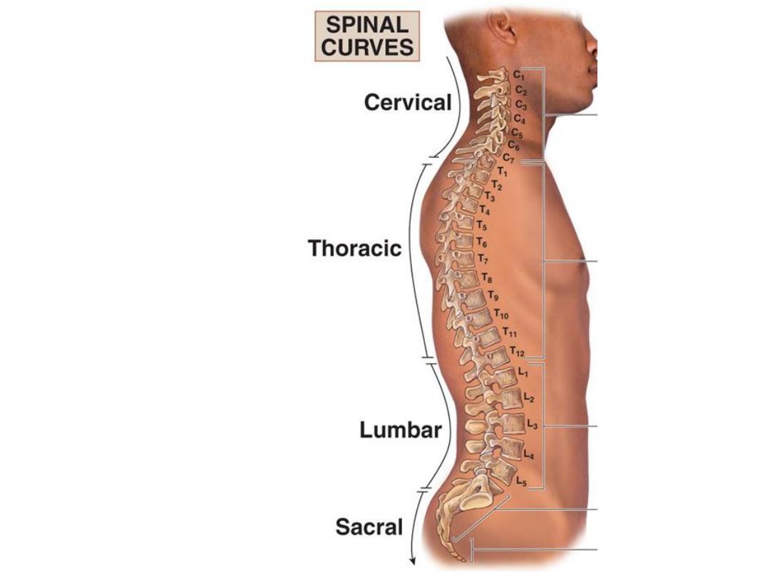
Curvatures:
Primary curvatures (Flexion):
1- Thoracic; T2-T12
2- Pelvic; LS joint-coccyx
Secondary curvatures (Extension):
1- Cervical; C2-T2
2- Lumbar; T12-LS joint
Articulations of the vertebral column:
1- A series of synovial joints between the
vertebral
arches
(between
articular
facets)
2- A series of secondary cartilagenous
joints
between
vertebral
bodies
(intervertebral discs)
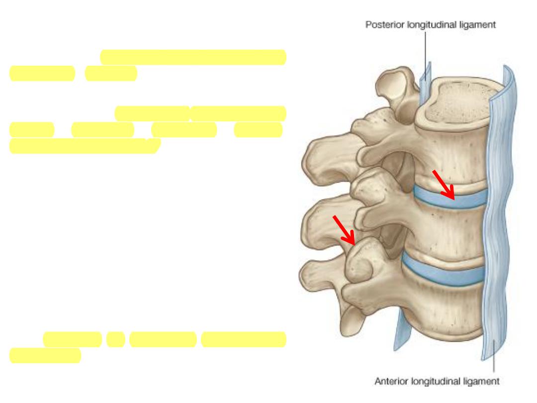
Articulations between vertebral bodies:
-Bodies of adjacent vertebrae are held to
each other by fibrous discs which
strongly adhere these vertebrae to each
other
-Movements at these joints is slight
though summative movements permits
considerable range
-Ligaments supporting these joints are
the anterior & posterior longitudinal
ligaments
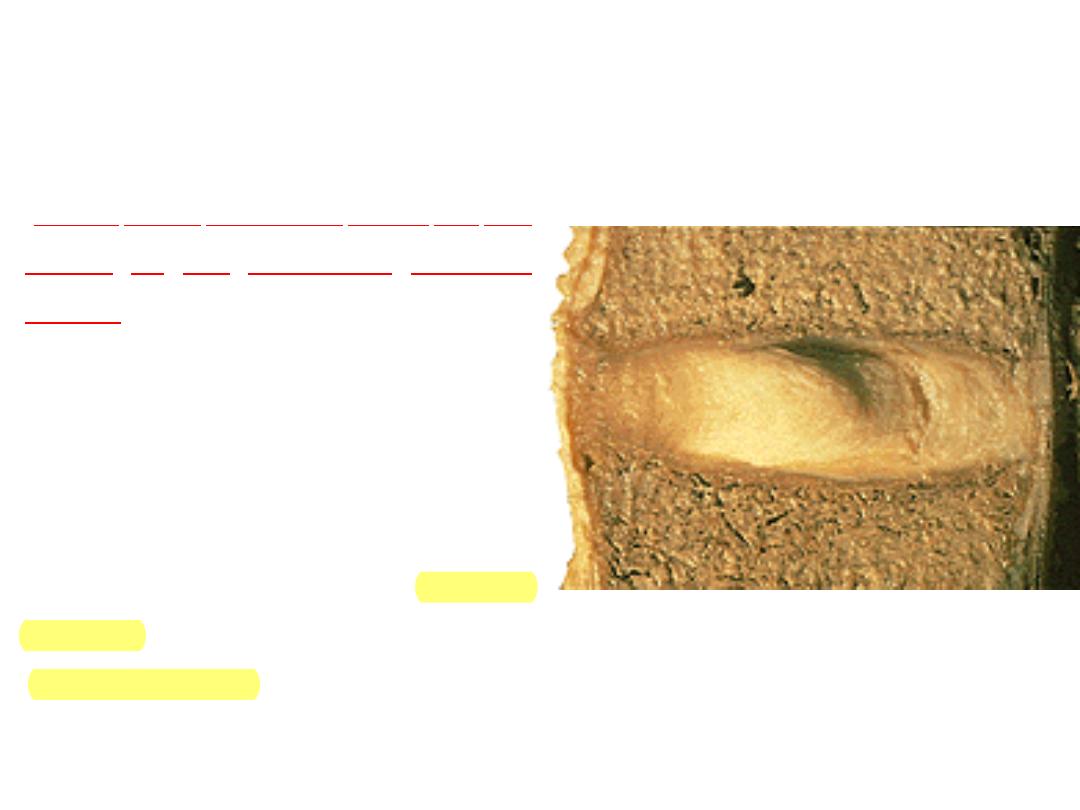
The intervertebral discs:
-These discs constitute about 1/4 the
length of the articulated vertebral
column
-They vary in shape, size, and
thickness, in different parts of the
vertebral column, correspond with the
surfaces of the adhering bodies
-Formed of periferal fibrous (annulus
fibrosus) zone & central gelatenous
(nucleus pulposis) zone
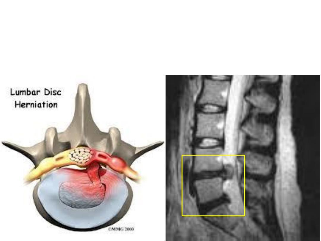
Prolapsed IVD:
-Herniation of nucleus pulposus into the vertebral canal compressing
on spinal nerve roots
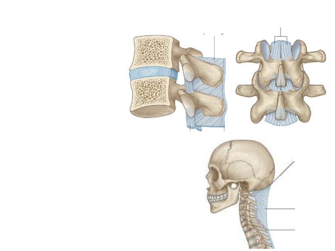
Other ligaments in the vertebral column:
1- Ligamenta flava:
-Elastic ligaments
-Between adjacent laminae
2- Interspinous ligaments:
Connects adjacent spines
3- Supraspinous ligaments:
Connects spines tips
4- Ligamentum nuchae:
-Triangular fibrous sheet
-Attached to cervical spines & skull
-Divides the back of neck into two halves
1
2
3
4
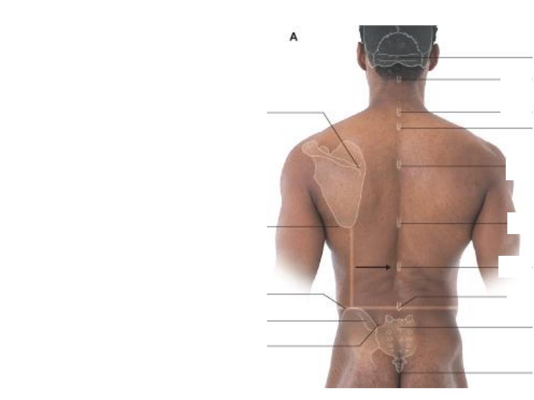
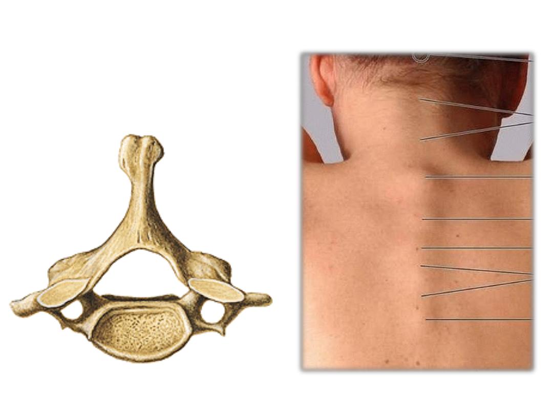
Surface localization of vertebrae:
Principles:
-The first palpable spine below the skull
is C2
-The next most prominent is C7
-T3 lies level with scapular spine
-T7 lies level with inferior scapular angle
-L4 lies level with iliac tubercle
-T12 midway between T7 & L4
-Coccyx is the lower end
C2
C7
T3
T7
L4
T12
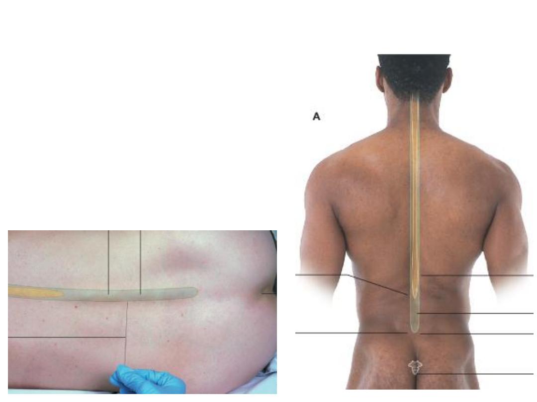
Surface localization of lower end of spinal cord:
Principles:
-Localize T12 & L4 as previously mentioned
-Spinal cord terminates midway between them (L1-2)
-Lumbar puncture is done at L3-4 level
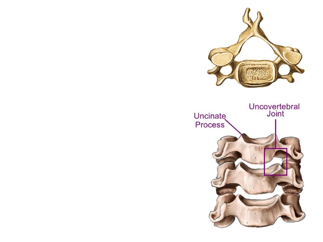
Characters of cervical vertebrae:
1-
Rectangular body
2- Transverse processes contain:
-Foramina transversaria (vertebral vessels)
-Anterior & posterior tubercles
3- Short bifid spine
4- Big triangular vertebral foramen
Joints of Luschka (uncovertebral joints):
-Synovial joints between the bodies
-Specific for cervical region
-Provide more freedom of movement
-Liable for more arthritic changes
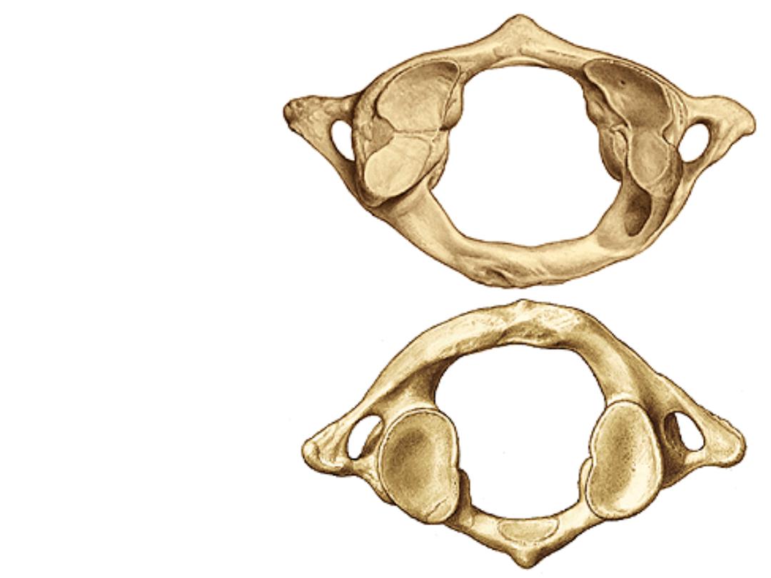
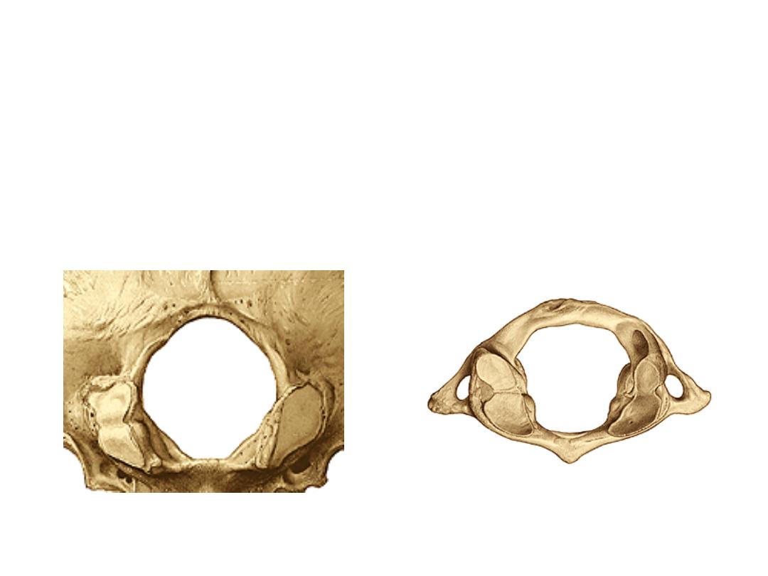
ATYPICAL vertebrae:
Atlas (C1):
No body
Shorter anterior than posterior arch
Deep kidney shape superior facet
Flat oval inferior facet
Facet on the back of anterior arch
for the dens
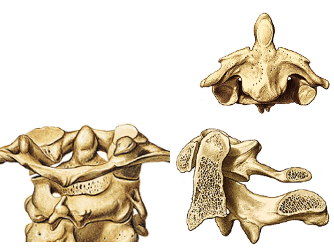
Axis (C2):
Dens (odontoid process)
Bulky body
Bulky spine
Vertebra prominence (C7):
Long non bifid spine
Atlanto-occipital joint:
-Represent incomplete single ellipsoid joint (deficient from its middle) with longer
transverse than AP diameter
-The general shape of the joint looks like an egg lies on its side in an egg-saucer
-Only permits flexion-extension (hinge joint)
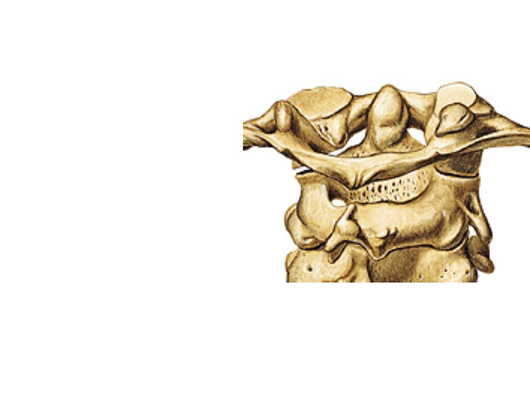
Atlanto-axial joints:
1- The lateral atlanto-axial joints:
Between articular facets
2- The median atlanto-axial joint:
A) Anterior:
Between anterior surface
of the dens & the back of the
anterior arch of atlas
B) Posterior:
Between the posterior
surface of the dens & transverse
limb of cruciate ligament
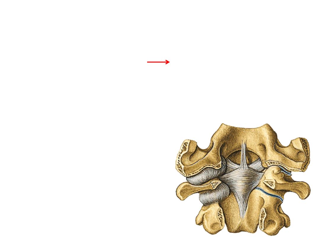
Stabilizing structures:
1- Cruciate ligaments (2 limbs)
Longitudinal limb: Occipital bone C2 body
Transverse limb: between C1 lateral masses (there is a small joint
between a cartilage on this limb & the back of the dens)
1
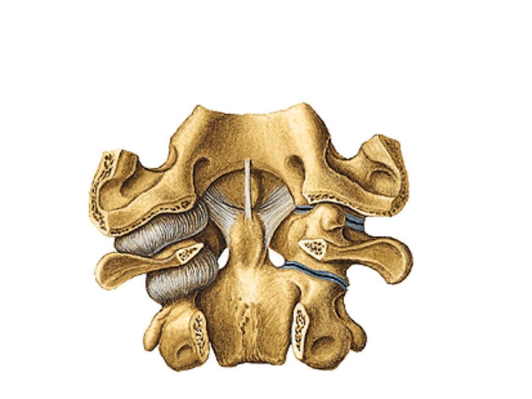
2- Apical ligament (single): dens
– occipital bone (midline)
3- Alar ligament (pair): dens
– occipital bone (lateral to the midline)
2
3
3
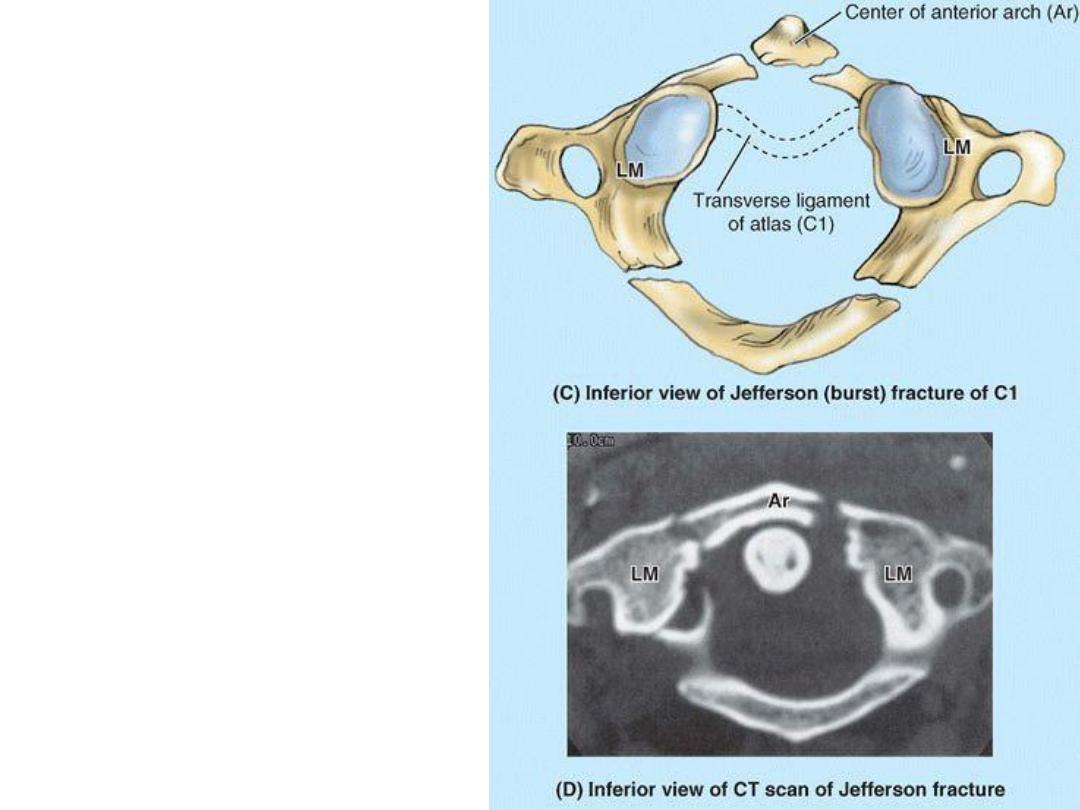
Jefferson fracture:
-Fracture of one or both arches
of C1 due to compression C1
between skull & C2 (in diving
accidents
with
striking
the
butom)
-If associated with tear in the
transverse band of cruciate
ligament leads to quadriplegia
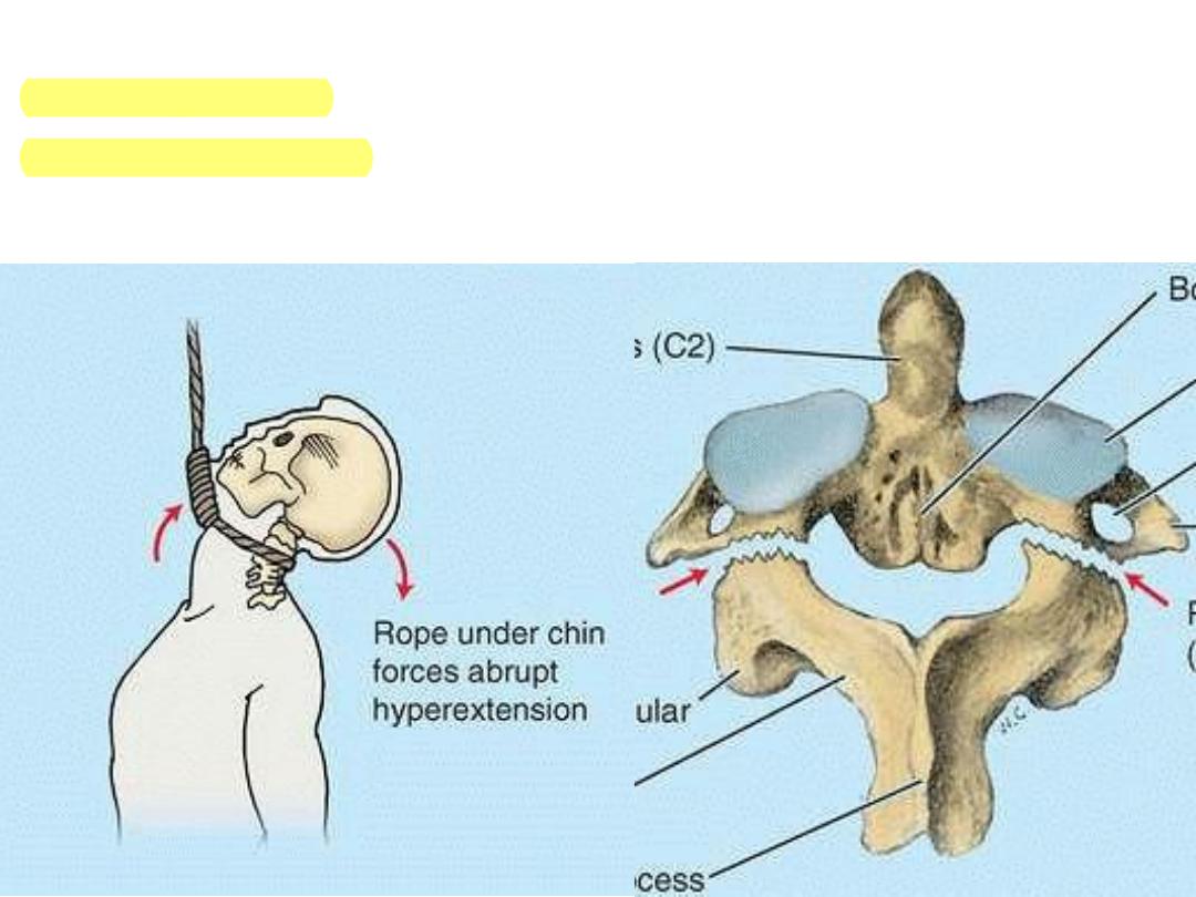
Fracture of vertebral arch of C2:
-Whiplash fractures
-
Hangman’s fractures
-The dens falls back pressing on the vital centers & causes death
Main characters
Region
-Additional joints of Luschka
-Vertebral vessels passing through foramina transversaria
-Seven vertebrae, eight spinal nerves
-Spinal nerve passes superior to the pedicle of its
numerically corresponding vertebra
Cervical
1
-Articulation by their bodies & transverse processes with
the ribs
-Spinal nerve passes inferior to the pedicle of its
numerically corresponding vertebra
-Mainly permit trunk rotation
Thoracic
2
-Giant, kidney shaped bodies
-Spinal nerve passes inferior to the pedicle of its
numerically corresponding vertebra
-Mainly permit trunk flexion-extensio & lateral flexion
Lumbar
3
-5 sacral segments fuse with each other
-Articulates with lower limb bone (the hip)
-Nerves leave through anterior & posterior sacral foramina
Sacral
4
Single triangular bone with no special feature
Coccyx
5
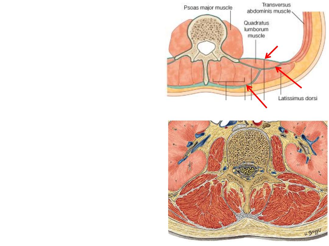
The thoracolumbar fascia:
-This strong fascial structure lies in the
posterior abdominal wall enclosing
muscular
compartments
&
gives
attachment to many other muscles.
-It is formed of 3 layers; anterior,
middle & posterior
-Anterior & middle layers are confined
to the abdomen
-The posterior one extends up in the
thoracic & cervical regions
-Quadratus lumborum is enclosed
between the anterior & middle layers
-Erector spinae is enclosed between
the middle & posterior layers
Back muscles:
1- Extrinsic:
-Form superficial & intermediate layers
-Involved with movements of the upper limbs
and thoracic wall
-Innervated by anterior rami of spinal nerves
2- Intrinsic:
-They lie deep in position
-They support and move the vertebral column
and participate in moving the head
-Innervated by the posterior rami of spinal
nerves
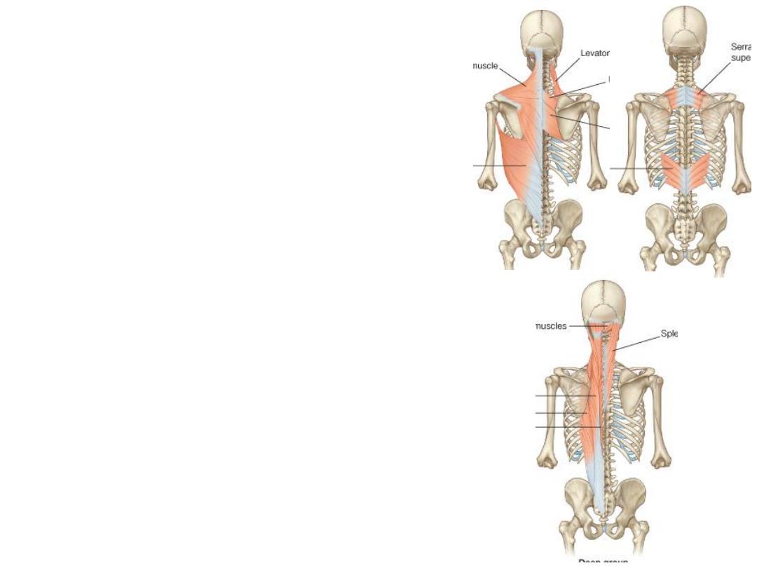
Muscles
Layer
-Trapezius
-Latissimus dorsi
-Levator scapulae
-The rhomboids
Superficial
1
-Serratus posterior superior & inferior
Intermediate
2
1- Splenius group:
-Capitis
-Cervicis
2- Erector spinae group:
-Iliocostais (external)
-Longissimus (intermediate)
-Spinalis (deep)
3- Semispinalis group:
-Semispinalis
-Multifidus
-Rotators
Deep
3
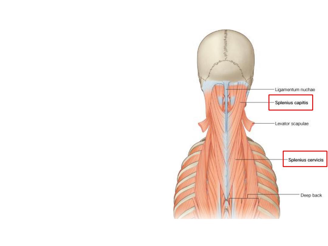
The splenius muscles:
-The two muscles run from the spinous
processes upward and laterally
-Splenius capitis
is a broad muscle
attached to the occipital bone and
mastoid process of the temporal bone
-Splenius cervicis
is a narrow muscle
attached to the transverse processes of
the upper cervical vertebrae
-Together they draw the head backward,
extending the neck.
-Individually, each muscle rotates the
head to the same side of the contracting
muscle
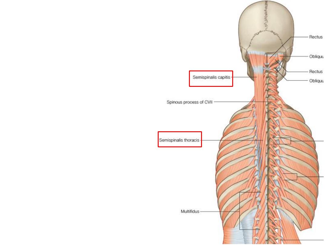
The semispinalis muscles:
-These muscles begin in the lower
thoracic region and end by attaching
to the skull
-Crossing between four and six
vertebrae from their point of origin to
point of attachment.
-Semispinalis muscles are found in
the thoracic region
(S. thoracis)
,
cervical region
(S. cervicis)
& attach
to the occipital bone
(S. capitis)
.
-They are prime extensors of the
vertebral column
The suboccipital muscles :
1- Rectus capitis posterior minor
2- Rectus capitis posterior major
3- Superior oblique
4- Inferior oblique
-These
muscles
are
skull
extensors
-All are supplied by C1, posterior
ramus
1
2
3
4
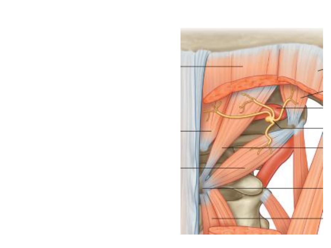
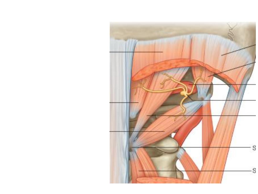
Suboccipital triangle:
-2, 3 & 4 form the boundaries of this
triangle
-The triangle is roofed by splenius capitis
-Floor is the back of atlas
-Contents:
1- In the triangle:
-Vertebral artery
-C1 posterior ramus
2- Passing in the roof:
1- Occipital artery
2- Great occipital nerve (C2)
2
3
4