The Skull 2

To place individual skull bones in position
To relate bones to regions
To relate certain parts to individual bones
To describe cranial fossae
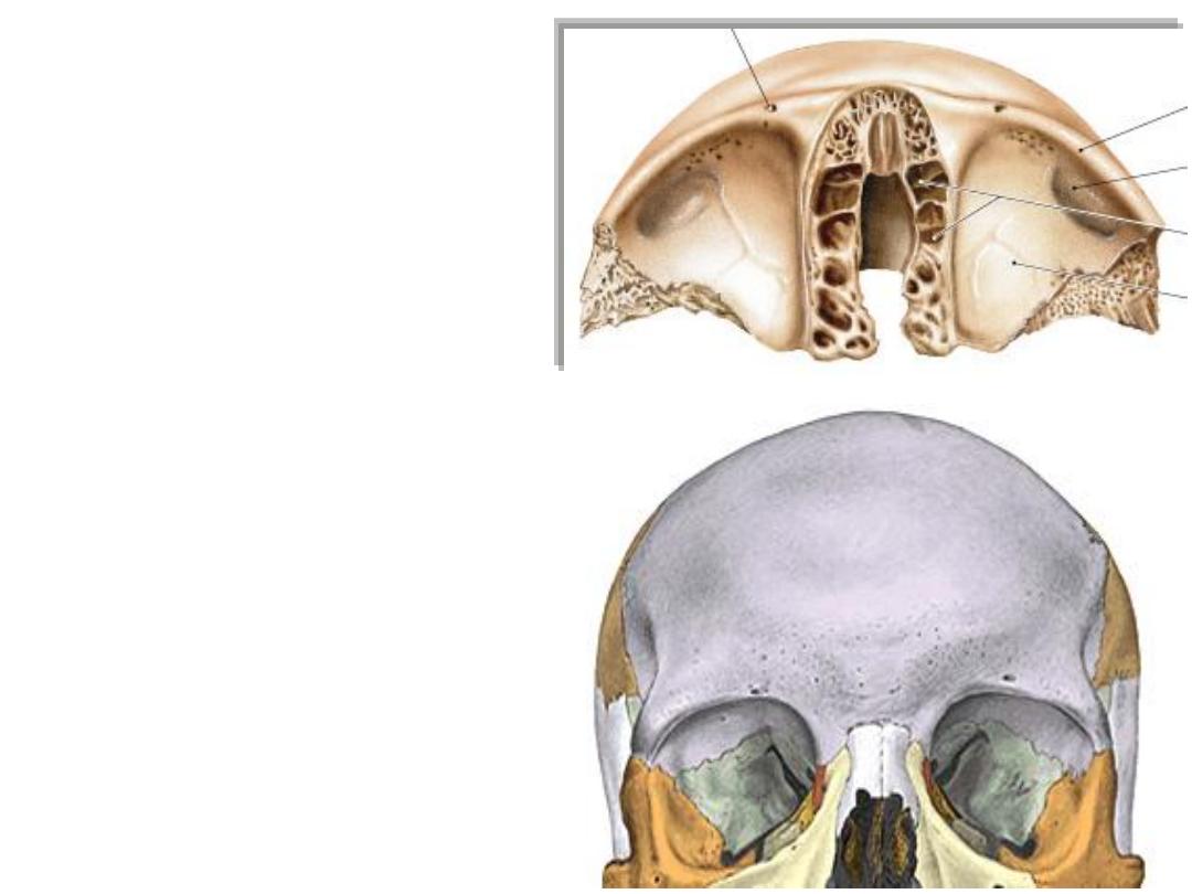
Frontal
Orbital plate
The frontal bone:
-The bone forms the forehead
-Has a posteriorly projecting orbital
plates which form the roof of the
orbit & floor of anterior cranial fossa
-The gap between the orbital plates
is filled by the ethmoid bone
-Other processes articulate with
facial bones
-The frontal sinuses are located in its
anteterior part
-The free posterior border articulates
with the 2 parietals forming the
coronal suture
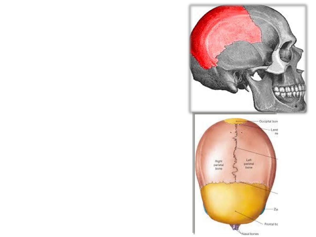
Parietal bones:
-Form most of the vault
-The sagittal suture separates them
from each other
-The coronal suture separates them
from the frontal
-The lambdoid suture separates them
from the occipital
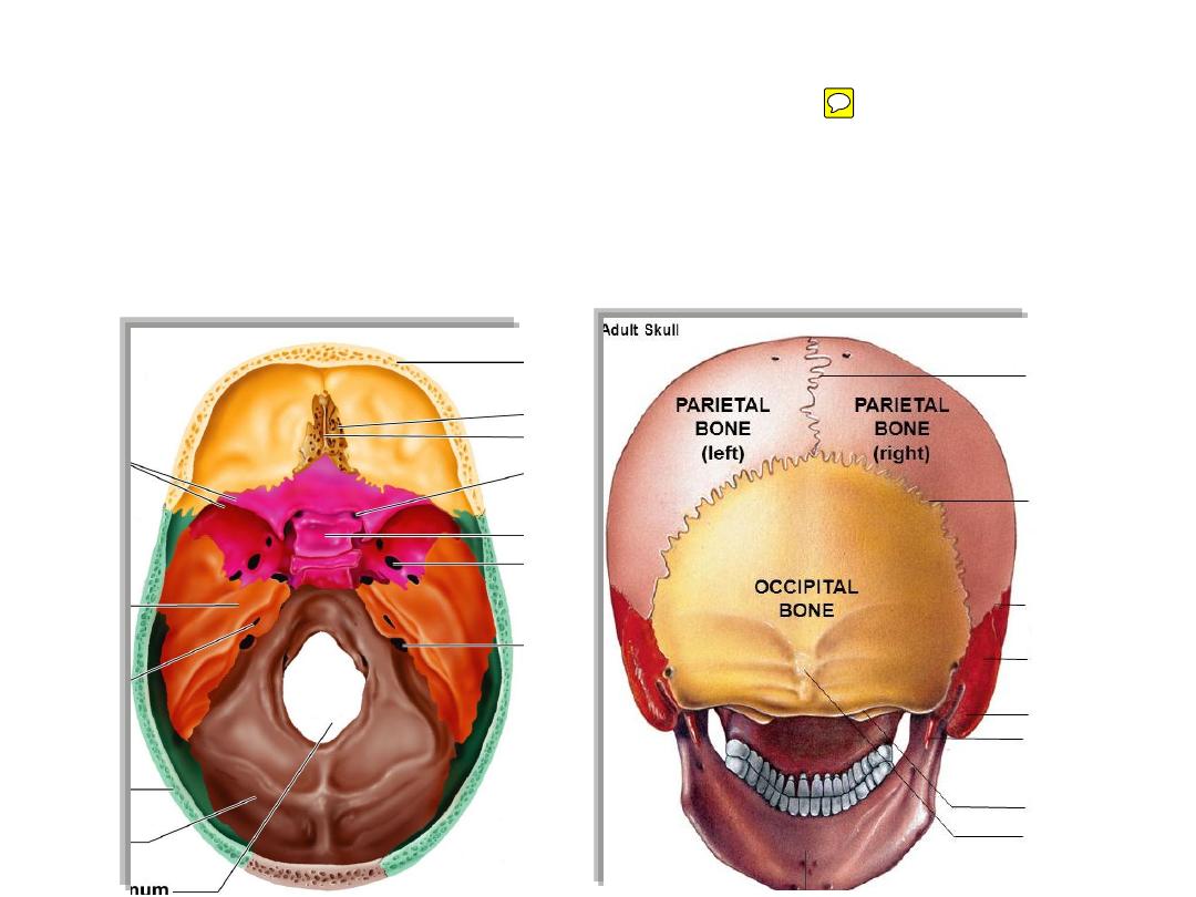
The occipital bone:
-Form the back of skull
-Forms the floor of posterior cranial fossa which holds the cerebellum
-Contains the largest skull foramen
-The site where the skull articulates on the cervical spines
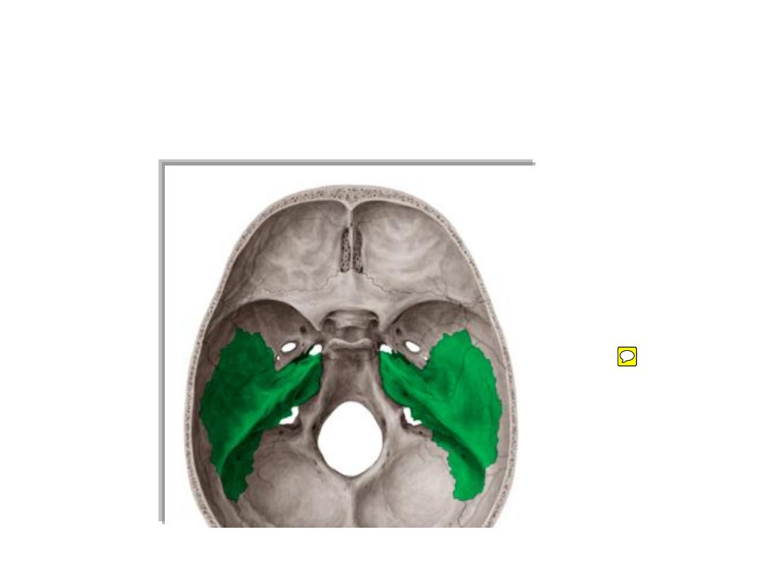
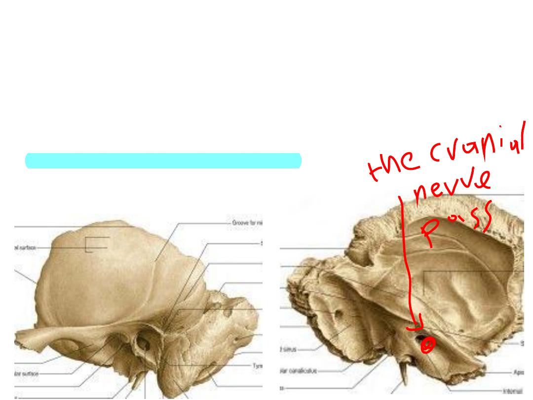
The temporal bone:
• Forms the side & part of the base of the skull
• Is a pathway for many important structures like facial nerve & ICA
• Contains vital organs like the ear
• Formed of 4 parts
1- Squamous;
- Forms the side of skull & floor of middle cranial fossa
- Contains mandibular fossa for TMJ
- Contains the zygomatic & styloid processes
2- Petrous:
- Forms part of the middle & posterior cranial fossae
- Contains the carotid & facial canals & the ear
3- Tympanic;
forms part of the middle & external ears
4- Mastoid process;
contains air cells
1
2
3
4
5
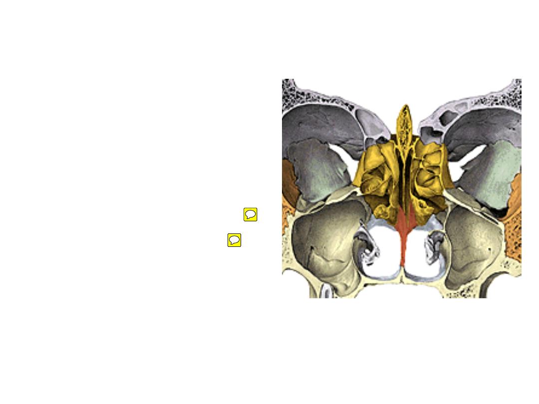
The ethmoid::
-Lies in the midline between both
orbits anterior to the sphenoid
-Forms the upper part of the nasal
cavity (roof, medial & lateral walls)
-Contains ethmoidal sinuses
Parts:
-Cribriform plate
(roof of nasal cavity)
-Crista galli & perpendicular plate
-Labyrinth (lateral mass):
lies between
the orbit & nasal cavity
labryinth contain ethmoidal sinus
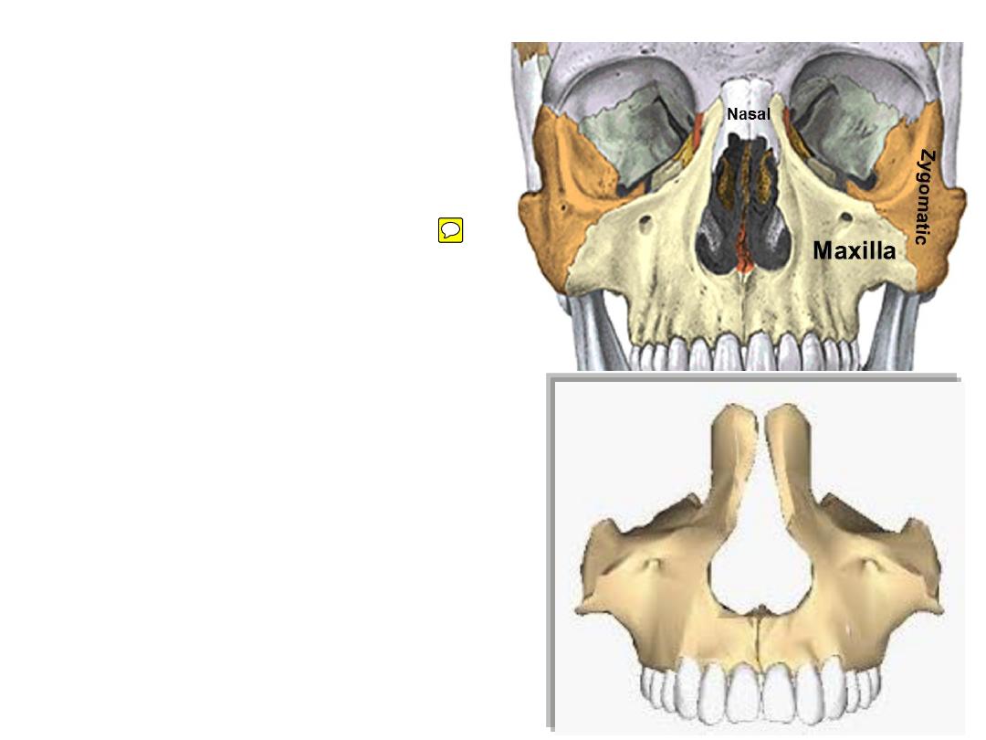
The maxilla:
-Maxilla forms the midface
-It forms the upper jaw & big portion of
the roof of the mouth
-It also forms the floor & sidewall of the
nose
-Contains the maxillary sinuses
-The two maxillae meet in the midline
below & are separated above by nasal
bones
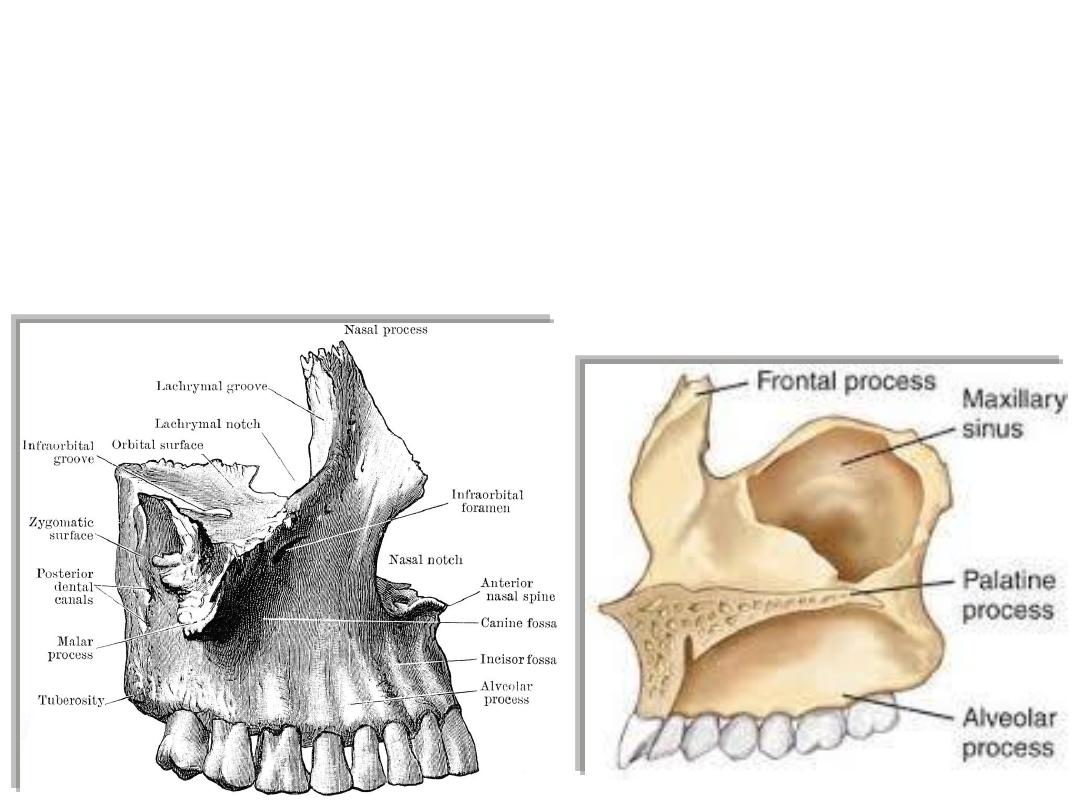
Processes:
-Frontal
-Palatal
-Alveolar
-Zygomatic
The sphenoid:
-Lies posterior to the ethmoid
-Forms the floor of MCF & roof
of ITF
-Contains
the
sphenoidal
sinuses
-Supports the pharyngeal wall
-Contains foramina & fissures
that communicate with different
parts of the head
-Looks like a butterfly
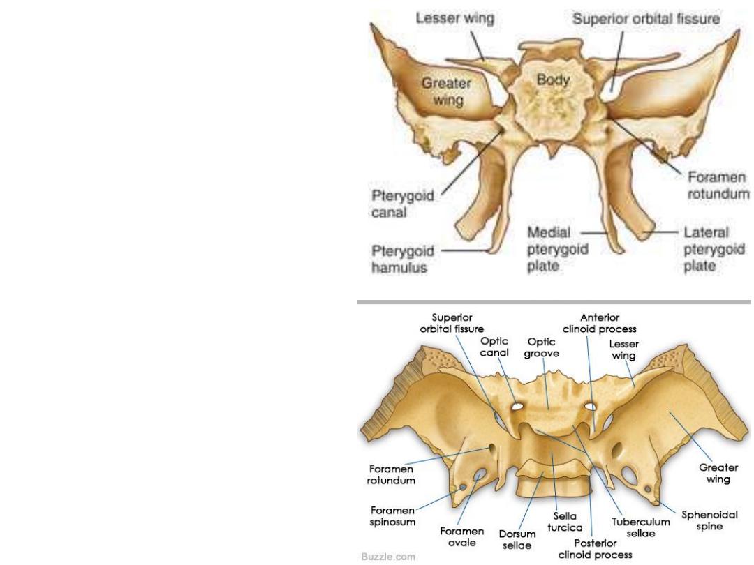
Parts:
-Body;
forms the sella turcica in
the middle cranial fossa
-The
lesser
wings
form
the
posterior part of orbital roof
-Greater wings
form the floor of
middle cranial fossa then ascend
to form the side of the skull
-Pterygoid
processes
project
inferiorly to be seen at the skull
base
-Between the two wings lie the
superior orbital fissure
leads to the
orbit
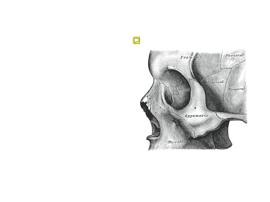
The zygomatic (malar) bone:
-Situated at the upper and lateral
part of the face
-Forms
the
prominence
of
the cheek, part of the lateral wall
and floor of the orbit, and parts of
the temporal and infratemporal
fossa
-Articulates anteriorly with maxilla
& posteriorly with the zygomatic
process of temporal bone
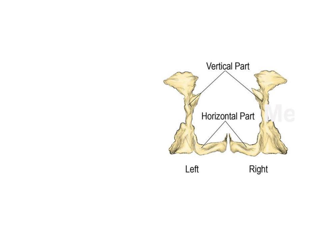
The palatine bone:
- Situated at the back part of
the
nasal
cavity
between
the maxilla and the pterygoid
process
- They contribute to the walls of
three cavities: the floor and
lateral walls of the nasal cavity,
the roof of the mouth, and the
floor of the orbits
- Palatine
bone
resemble
an
opposing L letters
- Consists of a horizontal & vertical
plates with a pyramidal process
in between
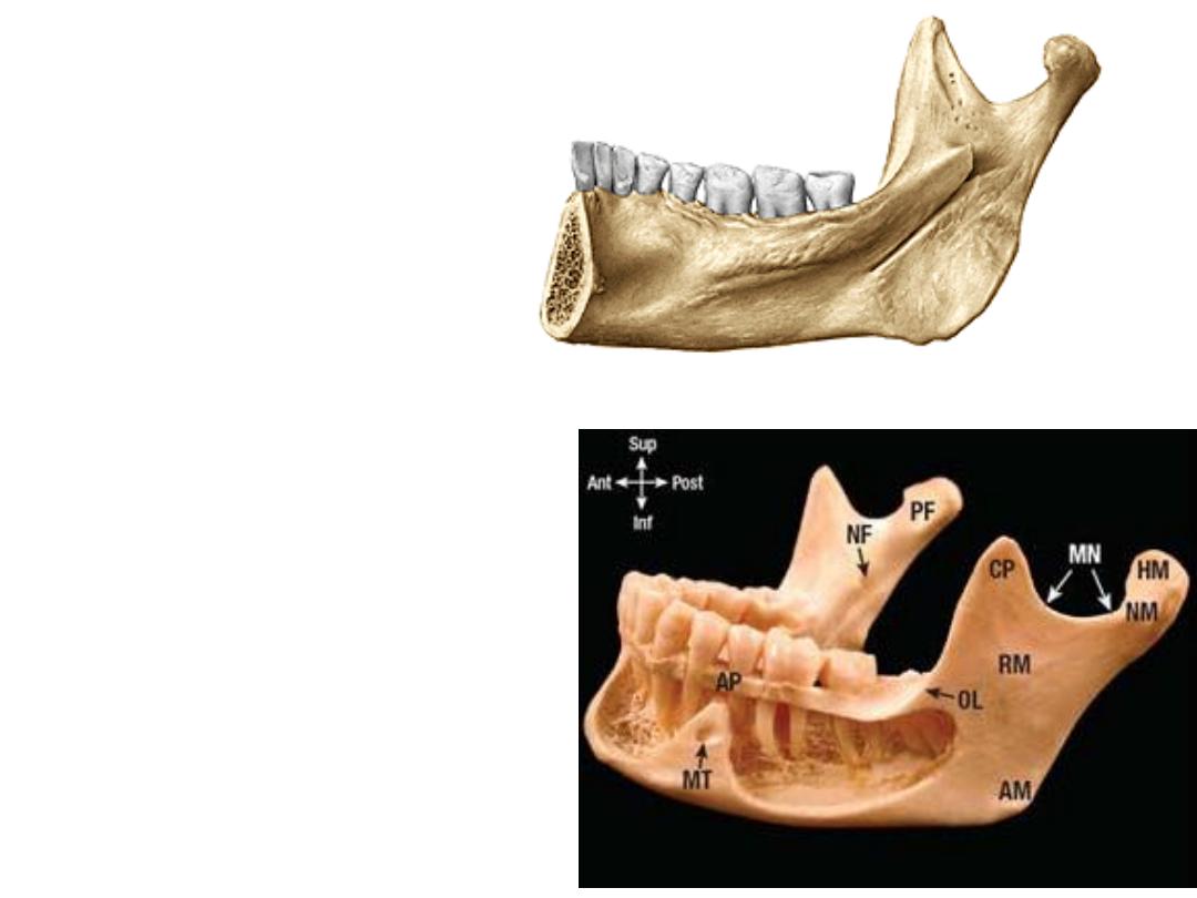
The mandible:
-The head
-The neck
-The coronoid process
-The mandibular notch
-The ramus
-The mandibular foramen
-The mandibular canal
-The body
-The mylohyoid line
-The genial tubercles
-The digastric fossa
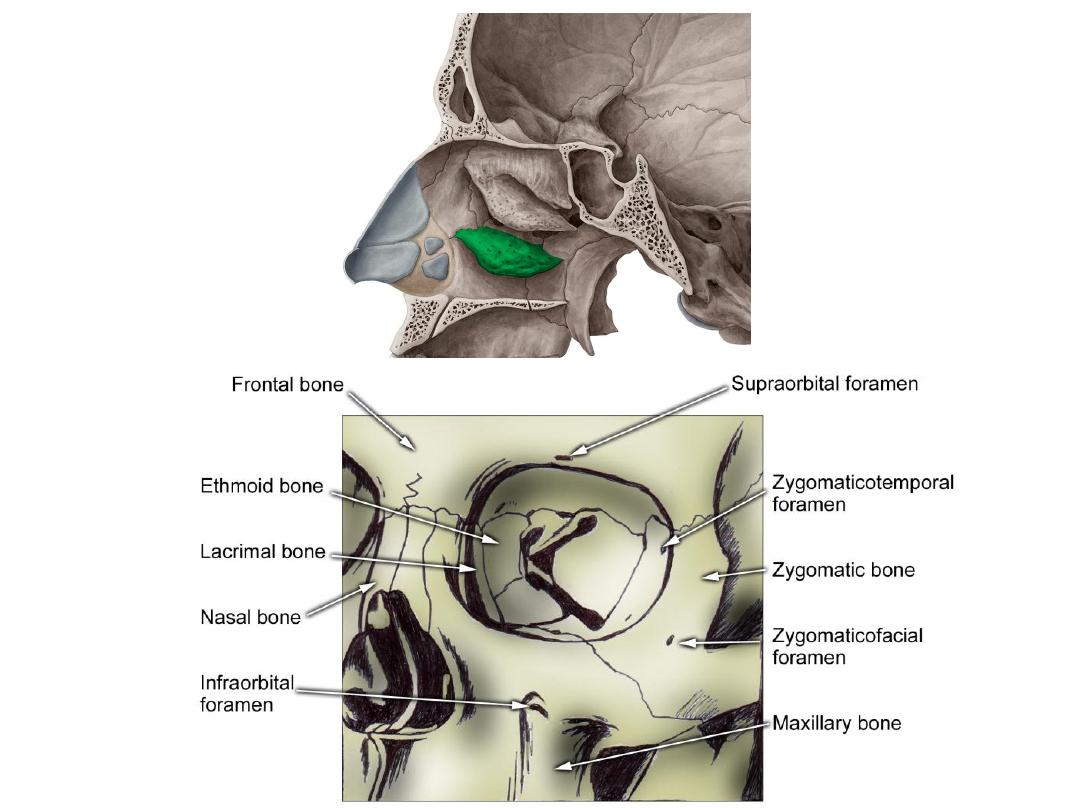
Other bones:
1- Nasal
2- Lacrimal
3- Vomer
4- Inferior concha
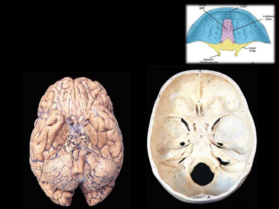
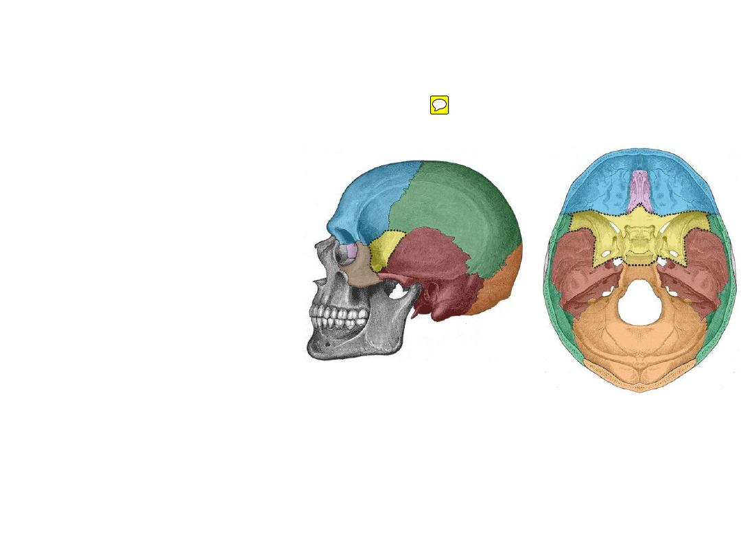
1- Anterior cranial fossa:
-
Made of: frontal bone + sphenoid
-
Overlies: the orbit, ethmoidal sinuses & nasal cavity
-
Lodges the frontal lobe of the brain
1
2
3
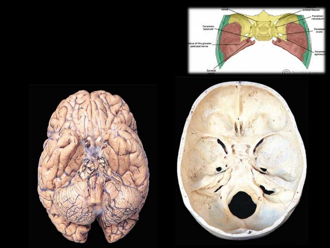
2- Middle cranial fossa:
-
Made of: temporal bone + sphenoid
-
Overlies:
the
para-pharyngeal
area,
nasopharynx
-
Lodges the temporal lobe of the brain
1
2
3
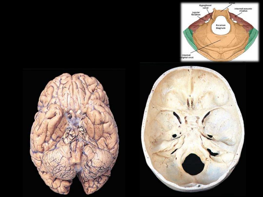
3- Posterior cranial fossa:
-
Made of: occipital bone, temporal bone & sphenoid
-
Ooverlies the neck & vertebral canal
-
Lodges the cerebellum
1
2
3