Joints disease
Imaging techniques :1.plain film examination
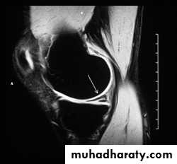
2.MRI
.Meniscal & ligamentous tears in the knee .
.Rotator cuff tears of the shoulder .
.Avascular necrosis of the hip.
.Septic arthritis.
3.Artherography.
Plain film sings of joint disease .
I.Signs indicating the presence of an arthritis.
1.Joint space narrowing .
2.Soft tissue swelling.
3.Osteoporosis.
II. Sings that point to the cause of an arthritis
1.Articular erosions: *Inflammatory overgrowth of the synovium ( pannus ) which occurs in
.Rheumatoid arthritis.
.Juvenile RA ( stills disease )
.Juvenile RA ( stills disease )
.Psoriasis .
.Reiter disease.
.Ankylosing spondylitis
.Tuberculosis .
Response to the deposition of urate crystals in gout .*
*Destruction caused by infection ( pyogenic & tuberculos arthritis ).
*Synovial over growth produced by repeated hemorrhage in hemophilia & relating bleeding disorders .*Newplastic overgrowth of synovium e.g synovial sarcoma.
2.Osteophytes ,subchondral sclerosis & cyst.
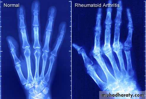
3.Alteration in the shape of the joint.Rheumatoid arthritis :
The changes seen in plain radiograph :Periarticular soft tissue swelling and osteoporosis.
Joint space narrowing .
Bony erosions.
Ulnar deviation.
With severe disease , there may be subluxation at the atlantoaxial joint.
Juvenile rheumatoid arthritis (still sdisease ; juvenile chronic poly arthritis ):
Shows many features similar to RA but erosions are less prominent . the knee , ankle and wrist are the joints most commonly affected .
psoriasis
There is an erosive arthreopathy with predominant involvement of the terminal interphalangeal joints.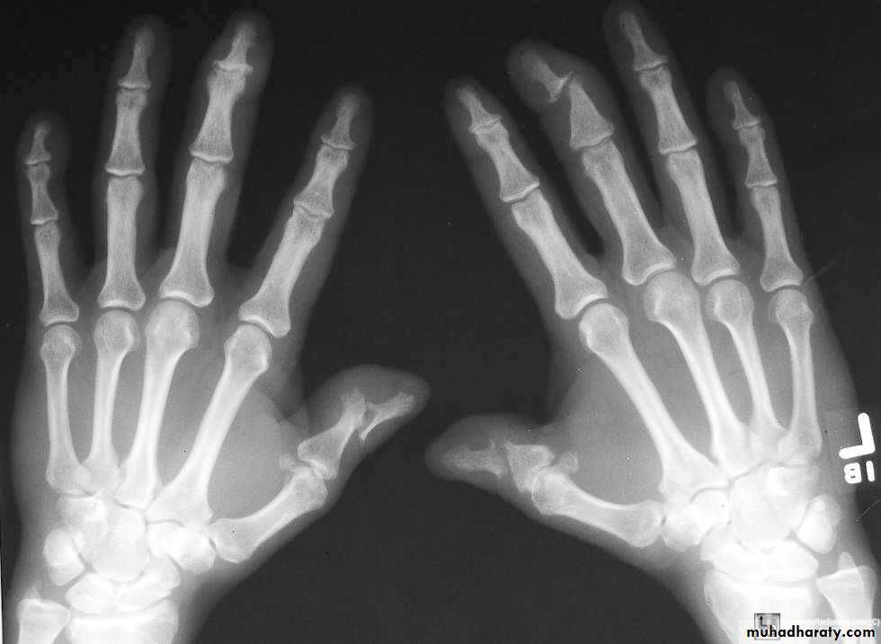
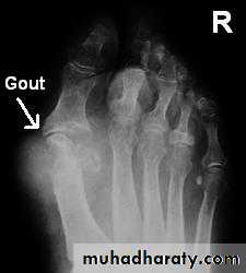
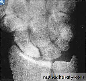
Gout :
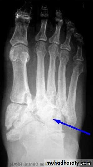
In gout ,the deposition of the urate crystals in the joint & in the adjacent bone gives arise to an arthritis which most commonly affects the metatarsophalangeal joint of the big toe .Radiological changes involving :
1.soft tissue swelling 2 . erosions 3 . usually no osteoporosis .4 . Tophi.
Pyogenic arthritis :
Usually caused by staphylococcus aureus , there is rapid destruction of the articular cartilage followed by desdtruction of the subchondral bone and soft tissue swelling seen around joint . Joint effusion is first sing which can be detected by U/S , MRI is often performed if diagnosis is still in doubt.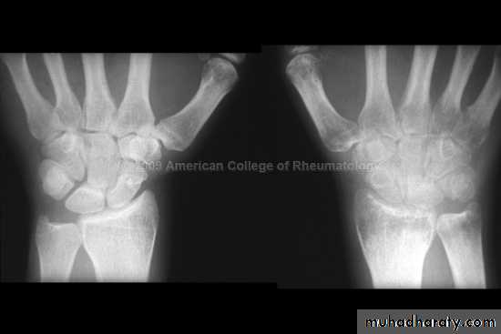
Tuberculous arthritis:
Hip & knee joint are most commonly affected joints radiological features1.Joint space narrowing 2.Erosions which may cause extensive destruction of the articular cortex 3. Severe osteoporosis 4. In later stages there may be gross disorganization of the joint with calcifiy debris near the joint.
Hemophilia & bleeding disrorder :
Repeated haemorrahge in to the joints result in soft tissue swelling , erosions and cyst in subchondral bone , the epiphysis may enlarge & fuse prematurely .
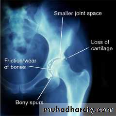
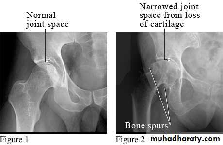
Osteoarthritis :
OA is the commonest form of arthritis , resulting from wear & teat of the articular cartilage .Features seen in OA :
1.joint space narrowing. 2. Osteophytes. 3. Subchondral sclerosis . 4. Subchondral cysts.
OA
RA
1.joint space narrowing maximal at weight- bearing site.
2. erosions don’t occur but crumbling of the joint surface may mimic erosions.
3.subchindral cyst & sclerosis may be seen.
4.sclerosis is a prominent feature.
5. no osteoporosis.
1.joint space narrowing uniform.2. Erosions a characteristic feature.
3. not a feature but erosion enface may mimic cysts.
4. sclerosis is not a feature unless there is secondary OA.
5. osteoporosis often present .
Neuropathic joint :
Changes are seen in the feet of diabetics with peripheral neuropathy.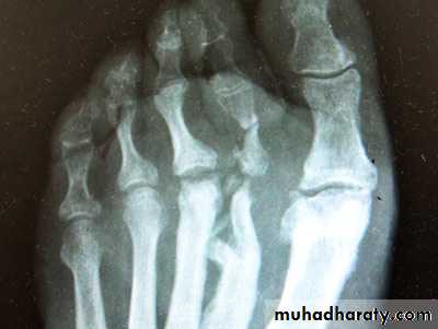
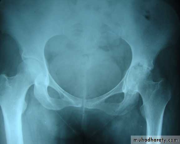
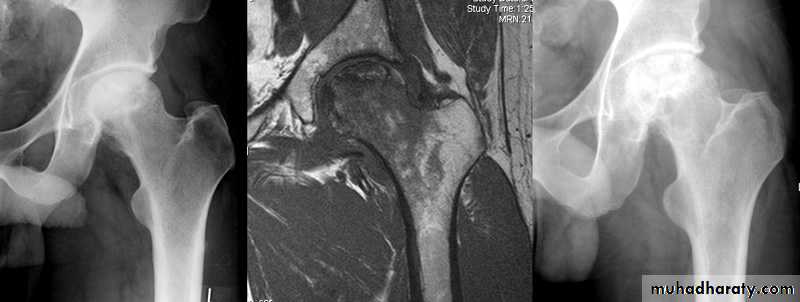
A vascular ( a septic ) necrosis :
Avascular necrosis , also known as osteonecrosis , is where there is death of bone due to interruption of the blood supply.The plain radiographic features including :
1. Increase density of the subchondral.
2. crescentic lucent line may be seen just beneath the articular cortex.
3.Articular cartilage space is preserved until degenerative changes super even .
4.In sub capital fracture of the femoral neck & fracture through the waist of the scaphoid bone , the femoral head & proximal pole of the scaphoid become fragmented & dense due to the ischemia .
MRI is imaging modality of choice for demonstrating a vascular necrosis and may show changes at time so early when radiograph may be normal .
Osteochondritis :
A group of conditions , in which no associated cause for a vascular necrosis can be found . But the osteochondritis are now regarded as being due to impaired blood supply associated with repeated trauma .A-Perth's disease :
A vascular necrosis of the femoral head in children , is the most important example of the osteochondritis . the plain radiograph changes :
1.the earliest change is increase in density and flattening of the femoral epiphyses which later on progress to collapse & fragmentation.
2.epiphysis widened & consequently the femoral neck enlarge and may contain small cyst .
3.joint space is widened but the accetabulum not affected .
4.with healing, the femoral head reforms but remain permanently flattened & therefore responsible for OA in later life .
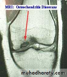
Osteochondritis dissecans :
It is localized form of the a vascular necrosis , small fragment of bone becomes separated from the articular surface of a joint leaving a defect , and the bony fragment can often be detected lying free within the joint.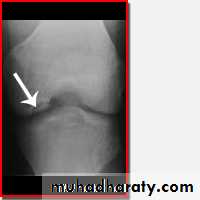
Slipped femoral epiphysis :
Occurs between the age of 9 and 17 years , and may present with pain in the hip or pain referred to the knee. the epiphysis slips posteriorly from normal position it is best appreciated on a lateral film of the hip , with a greater degree of slip , the condition can be recognized on the frontal view as down ward displacement of the epiphysis .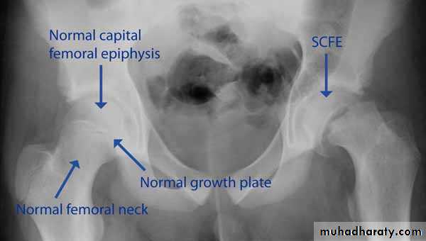
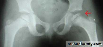
Developmental dysplasia of the hip (DDH ) :
Referring to the dislocation or subluxation of the hip in infant . Best imaging modality is U/S as the head still cartilaginous in this age group not seen in plain film , but later in life condition is easier to DX by plain film and the features are lateral and upper displacement of the head of the femur, increased slope to the acetabular roof is sometimes seen .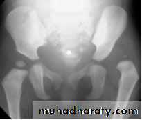
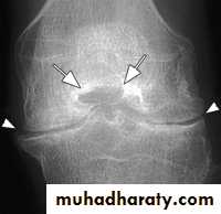
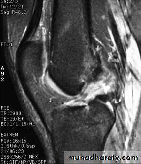
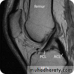
Internal derangements of the knee joint:
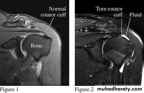
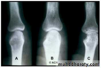
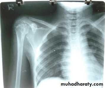
Shoulder rotator cuff degeneration /tear:
Is best evaluated by MRI , showing supraspinatus tear with high sensitivity.