Development of the Placenta
In human placenta the trophoblast erodes into the decidua, so that the endothelium of the maternal blood vessels is destroyed & maternal blood is in direct with the chorion, without the intervention of any decidual tissue (haemochorial placenta).The trophoblast soon arranged in trabeculae, which are covered by syncytiotrophoblast.When the embryonic mesoderm appears it extends into each of these trabecula & finally the vascularization of the mesoderm complete the formation of chorionic villi by about the 16th day after fertilization.
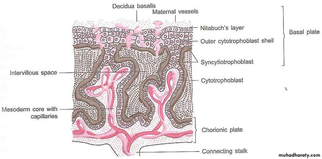
The trophoblast at some point comes into direct contact with the decidua, thus anchoring villi are formed, then by budding from both them & the chorion, true chorionic villi are formed.
The trophoblast extend for a variable distance into maternal spiral arterioles where they enter the intervillous space. After the 20th week the cytotrophoblast disappear & finally only thin layer of syncytium remains.
At first the villi are formed over all the surface of the gestational sac (at 4th week). Between 12th-16th week the villi on the capsular surface degenerate & become smooth called the chorion leave. In compensation the villi on the decidua basalis undergo great hypertrophy called chorion frondosum & its matted into solid disc which is the fully developed placenta (formed by the 12th week).
Placenta at Term-
FleshyWeight-500gm
Diameter- 15-20 cm
Thickness-2.5 cm
Spongy to feel
Occupies 30% of the uterine wall
Two surfaces- Maternal and fetal
4/5th of the placenta is of fetal origin and 1/5 is of maternal origin
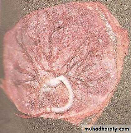
Fetal surface of the placenta
Covered by smooth and glistening
amnion overlying the chorion
Umbilical cord is attached
at or near its centreBranches of the umbilical vessels
are visible beneath the amnionas they radiate from the insertion
of the cord
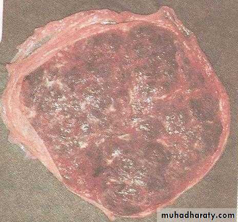
Maternal surface of the placenta
Rough and spongyMaternal blood gives it
dull red colour
Remanants of the decidua basalis
gives it shaggy appearance
Divided into 15-20 cotyledons
by the septa
Placental Function
-Transfer of gases ,nutrients and waste products , namelyRespiratory function
Nutritive function
Excretory function
-Endocrine and enzymatic function
-Barrier function
-Immulogical function
Normal placentation:
The maternal flow to the placenta increases throughout pregnancy from 50mL/min in the 1st trimester to 600mL/min at term. This increase in perfusion can only be accomplished by the anatomical conversion of the maternal spiral arteries by trophoblast, from narrow tortuous muscular vessels to wide-bored flaccid vessels.In the first 12 weeks the decidual segment of the spiral a. is invaded by trophoblast & fibrinoid. Following this, the trophoblast invasion of the intramyometrial segment of the spiral arteries which further reduces resistance to blood flow to the placenta.
This process should be complete by 20 week, also because they lack smooth muscle, they are less likely to respond to vaso-active compound
Abnormal placentation :
Pre-eclampsia.Intrauterine growth restriction(IUGR).
Abruptio placentae.
Collagen vascular disease.
Antiphospholipid syndrome.
Sever D.M.
Chronic hypertension.
All these are clinical manifestations of total or patchy failure of trophoblast invasion of the myo-metrial segments of the spiral arteries. All these result in a small placenta with gross morphological changes which are :
Infracts represents an area of ischemic necrosis of cotyledon resulting from spiral a. occlusion, usually by thrombosis.
Basal haematomas consist of a mass of blood in the centre of the cotyledon due to the rupture of the damaged spiral artery these pathological condition associated with increased perinatal mortality.
Anomalies in weight: In cases of diabetes & haemolytic disease of the newborn the placental wt may increase to up to half the wt of the fetus.
Site of implantation of the placenta: The placenta usually attached to the uterine wall near the fundus, to either the anterior or posterior surface. In about 1 in 250 pregnancies the placenta is implanted wholly or partially on the lower segment of the uterus (placenta previa). This is a serious abnormality which may cause severe haemorrahge in pregnancy or labour.
Bilobate & trilobate placenta: Instead of a single disc , it may consist of 2 or 3 lobes partly fused( of no clinical importance).
Morbid adherence of the placenta: In 3rd stage of labour the placenta normally separates through the stratum spongiosum of the maternal decidua( the superficial part of the decidua comes away with the placenta & the deeper part remains on the uterine wall), normally the chorionic villi only penetrate as far as this.
Morbid adherence of the placenta results from increased penetration of the decidua & myometrium by the villi. The degree of morbidity is determined by the depth of invasion.
1.Placenta accrete: the placenta is partially or completely adherent to the uterus with penetration of villi into the superficial part of the myometrium.
2.Placenta increta: the villi penetrate deeply through the decidua into the myometrium.
3.Placenta percreta : penetration can even be seen on the serosal surfacePlacenta previa, c.s, curettage are the most pre-disposing causes.
There is delay in the 3rd stage of labour with PPH & the abnormalities is only discovered when an attempt to remove the placenta manually & no plane of cleavage is found.Morbid adherence is of great importance clinically because it makes it impossible to remove the placenta completely thus exposing the mother to risk of sever PPH & it may end with hysterectomy.
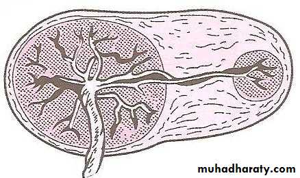
Placenta succenturiata (3%)
One or more small lobe or cotyledon
of placenta may be placed at a varying
distance from the main placental margin
A leash of vessels connecting the main
to the small lobe traverse throughthe membranes
Clinical significance-
If succenturiate lobe is retained following birth of placenta it may lead toPPH
Subinvolution
Uterine sepsis
Treatment- exploration of the uterus and removal of the lobe.
Circumvallate placenta:
Development-Due to smaller chorionic plate than the basal plate
The chorionic plate does not extend into the placenta margin
Morphology
Fetal surface has a central depressed zone surrounded by a usually complete thickened white ring made up of double fold of amnion and chorion
Branching vessels radiate from the cord insertion up to ring only
Area outside the ring is thicker, elevated and rounded
Clinical significance
There are more chances of –
Miscarriage
Hydrorrhoea gravidarum
Antepartum haemorrhage
Preterm delivery
Fetal growth restriction
Retained placenta or membrane
Chorioangioma:
Are the most common benign tumors of the placenta and are hamartomas of primitive chorionic mesenchymeSmall tumors may be asymptomatic but large tumors may be associated with hydroamnios and antepartum haemorrhage .
Hydropic placenta: In sever cases of isoimmunization (hydrops fetalis) the placenta show the same changes of fetus, being enlarged, pale & oedematous with a marked increase in weight.
The umbilical cord:
-Abnormal length: The usual length, same as fetus at term 50cmExcessive length .
Short cord.
-Knots in the cord: These may be formed by fetal movement.
-Abnormal insertion of the cord: The cord usually attached to the centre of the placenta, but sometimes attached to the edge of placenta ( squash racket placenta) of no clinical importance. In very rare cases the cord is attached to the membrane at some distance from the edge of the placenta, at this point the vessel may divide into branches which run on the membrane before reaching the placenta (velamentous insertion of the cord ).
-This can be dangerous to the fetus if the vessels happen to pass across part of the chorion that lies below the presenting part( vasa previa), as a branch may be torn when the membrane rupture, leading to fetal blood loss.
-Single umbilical artery: This is uncommon, but can be associated with other abnormalities of the fetus, notably those of the kidneys, ureters or bladder.