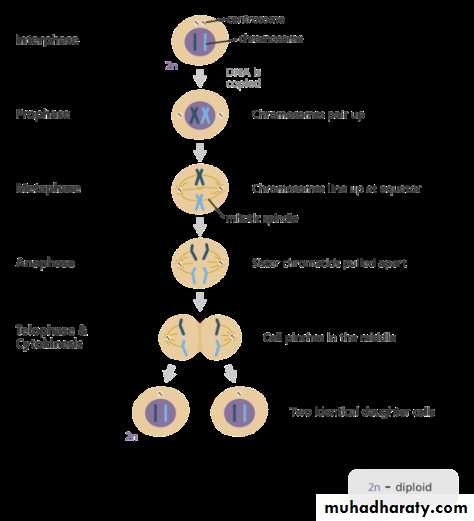
Cell cycle
MitosisMitosis is a process where a single cell divides into two identical daughter cells (cell division). During mitosis one cell divides once to form two identical cells and the DNA of the cell's nucleus is split into two equal sets of chromosomes.. The major purpose of mitosis is for growth and to replace worn out cells with new ones.
If not corrected in time, mistakes made during mitosis can result in changes in the DNA, that can potentially lead to genetic disorder.
Phases of mitosis
After interphase of cell cycle, mitosis consists of four basic phases: prophase, metaphase, anaphase, and telophase. Some textbooks list five, breaking prophase into an early phase (called prophase) and a late phase (called prometaphase). These phases occur in strict sequential order, and cytokinesis ( the process of dividing the cell contents to make two new cells) starts in anaphase or telophase.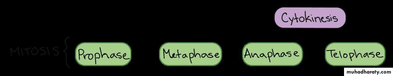
Prophase:
The chromosomes condense into X-shaped structures that can be easily seen under a microscope.Each chromosome is composed of two sister chromatids, containing identical genetic information. The chromosomes pair up so that both copies of each chromosome are together.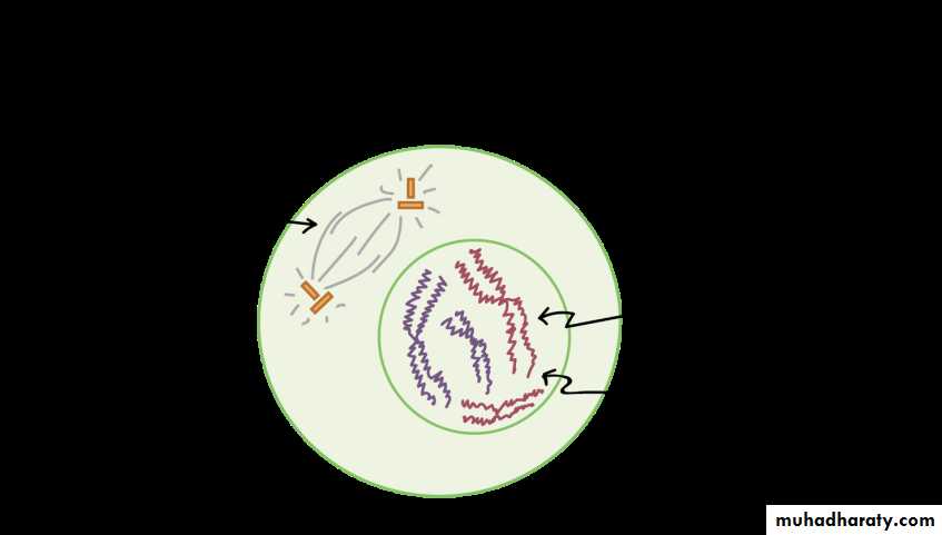
At the end of prophase the membrane around the nucleus in the cell dissolves away releasing the chromosomes.The mitotic spindle, consisting of the microtubules and other proteins, extends across the cell between the centrioles as they move to opposite poles of the cell and the mitotic spindle begins to capture and organize the chromosomes.
Metaphase:
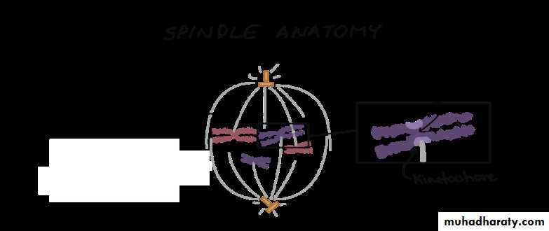
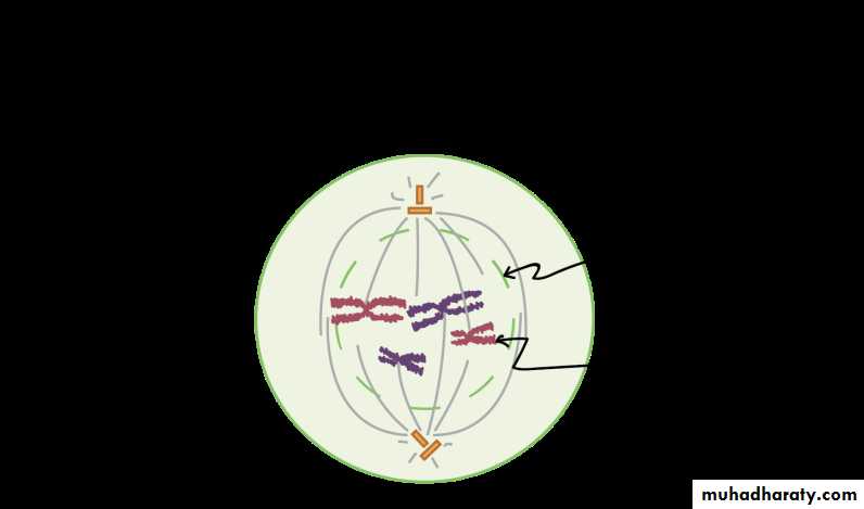
The chromosomes line up neatly end-to-end along the center (equator) of the cell. The centrioles are now at opposite poles of the cell with the mitotic spindle fibers extending from them.The mitotic spindle fibers attach to each of the sister chromatids.
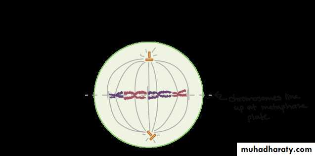
In metaphase, the spindle has captured all the chromosomes and lined them up at the middle of the cell, ready to divide.
All the chromosomes align at the metaphase plate (not a physical structure, just a term for the plane where the chromosomes line up).
At this stage, the two kinetochores of each chromosome should be attached to microtubules from opposite spindle poles.
Before proceeding to anaphase, the cell will check to make sure that all the chromosomes are at the metaphase plate with their kinetochores correctly attached to microtubules. This is called the spindle checkpoint and helps ensure that the sister chromatids will split evenly between the two daughter cells when they separate in the next step. If a chromosome is not properly aligned or attached, the cell will halt division until the problem is fixed.
Anaphase:
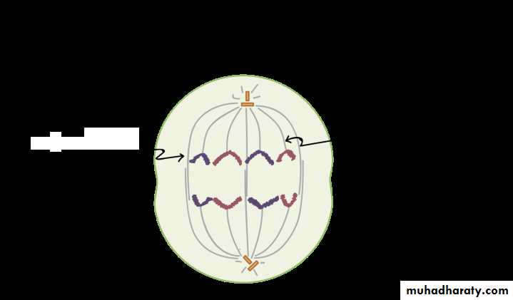
The sister chromatids are then pulled apart by the mitotic spindle which pulls one chromatid to one pole and the other chromatid to the opposite pole.
In anaphase, the sister chromatids separate from each other and are pulled towards opposite ends of the cell.
The protein “glue” that holds the sister chromatids together is broken down, allowing them to separate. Each is now its own chromosome. The chromosomes of each pair are pulled towards opposite ends of the cell.
Microtubules not attached to chromosomes elongate and push apart, separating the poles and making the cell longer.
All of these processes are driven by motor proteins, molecular machines that can “walk” along microtubule tracks and carry a cargo. In mitosis, motor proteins carry chromosomes or other microtubules as they walk.
Telophase:
At each pole of the cell a full set of chromosomes gather together.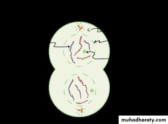
A membrane forms around each set of chromosomes to create two new nuclei.The single cell then pinches in the middle to form two separate daughter cells each containing a full set of chromosomes within a nucleus. This process is known as cytokinesis.
In telophase, the cell is nearly done dividing, and it starts to re-establish its normal structures as cytokinesis (division of the cell contents) takes place.
The mitotic spindle is broken down into its building blocks.
Two new nuclei form, one for each set of chromosomes. Nuclear membranes and nucleoli reappear.
The chromosomes begin to decondense and return to their “stringy” form.
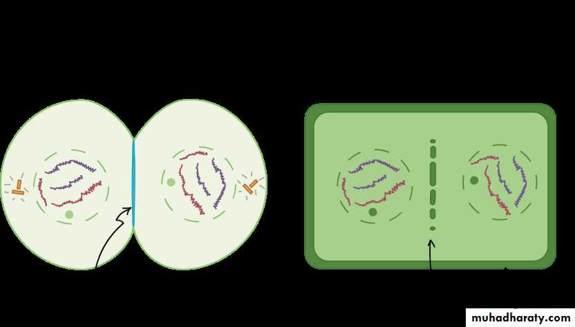
Cytokinesis, the division of the cytoplasm to form two new cells, overlaps with the final stages of mitosis. It may start in either anaphase or telophase, depending on the cell, and finishes shortly after telophase.
In animal cells, cytokinesis is contractile, pinching the cell in two like a coin purse with a drawstring. The “drawstring” is a band of filaments made of a protein called actin, and the pinch crease is known as the cleavage furrow. Plant cells can’t be divided like this because they have a cell wall and are too stiff. Instead, a structure called the cell plate forms down the middle of the cell, splitting it into two daughter cells separated by a new wall.
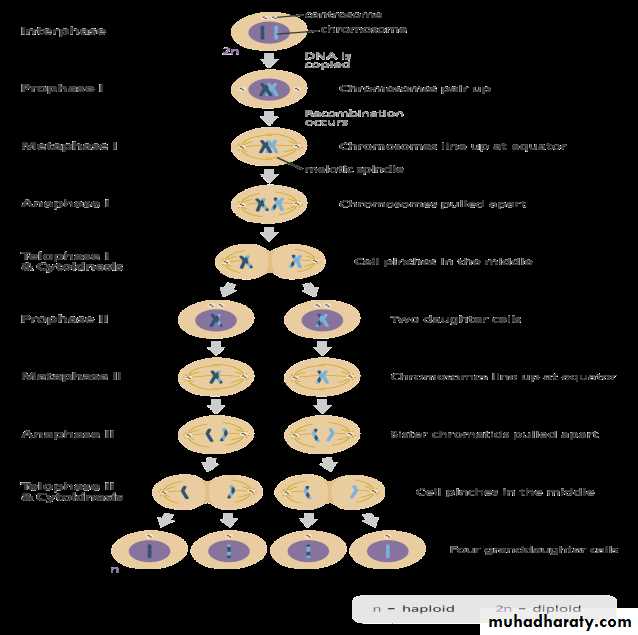
Meiosis
Meiosis is a process where a single cell divides twice to produce four cells containing half the original amount of genetic information. These cells are sex cells – sperm in males, eggs in females.During meiosis one divides twice to form four daughter cells.
These four daughter cells only have half the number of chromosomes of the parent cell – they are haploid.
Meiosis can be divided into nine stages. These are divided between the first time the cell divides (meiosis I) and the second time (meiosis II):
Meiosis I
Interphase:
The DNA in the cell is copied resulting in two identical full sets of chromosomes.
Outside of the nucleus are two centrosomes, each containing a pair of centrioles, these structures are critical for the process of cell division.
During interphase, microtubules extend from these centrosomes.
Prophase I:
The copied chromosomes condense into X-shaped structures that can be easily seen under a microscope.
Each chromosome is composed of two sister chromatids containing identical genetic information.
The chromosomes pair up so that both copies of each chromosome.
The pairs of chromosomes may then exchange bits of DNA in a process called recombination or crossing over.
At the end of Prophase I the membrane around the nucleus in the cell dissolves away, releasing the chromosomes.
The meiotic spindle, consisting of microtubules and other proteins, extends across the cell between the centrioles.
Metaphase I:
The chromosome pairs line up next to each other along the center (equator) of the cell.
The centrioles are now at opposites poles of the cell with the meiotic spindles extending from them.
The meiotic spindle fibers attach to one chromosome of each pair.
Anaphase I:
The pair of chromosomes are then pulled apart by the meiotic spindle, which pulls one chromosome to one pole of the cell and the other chromosome to the opposite pole.
In meiosis I the sister chromatids stay together. This is different to what happens in mitosis and meiosis II.
Telophase I and cytokinesis:
The chromosomes complete their move to the opposite poles of the cell.
At each pole of the cell a full set of chromosomes gather together.
A membrane forms around each set of chromosomes to create two new nuclei.
The single cell then pinches in the middle to form two separate daughter cells each containing a full set of chromosomes within a nucleus. This process is known as cytokinesis.
Meiosis II
Prophase II:
Now there are two daughter cells, each with 23 chromosomes (23 pairs of chromatids).
In each of the two daughter cells the chromosomes condense again into visible X-shaped structures that can be easily seen under a microscope.
The membrane around the nucleus in each daughter cell dissolves away releasing the chromosomes.
The centrioles duplicate.
The meiotic spindle forms again.
Metaphase II:
In each of the two daughter cells the chromosomes (pair of sister chromatids) line up end-to-end along the equator of the cell.
The centrioles are now at opposites poles in each of the daughter cells.
Meiotic spindle fibers at each pole of the cell attach to each of the sister chromatids.
Anaphase II:
The sister chromatids are then pulled to opposite poles due to the action of the meiotic spindle.
The separated chromatids are now individual chromosomes.
Telophase II and cytokinesis:
The chromosomes complete their move to the opposite poles of the cell.
At each pole of the cell a full set of chromosomes gather together.
A membrane forms around each set of chromosomes to create two new cell nuclei.
This is the last phase of meiosis, however cell division is not complete without another round of cytokinesis.
Once cytokinesis is complete there are four granddaughter cells, each with half a set of chromosomes (haploid):
in males, these four cells are all sperm cells
in females, one of the cells is an egg cell while the other three are polar bodies (small cells that do not develop into eggs).
Types of Genetic Diseases
There are several types of genetic disorders. The way in which the disorder is inherited can help determine the risks it will have on a pregnancy and the risk of recurrence it will recur in future children. Risks for having a baby with a birth defect from a genetic abnormality may be increased when:The parents have another child with a genetic disorder.
There is a family history of a genetic disorder.
One parent has a chromosomal abnormality.
The fetus has abnormalities seen on ultrasound.
The types of genetic disease:
Chromosomal abnormalities
Single gene defects
Multifactorial problems
Teratogenic problems
Chromosomal abnormalities:
Chromosomal abnormalities in the baby may be inherited from the parent or may occur with no family history. It is include:
Aneuploidy. More or fewer chromosomes than the normal number, including:
Down syndrome (trisomy 21). Cells contain three #21 chromosomes.
Turner syndrome. One of the two sex chromosomes is not transferred, leaving a single X chromosome, or 45 total.
Deletion. Part of a chromosome is missing, or part of the DNA code is missing.
Inversion. When a chromosome breaks and the piece of the chromosome turns upside down and reattaches itself. Inversions may or may not cause birth defects depending on their exact structure.
Translocation. A rearrangement of a chromosome segment from one location to another, either within the same chromosome or to another.
Balanced translocation. The DNA is equally exchanged between chromosomes, and none is lost or added. A parent with a balanced translocation is healthy, but he or she may be at risk for passing unbalanced chromosomes in a pregnancy.
Robertsonian translocation. A balanced translocation in which one chromosome joins the end of another.
Mosaicism. The presence of two or more chromosome patterns in the cells of a person, resulting in two or more cell lines (for example, some with 46 chromosomes, others with 47).
Single gene disorders:
These are also known as Mendelian inheritance disorders. In these disorders, a single gene is responsible for a defect or abnormality. Single gene disorders usually have greater risks of inheritance. Single gene disorders can be:
Dominant. An abnormality occurs when only one of the genes from one parent is abnormal. If the parent has the disorder, the baby has a 50 percent chance of inheriting it. It is include the following:
Achondroplasia. Imperfect bone development causing dwarfism.
Marfan syndrome. A connective tissue disorder causing long limbs and heart defects.
Recessive. An abnormality only occurs when both parents have abnormal genes. If both parents are carriers, a baby has a 25 percent chance of having the disorder. Examples include the following:
Cystic fibrosis. A disorder of the glands causing excess mucus in the lungs and problems with pancreas function and food absorption.
Sickle cell disease. A condition causing abnormal red blood cells.
Tay-Sachs disease. An inherited autosomal recessive condition that causes a progressive degeneration of the central nervous system, which is fatal (usually by age 5).
X-linked. The disorder is determined by genes on the X chromosome. Males are mainly affected and have the disorder. Daughters of men with the disorder are carriers of the trait and have a one in two chance of passing it to their children. Sons of women who are carriers each have a one in two chance of having the disorder. Examples include the following:
Duchenne muscular dystrophy. A disease of muscle wasting.
Hemophilia. A bleeding disorder caused by low levels, or absence of, a blood protein that is essential for clotting.
Multifactorial problems:
Some birth defects do not follow a single gene or chromosomal abnormality pattern. They may be due to several problems, or a combined effect of genes and the environment. It is difficult to predict inheritance of abnormalities caused by multiple factors. Examples include heart defects, cleft lip or cleft palate, and neural tube defects (defects in the spine or brain).
Teratogenic problems:
Certain substances are known to cause abnormalities in babies. Many birth defects occur when the fetus is exposed to teratogens (substances that cause abnormalities) during the first trimester of pregnancy when organs are forming. Some known teratogens include the following:
Certain medications (always consult your doctor before taking any medications during pregnancy).
Alcohol.
High level of radiation exposure.
Lead.
Certain infections (such as Rubella).