Types of tissues
The human tissues can be divided into 4 types:Connective tissue.
Muscular tissue.
Nervous tissue.
Epithelial tissue.
Connective tissue
It is a specialized cells, ground substance and protein fibers. Originated its name because it connect between tissues, The role of connective tissue is to protect, support, and bind together parts of the body.
Connective tissues tend to be very vascular (have a rich blood supply). Some exceptions, such as tendons, ligaments, and cartilages, are less vascularized, but overall, connective tissues possess a great blood supply than the epithelial tissue previously discussed.
Connective tissues are made up of many types of specialized cells.
Connective tissues contain a large amount of non-living material referred to as the matrix (composed of ground substance and fibers
There are four types of connective tissues found in the human body:
Connective tissue proper
Loose Connective Tissue
Areolar : cushion around organs, loose arrangement of cells and fibers.
Adipose: Adipose tissue , in which adipocytes ( crowded and filled with liquid fat), use for energy , insulation , organ protection and release of leptin to regulates appitite-control centers in the brain. It lies under the skin, around the kidneys and on the heart surface.
Reticular: internal supporting framework of some rgans, delicate network of fibers and cells
Dense Connective Tissue
Dense regular : tendons and ligaments, regularly arranged bundles packed with fibers running same way for strength in one direction.
Dense irregular : skin, organ capsules, irregularly arranged bundles packed with fibers for strength in all directions.
Elastic
Cartilage
Cartilage in which the cells found in lacunae in solid flexible matrix produced by chondroblasts and chondrocytes. According to the type of fiber found in matrix the cartilage can be divided into:
Hyaline cartilage ( fine collagen fibers) found in nose , ends of long bone, ribs and ring of respiratory passages.
Elastic cartilage ( elastic fibers) found in the framework of outer ear.
Fibrocartilage (strong collagen fibers) found in intervertebral disc and cushions of the knee joint.
Bone (osseous tissue) : rigid connective tissue, composed of calcium salts, collagen fibers and osteoblasts and osteoclasts which produce of hard matrix. According to rigidity the bone divided into :
Compact bone ( long bone) it composed of cylindrical structure (osteon) surrounded by hard matrix, bone cells and central canal which carry nerves and blood vessel.
Spongy bone ( ends of long bone) it composed of bony latticework
Fluid connective tissue
Blood which composed of elements and plasma, found in the blood vessels, it is include:
RBCs.
WBCs.
Platelets.
Lymph which is clear-yellow fluid derived from surrounding the tissues , it composed of WBCs.
Muscular tissues
It is composed of muscle fibers which contain actin and myosin filaments.
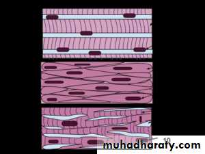
Skeletal muscle ( voluntary muscle) it is striated by cylindrical, long fibers , fused cells forming one fiber with multiple nuclei.
Smooth muscle ( involuntary muscle) it is smooth (lack of striation), composed of spindle-shaped cells with single nucleus. It found in the walls of viscera.
Cardiac muscle (involuntary muscle ) which found in the heart , it have features of both skeletal and smooth muscle. It is characterized by intercalated disk.
Smooth muscle
Smooth muscleNervous tissues
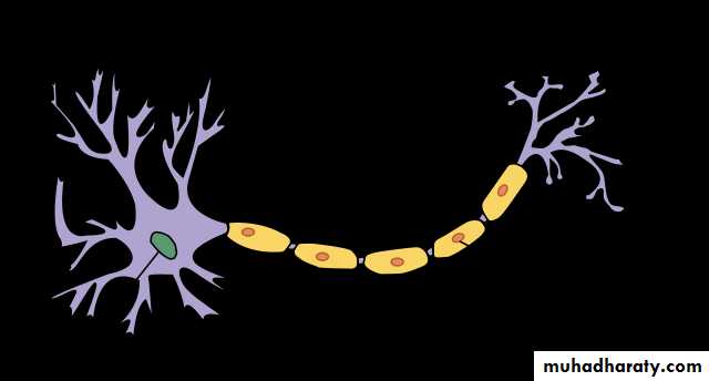
It is composed of nerves cells (neurons) and neuroglia.Neurons which have dendrites, cell body and axon. The dendrite for receive a signal from other neuron , cell body have cytoplasm and nucleus, the axon for conducting of impulses. Fiber is used to subject of axon + myelin sheath.
Neuroglia ( microglia, astrocytes and oligodendrocytes) is a cell that support and nourish the neuron, it have no axon or dendrites.
Epithelial tissues
Epithelium forms the coverings of surfaces of the body. As such, it serves many purposes, includingIt protects us from the outside world like skin.
Absorbs – stomach and intestinal lining like gut.
Filters like kidney.
Secretes like glands.
Important notes:
Polarity- Epithelium is arranged so there is one free surface (apical surface) and one attached surface (basal surface)
Cellular nature- Cells in epithelium fit closely together side by side and sometimes atop each other to form sheets of cells. These sheets are held together by specialized junctions.
Supported by connective tissue- Attachment to a layer of connective tissue at the basal surface forms a layer called the basement membrane, an adhesive layer formed by secretions from the epithelial cells and the connective tissue cells.
Avascular- Epithelium typically lacks its own blood supply.
Regeneration- Epithelium cells can regenerate if proper nourished.
Classification according to the following criteria:
Arrangements:Simple- Cells are found in a single layer attached to the basement membrane
Stratified- Cells are found in 2 or more layers stacked atop each other
Pseudostratified- a single layer of cells that appears to be multiple layers due to variance in height and location of the nuclei in the cells.
Transitional- cells are rounded and can slide across one another to allow stretching
Shapes:
Squamous- (Latin, squama- scale)- flat, thin, scale-like cells
Cuboidal- cells that have a basic cube shape. Typically the cell's height and width are about equal.
Columnar- tall, rectangular or column-shaped cells. Typically taller than they are wide.
Special Features of Epithelium:
Cilia- (singular= cilium, Latin= eyelash)- hair-like appendages attached to the apical surface of cells that act as sensory structures or to produce movement.Goblet cells- specialized cells that produce mucus to lubricate and protect the surface of an organ
Villi- (singular= villus, Latin= shaggy hair)- finger-like projections that arise from the epithelial layer in some organs. They help to increase surface area allowing for faster and more efficient adsorption.
Microvilli- smaller projections that arise from the cell's surface that also increase surface area. Due to the bushy appearance that they sometimes produce, they are sometimes referred to as the brush border of an organ.
The Integument:
The body is protected externally by one of its largest organs, the skin or integument. While protection is the main function of the skin, it performs main other functions, such as providing insulation, helping with temperature regulation, and provides tactility (sense of touch). It evens functions in the production of vitamin D needed for proper body function.Skin Composition:
The skin is formed by three distinctive layers; the epidermis (outer layer), the dermis (middle layer), and hypodermis {subcuntaneous layer} (innermost layer).Epidermis- composed of keratinized stratified squamous epithelium ,
There are two types of specialized cells:Langerhans cells: are macrophages, WBCs that phagocytize infectious agents and then travel to lymphatic organs.
Melanocytes: which produce melanin pigment that responsible for skin color. Another pigment called carotene is present in epidermis and dermis which give yellowish form (Asian peoples).
Epidermis arranged into five layers called strata (singular = stratum)
Stratum corneum- outermost layer of flattened, dead cells.
Stratum lucidum- thin, translucent layer found only in thick areas of the skin.
Stratum granulosum- names for the abundance of granules present. Upper boundary of this layer is where cells begin to die.
Stratum spinosum- layer where cells divide rapidly. Usually one of the thicker layers of the epidermis.
Stratum basale- the lowest layer of the skin. Attached to the dermis where it forms a basement membrane. Cells are constantly dividing to produce new cells.
Dermis- composed of dense irregular connective tissue.
Dermal papillae- projections or ridges that arise from the dermis that serve as attachment points for the epidermis
Hypodermis- composed of adipose tissue .
Skin also has unique structures that perform various functions:
Gland Structures
Sweat gland (sudoriferous gland)- produces sweat (mixture of water, salts, and urea) that acts to cool the body.
Sebaceous gland- produce sebum (oil) to help keep the skin soft and pliable.
Nervous Structures
Free nerve endings- associated with pain sensation; located in near dermal papillae .
Meissner’s corpuscle- touch receptors- associated with tactility; located in near dermal papillae.
Pacinian corpuscle- pressure receptors; located deep within dermis at the boundary of the dermis and hypodermis.
Muscle Structures
Arrector pili- muscle that pulls up hair follicle leading to goose flesh or “goose bumps” .Appendages
Hair shaft .
Hair follicle.
Cellular respiration
Mitochondria is the main site of:Energy metabolism and respiration : the major function of mitochondrion is produce chemical energy (ATP) from food stuffs.
Heat production : energy of oxidation is dissipated as heat instead of being converted into ATP, so mitochondrion is the site to produce heat to maintain body temperature.
Amine metabolism : some amines are metabolized in mitochondria
It's structure are well organized to do this function.The inner membrane is folded to form shelves projected into matrix called cristae, the inner space are filled with a gel-like fluid. The matrix composed of several types of enzymes for glucose breakdown. The outer membrane is a double membrane. Glucose is the main source of energy in our body, it needs to breakdown to form as energy, when the glucose molecule reach to the blood , it enters to the cells via insulin hormone.Once the glucose become inside a cell, cellular respiration is begin. There are three pathways for glucose breaking down:
1. Glycolysis : it is an anaerobic pathway which occur in the cytoplasm without oxygen by breaking the glucose molecules (C6) into two pyruvate molecules +2 ATP molecules.
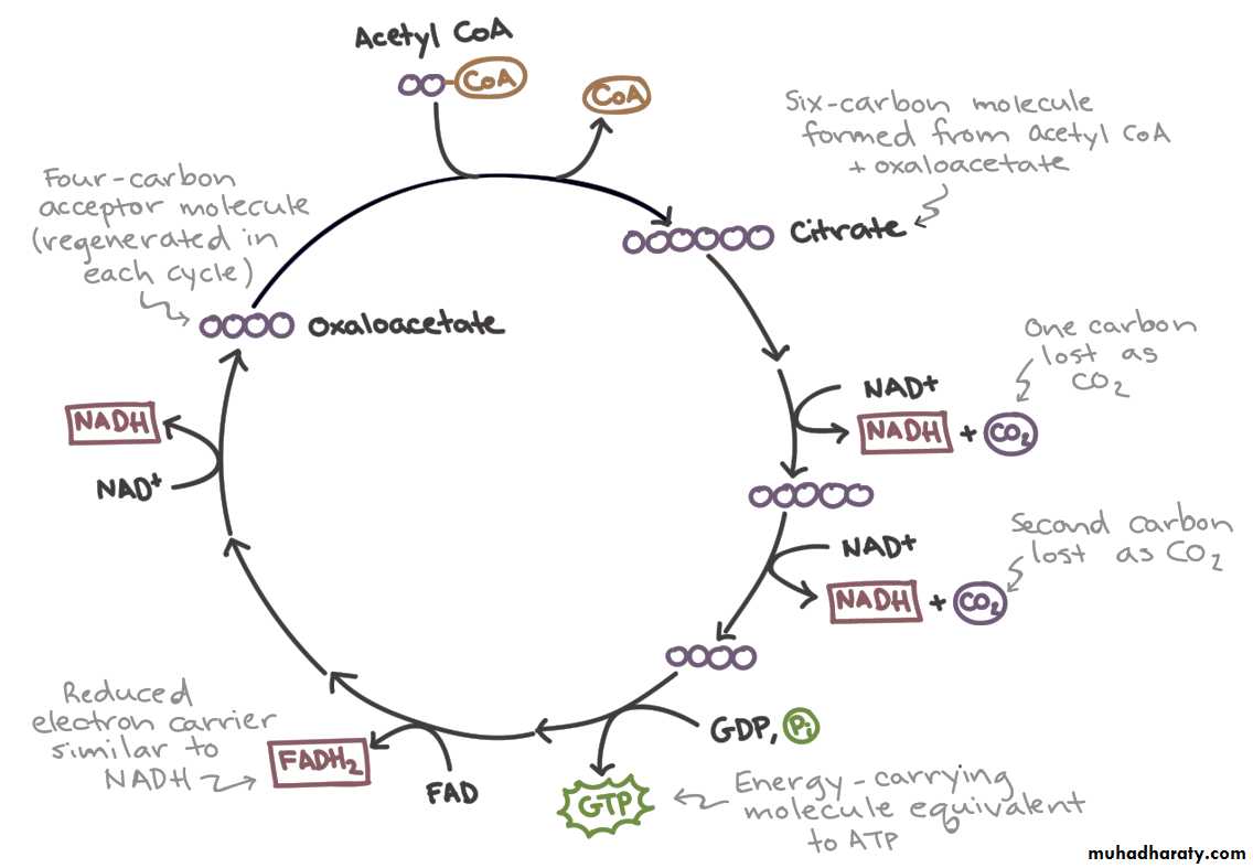
2. Citric acid cycle ( tri-carboxylic acid cycle or Krebs cycle) : it is an aerobic pathway that occur in the matrix of mitochondria to complete breakdown of glucose molecule started by enter the pyruvate molecule as acetyl CoA to produce, 4CO2 + 6 NADH +2 ATP.
3. Chemi -osmotic mechanism (Electron transport chain, ionization of hydrogen and water formation) : it is an aerobic pathway that occur in the cristae of mitochondria via a members of carrier proteins complexes that embedded in the cristae. NADH transfer electrons, from glycolysis and citric acid cycle, into electron transport chain. During the glucose breakdown a total of 24 hydrogen atoms are released during glycolysis and citric acid cycle, twenty of these atoms are oxidized with release of three ATP molecules/ two hydrogen atoms, this process gives 30 ATP. The remaining 4 hydrogen atoms are released by dehydrogenase into the electron transport chain then two ATP molecules are usually released from every two hydrogen molecules, thus giving a total of four ATP molecules. High –energy Electrons enter the chain and transport from carrier to carrier until become low- energy. The electrons finally received by oxygen molecule.
Total ATP molecules are 38 molecules for each glucose molecule degraded into CO2 +water.
4. Fermentation : it is an anaerobic pathway, in which the citric acid cycle and electron transport chain are not working because absence of oxygen molecules.
It Occurs without oxygen by recycled of NADH passes it's hydrogen and electrons to pyruvate molecules to produce lactic acid or lactate. In case of oxygen available again the lactate can be converted into pyruvate and the cellular respiration can proceed as usual. The human cell fermentation finally produce lactate , while in yeast cell fermentation finally produce alcohol and carbon dioxide.