Glaucoma:
Is
damage to the optic nerve (optic neuropathy) usually caused by raise
intraocular pressure
Intra ocular pressure depend on production and removal of aqueous humour
1. conventional pathway 96%
Cilliary process posterior chamber pupil anterior
chamber trabecular meshwork Schlemm chanaal episcleral
veins
2. uveoscleral pathway 4 % drain directly by diffusion to suprachoroidal
space to venous circulation
The mechanism by which an elevated intraocular pressure damages nerve
fibres
:
mechanical damage to the optic nerve axons.
ischemia of the nerve axons by reducing blood flow
Ophthalmology
Zakho hospital
Glaucoma
20 May 2017
2
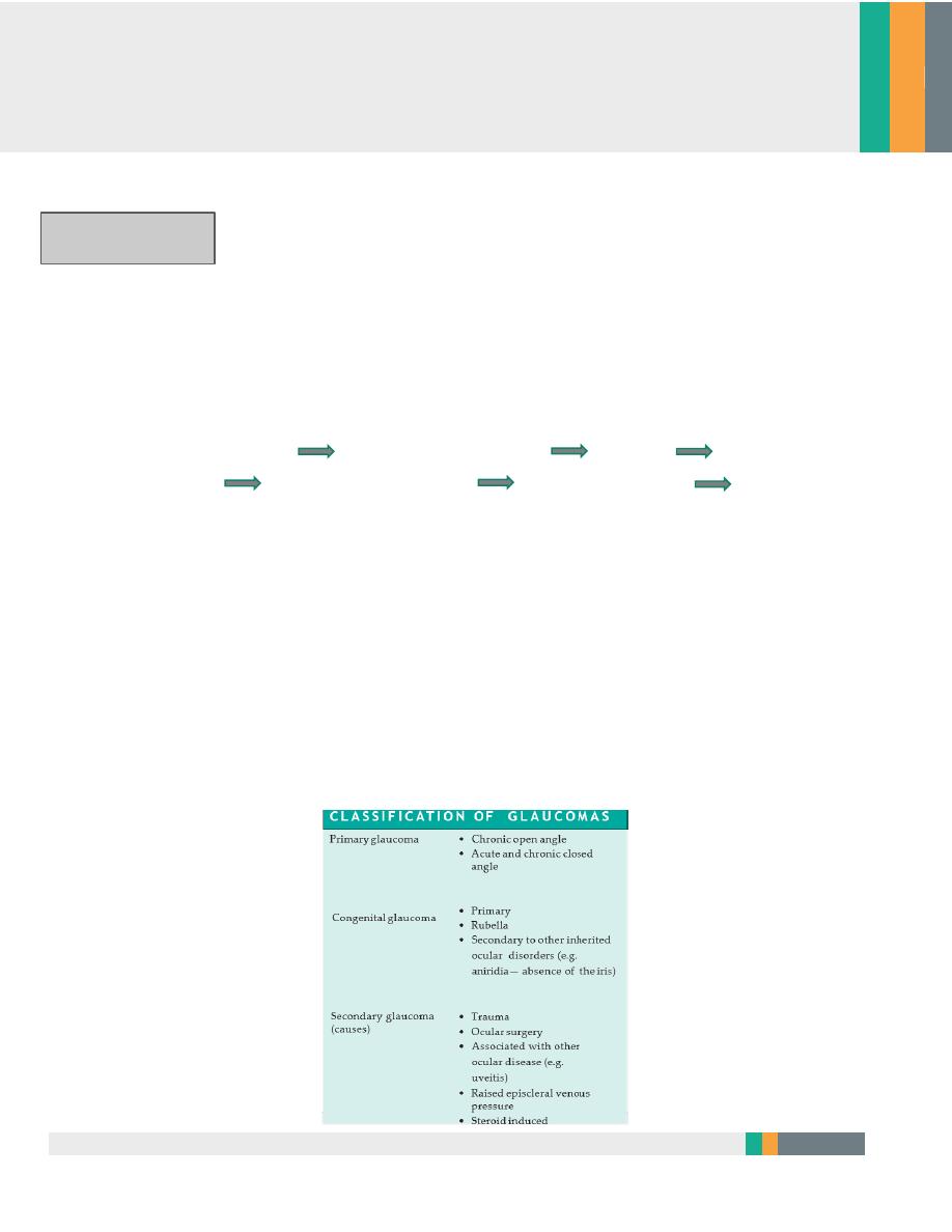
Primary open angle glaucoma
Diagnosis of glaucoma require
1. 1 increase IOP
2. 2 Abnormal cupping of optic disc
3. 3 visual field defect
Note:
Some patient have raised IOP but normal cupping and V field (not
glaucoma)
Some patient have normal IOP but cupping and visual field defect (normal
tension glaucoma )
affects 1 in 200 of the population over the age of 40,
a males= females
The prevalence increases with age
a family history
History or (symptom )
It is symptomless unless in advanced cases of visual field defect
Examination
1. Using a special lens (Gonioscopy lens ) with slit lamp We see normal
trabecular meshwork
2. Tonometer to measure IOP >>may reach 22-40 mmHg (normally 11-21)
3. Optic disc : pathologic cupping cup disc ratio greater than 0.4
Neuro retinal rim thinning
Nerve head atrophy
Notching of the rim
4. The visual field (perimetry) visual filed defect scotom

3
Treatment
Is aimed at reducing intraocular pressure
Careful monitoring in the outpatient clinic
1. Medical treatment;
2. Laser treatment;
3. Surgical treatment
1. Medical :
Prostaglandin analogue
topical adrenergic beta- blockers are the usual first line treatment
2. LASER TRABECULOPLASTY
3. Drainage surgery (trabeculectomy) relies on the creation of a fistula between
the anterior chamber and the subconjunctival space
Complications of surgery include:
Shallowing of the anterior chamber in the immediate postoperative period
risking damage to the lens and cornea;
Intraocular infection;
Possibly accelerated cataract development;
Failure to reduce intraocular pressure adequately

4
Closed angle glaucoma
The condition occurs in small eyes (i.e. often hypermetropic) with shallow
anterior chambers.
In angle closure glaucoma, sometimes in response to pupil dilation, this
resistance is increased and the pressure gradient created bows the iris forward
and closes the drainage angle
Aqueous can no longer drain through the trabecular meshwork and ocular
pressure rises, usually abruptly.
• affects 1 in 1000 subjects over 40 years old,
• females more commonly affected than males.
• Patients with angle closure glaucoma are likely to be long-sighted
HISTORY (Symptoms )
1. abrupt increase in pressure and the eye becomes
2. very painful and
3. photophobia.
4. watering of the eye
5. Loss of vision. The patient may be
6. systemically unwell with nausea and abdominal pain,
EXAMINATION
On examination visual
1. acuity is reduced,
2. the eye red, the
3. cornea cloudy and the
4. pupil oval, fixed and dilated.

5
TREATMENT
It need urgently treatment to prevent permanent damage to the vision.
1. Acetazolamide is administered I.V and subsequently orally
2. topical pilocarpine (constricts the pupil)
3. beta-blockers.
4. . Subsequent management requires that a small hole (iridotomy or
iridectomy) is made in the peripheral iris to prevent subsequent attacks
This can be done with a YAG laser or surgically
Secondary glaucoma
Intraocular pressure usually rises in secondary glaucoma due to blockage of the
trabecular meshwork. i.e (open angle) the trabecular meshwork may be blocked
by:
1. Blood (hyphaema), following blunt trauma.
2. Inflammatory cells (uveitis).
3. Pigment from the iris (pigment dispersion syndrome).
4. Deposition of material produced by the epithelium (pseudoexfoliative
glaucoma).
5. Drugs increasing the resistance of the meshwork (steroid-induced glaucoma).
6. Secondary glaucoma may also result from blunt trauma to the eye damaging
the angle (angle recession).
Angle closure may also account for some cases of secondary glaucoma:
Abnormal iris blood vessels
A large choroidal melanoma.
A cataract may swell, pushing the iris forward and closing the drainage
angle.
Uveitis may cause the iris to adhere to the trabecular meshwork.
A&b&p&s
ا
ن
ز
ل
ض
غ
ط
ا
ل
ع
ي
ن
ق
ب
ل
م
ا
ا
ف
ت
ح

6
• The symptoms and signs depend on the rate at which intraocular pressure rises;
most are again symptomless.
• Treatment to treat any underlying cause, e.g. uveitis, which may be
responsible for the glaucoma.
• In particularly difficult cases it may be necessary to selectively ablate the ciliary
processes
This is done by application of a laser or cryoprobe to the sclera
Congenital glaucoma
It may present at birth or within the first year.
Symptoms and signs include:
• Excessive tearing;
• An increased corneal diameter (buphthalmos);
• A cloudy cornea due to epithelial oedema;
• Splits in Descemet’s membrane.
Congenital glaucoma is usually treated surgically.
An incision is made into the trabecular meshwork (goniotomy) to increase
aqueous drainage
or a direct passage between Schlemm’s canal and the anterior chamber is
created trabeculotomy
Summery
•
Glaucoma is an optic neuropathy caused by an elevation of intraocular pressure.Y
POINTS
•
Primary glaucoma is classified according to whether the trabecular meshwork is
obstructed by the peripheral iris (angle closure) or not (open angle glaucoma).
•
Treatment of glaucoma relies on lowering ocular pressure to reduce or prevent further
visual damage.
•
Ocular pressure can be reduced with topical and systemic medications, laser treatment
and surgery.
•
Beware patients who are acutely debilitated with a red eye; they may have acute angle
closure glaucoma.
•
If the diagnosis is made late arresting the glaucoma completely may still result in visual
loss during the patient’s lifetime. This emphasizes the need for early diagnosis
A.L.Y
immature increase in size
dx
iop
corneal diameter
fundus examination
all of these undre GA
TX:emergancy surgery