
1
What is surgical site infection? Discuss the methods to reduce surgical site infection.
Enumerate the factors responsible for surgical site infection.
NosocomialInfection
An infection acquired in hospital by a patient who was admitted for a reason other
than that infection . Infections occurring for more than 48 hours after admission are
usually considered nosocomial .Amongst surgical patients, SSI are the most
common nosocomial infections.
Classes of SSI
Superficial incisional SSI: Infection occurs within 30 days after the operation and
infection involves only skin of subcutaneous tissue of the incision and at least one
of the following:
1–Purulent drainage with or without laboratory confirmation from the superficial
incision
2–Organisms isolated from an aseptically obtained culture of fluid or tissue from
the superficial incision
3–At least one of the following signs or symptoms of infection:
--Pain or tenderness
--Localised swelling
--Redness
--Heat
--And superficial incision deliberately opened by a surgeon, unless incision is
culture negative
Deep incisional SSI: Infection occurs within 30 days after the operation if no implant
is left in place or within one year if implant is in place and the infection appears to
be related to the operation and infection involves deep soft tissues (e.g. fascial and
muscle layers) of the incision and at least one of the following:
1–Purulent drainage from the deep incision but not from the organ/space
component of the surgical site
2–A deep incision spontaneously dehisces or is deliberately opened by a surgeon
when the patient has at least one of the following signs or symptoms:
--Fever (>38°C)
--Localized pain
Surgical Site Infection

2
--Tenderness unless site is culture-negative
3–An abscess or other evidence of infection involving the deep incision is found on
direct examination, during re-operation or by histopathological or radiological
examination.
Organ/space SSI: Infection occurs within 30 days after the operation if no implant
is left in place or within one year if implant is in place and the infection appears to
be related to the operation and infection involves any part of the anatomy (e.g.
organs or spaces), other than the incision, which was opened or manipulated during
an operation and at least one of the following:
1–Purulent discharge from a drain that is placed through a stab wound into the
organ/ space
2–Organisms isolated from an aseptically obtained culture of fluid or tissue in the
organ/space
3–An abscess or other evidence of infection involving the organ/space that is found
on direct examination, during re-operation, or by histopathologic or radiological
examination
Strategies to prevent SSI
Objectives
1-Reduce the inoculum of bacteria at the surgical site
2-Surgical site preparation
3-Antibiotic prophylaxis strategies
4-Optimize the microenvironment of the surgical site
5-Enhance the physiology of the host (host defenses)
Risk factors of SSI classified as:
1-Patient-related (intrinsic)
2-Preoperative
3-Operative
Patient-related factors
a-Diabetes—recommendation Preoperative Control serum blood glucose reduce
HbA1C levels to <7% before surgery if possible Maintain the postoperative blood
glucose
level
at
less
than
200
mg/dL
b-Smoking, anemia, malnutrition
c-Hypoalbuminemia, jaundice
d-Obesity, hyperlipidemia

3
e-Ascites, PVD
f-Immunosupression.
2-Procedure-related risk factors
a-Hair removal technique (clipping> on table shaving > previous night shaving)
b-Preoperative infections control and bath
c-Surgical scrub
d-Skin preparation
e-Antimicrobial prophylaxis
f-Surgeon skill/technique/instruments
g-Asepsis
h-Operative time (should be within 1.5 times the normal)
j-Operating room characteristics/OT sterility.
Surgeon skill and technique
Excellent surgical technique reduces the risk of SSI
Includes
a-Gentle traction and handling of tissues
b-Effective hemostasis
c-Removal of devitalized tissues
d-Obliteration of dead spaces
e-Irrigation of tissues with saline during long procedures
f-Use of fine, nonabsorbed monofilament suture material
h-Wound closure without tension.
Cellulitis
Cellulitis is a common infection of skin and subcutaneous tissues, most frequently
caused by Streptococcus pyogenes and occasionally Staphylococcus species.
Infection
occurs
after
the
skin
is
breached
(e.g.
insect
bite,
scratching, skin rash, minor trauma).
Cellulitis may seem to occur spontaneously, although careful inspection
reveals a break in the skin After subcutaneous inoculation, streptococci release
toxins which permit rapid spread of organisms. The acute inflammatory response
results in the clinical features of warmth, pain and tenderness, erythema, and
oedema. Severe cellulitis may progress to suppuration and skin necrosis.
Differential diagnosis includes other causes of limb swelling, deep venous
thrombosis, rupture of a Baker’s cyst, calf haematoma and erythematous skin
conditions.

4
excretion of penicillin. Erythromycin or a third generation cephalosporin is used in
patients with penicillin allergy. Any predisposing cause (e.g. tinea pedis) is treated
vigorously. If cellulitis does not resolve rapidly, the antibiotic is increased or
changed.
Lymphangitis
Lymphangitis is associated with bacterial infections of extremities where the
inflamed lymphatic vessels appear as several thin, red, tender lines on the slightly
oedematous skin progressing towards the regional lymph nodes which are enlarged
and tender (lymphadenitis).
Cellulitis of an extremity is treated by elevation and immobilisation with a splint or
plaster ‘back slab’, and antibiotics. Penicillin (2 million units every 6 hours) or
flucloxacillin (1–2 g every 6 hours) is given intravenously for 3–5 days and then
continued orally for a further 10 days. Blood levels of penicillin may be increased by
oral probenecid, which reduces renal
Lymphangitis usually is caused by streptococci and staphylococci. Chemical
lymphangitis may result from irritative compounds used for lymphangiography.
Treatment is the same as for cellulitis, consisting of rest and elevation of the
extremity and antibiotics. Rarely, suppurative regional lymph nodes require
Surgical drainage.
Folliculitis, furuncles and carbuncles
‘Folliculitis’ refers to infection with pus formation within a hair follicle and is
limited to the dermis. It may be extensive if many follicles are infected over a wide
area, such as the face.
A ‘furuncle’ is infection of a small number of hair follicles within a small confined
area. A ‘carbuncle’ is an abscess involving a number of adjacent hair follicles
where the infection has penetrated through the dermis and formed a
multiloculated subcutaneous abscess between the fibrous septa which anchor the
skin to the deep fascia
Furuncles and carbuncles occur most frequently on the back of the neck, lower
scalp, and the torso. Abscesses on the upper part of the body are usually caused
by staphylococci, while infections below the umbilicus are due largely to aerobic
and anaerobic coliform organisms.
Local hygiene is usually sufficient to treat folliculitis, although antibiotics are
required for extensive infections. Furuncles and carbuncles require incision and
drainage. Fibrous tissue septa must be broken down so that all pockets of pus can
be drained completely. Antibiotics are indicated for severe and spreading

5
infections, and in immunocompromised patients.
Hidradenitis suppurativa
Hidradenitis suppurativa refers to infection of apocrine sweat glands, and occurs
in the axillae, around the external genitalia, and the inguinal and perianal regions
Apocrine sweat glands have tortuous secretory ducts within the skin and produce
thick secretions, and infection occurs when ducts become
blocked, most commonly during excessive glandular activity at
adolescence.Staphylococci or Gramnegative bacilli and anaerobes are causative
organisms..
Patients present with multiple small but painful abscesses and sinuses, often
bilaterally. Repeated or long-standing infection results in considerable scarring,
Antibiotic therapy alone is often inadequate, although long-term antibiotic
therapy may be useful in suppressing acute infections. Abscesses require incision
and drainage. Excision of the affected hair-bearing area and the subcutaneous fat
usually is required, and results in good symptomatic relief.
Synergistic gangrene
‘Synergistic gangrene’ refers to a group of soft tissue infections characterised by
tissue necrosis and caused by several species of microorganisms acting
synergistically. Previous nomenclature (necrotising fasciitis, necrotizing erysipelas,
Meleney’s gangrene, Fournier’s gangrene, non-clostridial gangrenous cellulitis)
Clinical features
Synergistic gangrene is caused by micro-aerophilic streptococci acting
synergistically with aerobic staphylococci, with or without Gram-negative bacilli. It
usually occurs in debilitated patients with other disorders (e.g. diabetes,
malnutrition, alcoholism, liver disease, renal failure, malignant disease, immune
compromise).
Synergistic gangrene presents initially as cellulitis with severe pain which is out of
keeping with the minor local clinical signs but consistent with the seriousness of
the condition. Infection spreads rapidly along fascial and subcutaneous planes
without a severe inflammatory reaction.
Bacterial toxins cause tissue and skin necrosis. Crepitus occurs when gas-forming
organisms are present. Signs of systemic sepsis and toxaemia occur quickly.
‘Fournier’s gangrene’ is the name given to synergistic gangrene involving the
perineum and scrotum. It may be extensive and involve the abdominal wall and
buttocks, and is a rare complication of anorectal and perineal surgery, trauma or
minor infection.
6
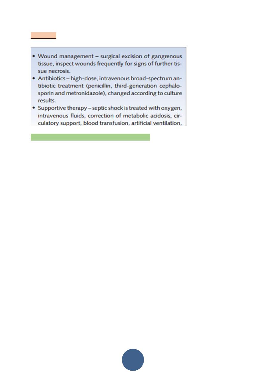
Treatment
Synergistic gangrene must be treated urgently by
Select the single correct answer to each question.
1:Cellulitis:
a-is occasionally caused by Gram-negative coliforms
b-often occurs spontaneously without any apparent
cause or organism
c- is treated with rest, immobilisation and high-dose
penicillin
d-frequently requires surgical drainage
e-is often complicated by suppuration and skin necrosis
2:Fournier’s gangrene:
a-is a form of pyomyositis
b-occurs mainly in debilitated patients and can be
life-threatening
c -is usually due to stapylococcal infection
d-can be treated by hyperbaric oxygen alone
e-is seldom managed surgically.