
Fertilization, Implantation,
early fetal development &
placental development
By
Dr. Afraa Mahjoob Al-Naddawi

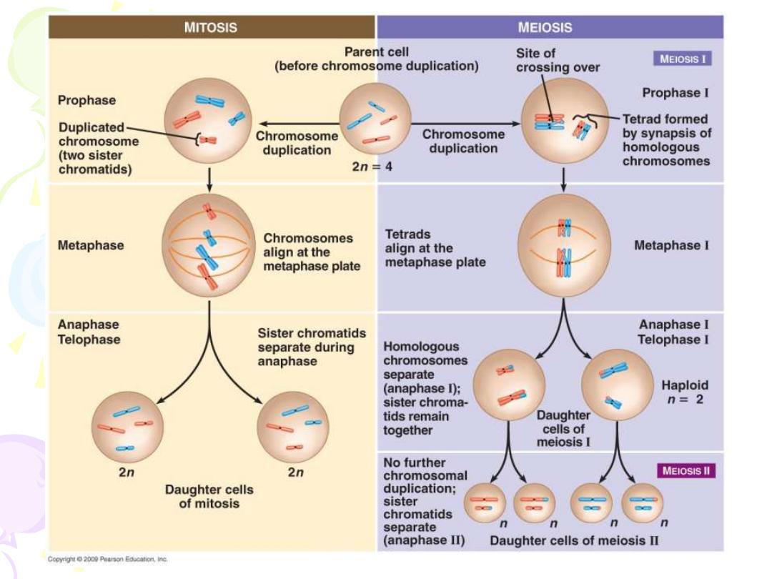
The human body has two types of cells:
1) Somatic cells: contain 23 pairs of chromosomes
(diploid), one of each pair originally is derived from
the mother and the other is from the father.
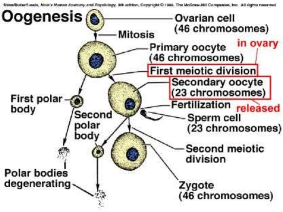
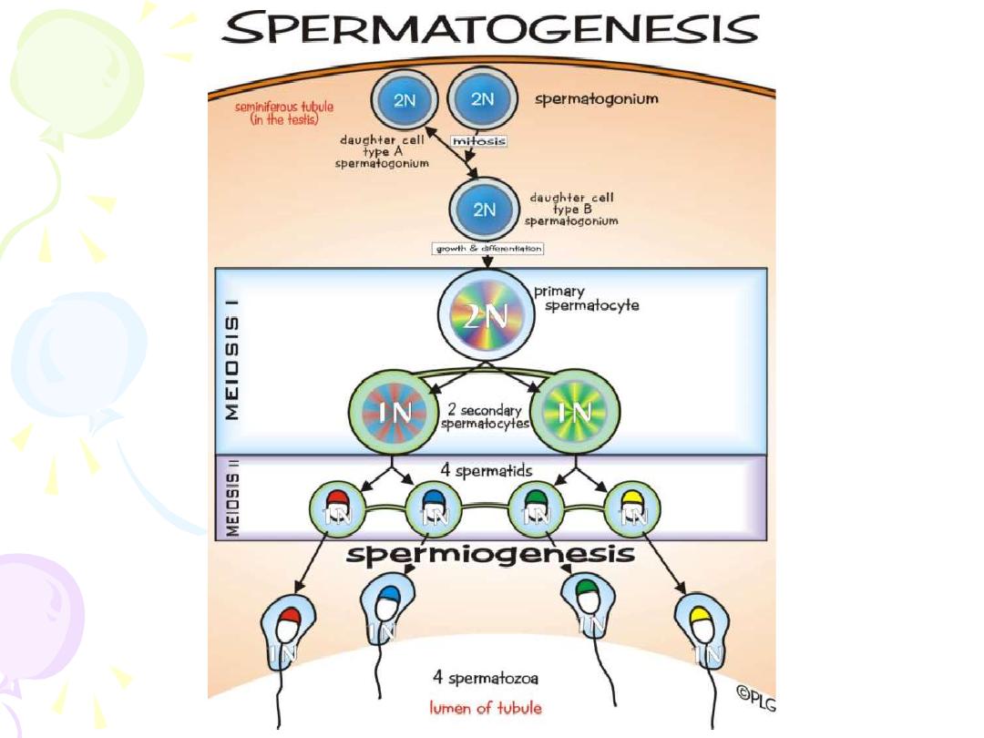
2) Germ cells: mature germ cells contain only one copy
of each chromosome, i.e. they are haploid in number
of chromosomes, these are found in the gonads
(testes in male, ovaries in female).
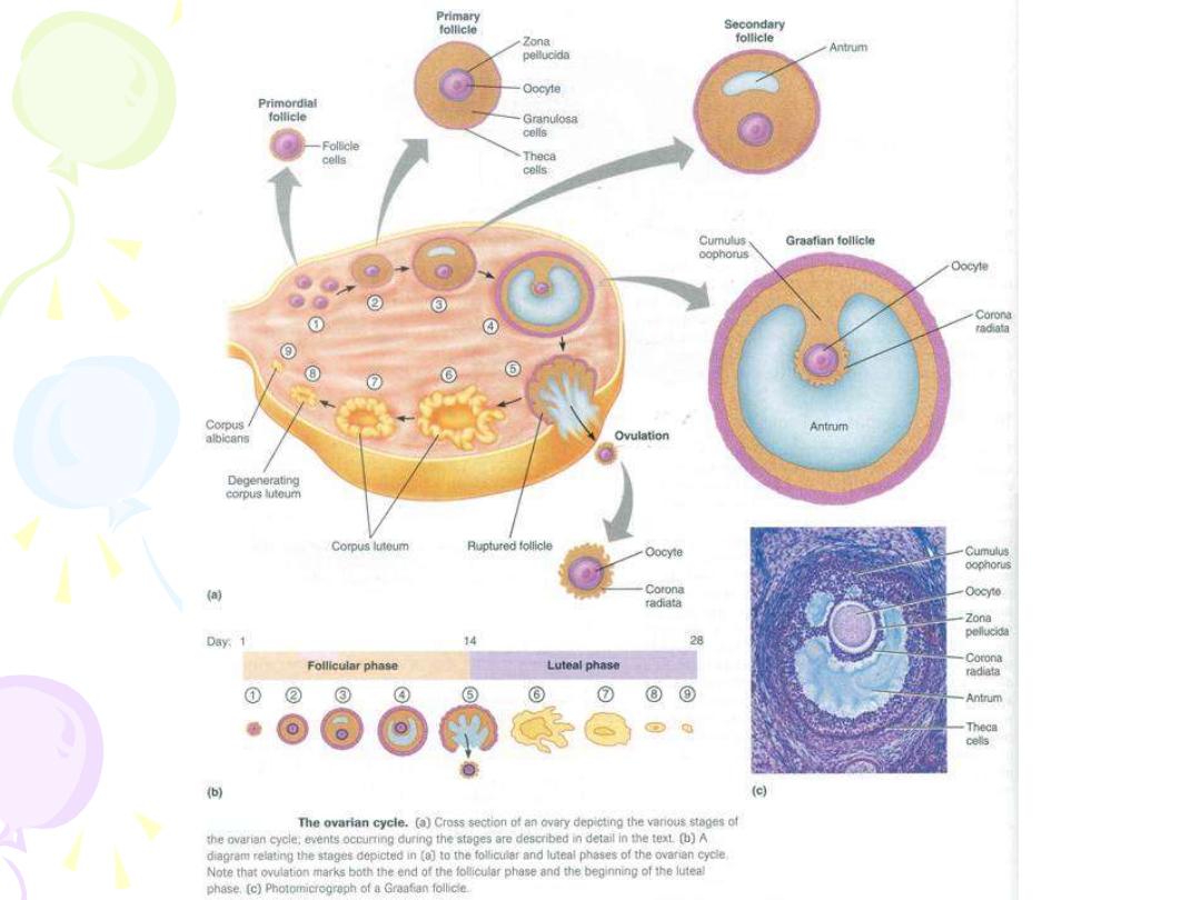

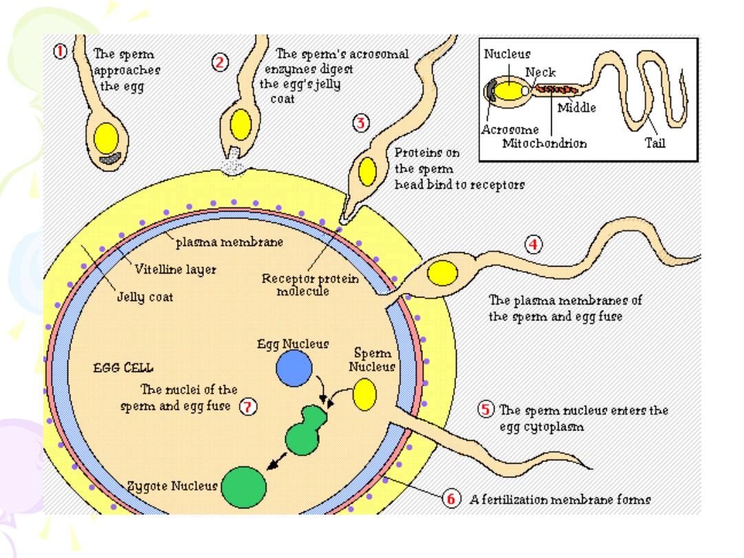
Fertilization is the fusion of ovum and sperm to form a
new microorganism (called zygote), this process
usually takes place in the ampullary region of
fallopian tube and it is composed of:
1. Penetration of
corona radiata.
2. Penetration of
zona pellucida
.
3. Sperm head attaches to ovum membrane.
4. The ovum reacts to sperm contact by zonal reaction
and completion of the second meiotic division from
metaphase II and expulsion of second polar body,
thus forming the female haploid pronucleus.

5. Once within the oocyte, the sperm tail degenerates
and the head enlarges to be transformed to male
pronucleus.
6. Male and female pronuclei approximate and fuse
forming the zygote which returns to diploid genetic
constitution.

• Fertilization of the ovum must occur a few hours and no
more than a day after ovulation
• 1 week postfertilization corresponds to approximately 3
weeks from the last menstrual period in women with regular
28-day cycles
Fertilization in the fallopian tube
zygote
a diploid cell with 46 chromosomes
blastomeres
morula
(solid mulberry-like ball of cells)
enters the uterine cavity about 3 days after fertilization
gradual accumulation of fluid between the cells of the
morula
early blastocyst
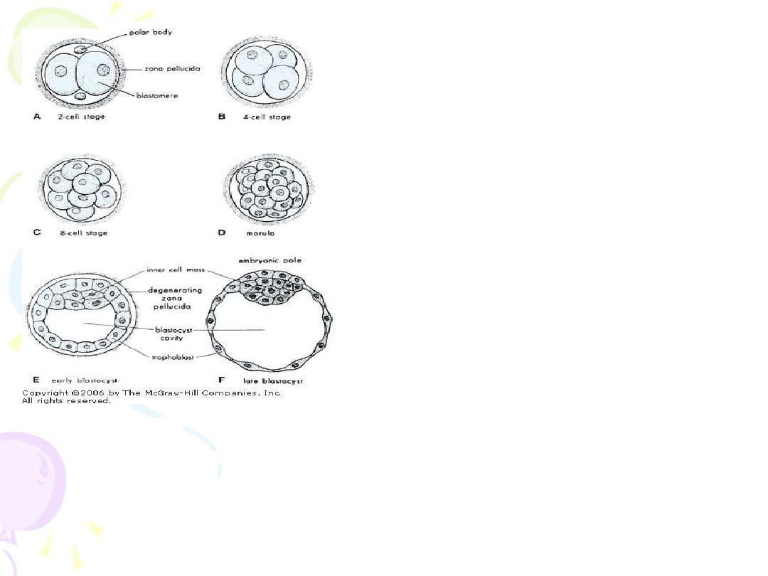
Cleavage of the zygote and
formation of the blastocyst.
A through D show various stages
of cleavage. The period of the
morula begins at the 12- to 16-
cell stage and ends when the
blastocyst forms, which occurs
when there are 50 to 60
blastomeres present.
E
and
F
are
sections
of
blastocysts. The zona pellucida
has disappeared by the late
blastocyst stage (5 days).
The polar bodies shown in A are
small, nonfunctional cells that
soon degenerate.

• In a 58-cell blastocyst,
– differentiates into: (4 to 5 days after fertilization)
– inner cell mass - five embryo-producing cells
– trophoblasts - 53 cells
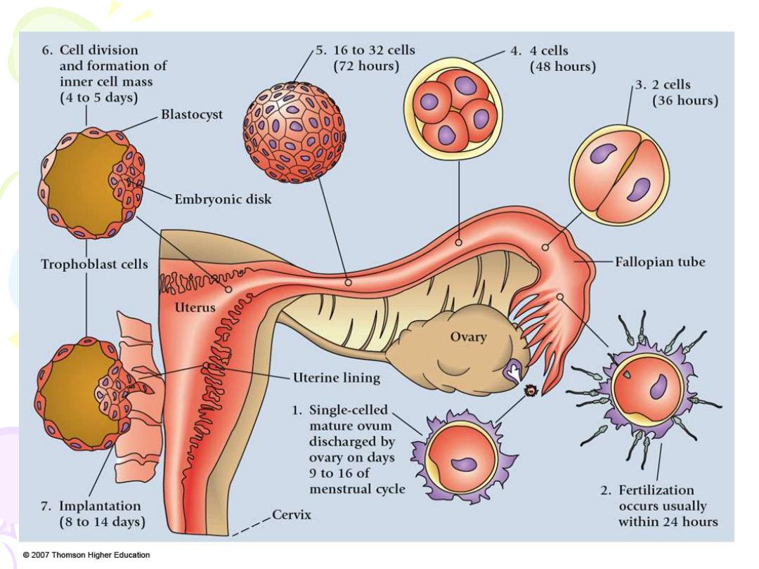

Implantation: a process by which the blastocyst attaches
to the uterine wall and penetrates first the
endometrium then the circulation of the mother to form
the placenta.
It is divided into 3 phases:
1. Apposition.
2. Adhesion.
3. Invasion: penetration of the syncytiotrophoblast and
cytotrophoblast into the endometrium, inner third of the
myometrium and uterine vasculature.

• Occurs 6 or 7 days after fertilization
• Uterine receptivity limited to days 20-24
• Apposition – blastocyst loosely adheres to the
endometrial epithelial, most commonly on the upper
posterior wall of the uterus
• Integrins - mediate the adhesion of cells to
extracellular matrix proteins
First weeks of human development:
Blastocyst embedded in endometrium.
TROPHOBLAST DIFFERENTIATION
(8th DAY POSTFERTILIZATION)
• cytotrophoblasts
– inner layer of primitive mononuclear
– germinal cells for the syncytium
– primary secretory component within the placenta
• syncytiotrophoblast
– amorphous cytoplasm
– without cell borders
– multiple and diverse in size and shape of nuclei
FURTHER TROPHOBLAST DIFFERENTIATION (2 MAIN
PATHWAYS):
• Villous trophoblast
– gives rise to chorionic villi of the placenta
– transports O2 and nutrients between fetus and mother
• Extravillous trophoblast
– migrates into the decidua and myometrium
– penetrates maternal vasculature
CLASSIFICATIONS OF
EXTRAVILLOUS TROPHOBLAST
• Interstitial
– invades maternal deciduas penetrates myometrium
form placental bed giant cells
– Surrounds maternal spiral arteries
• Endovascular – penetrates the lumen of spiral arteries
EARLY TROPHOBLAST INVASION
• Day 10 – blastocyst totally encased in the endometrium
• Mechanisms leading to trophoblast invasion into the
endometrium
– similar to characteristics of metastasizing
malignant cells
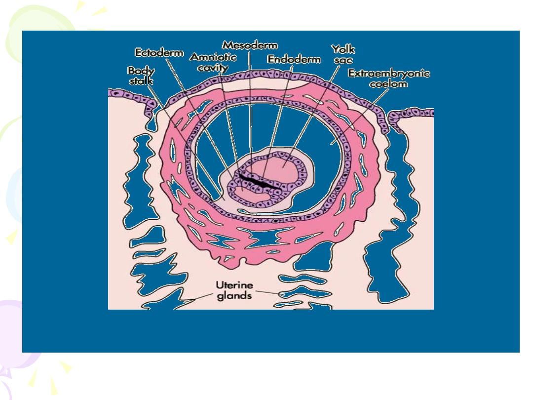
• 7 ½ days after fertilization
– Inner cell mass
– embryonic disc
– Differentiate into a thick plate of primitive ectoderm
and an underlying layer of endoderm
• Chorionic vesicle – cavity completely lined with
mesoderm
• Chorion – chorionic vesicle membranes, composed of
trophoblasts and mesenchyme
• Body stalk – join the embryo to nutrient chorion, later
develops into umbilical cord
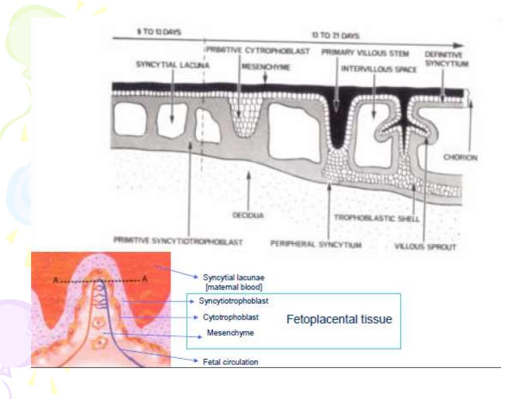
LACUNAE FORMATION WITHIN THE
SYNCYTIOTROPHOBLAST
• 12 days after conception
• Syncitiotrophoblast of the trophoblast shell is permeated
by a system of intercommunicating channels of
trophoblastic lacunae
• As embryo enlarges, the lacunae become filled with
maternal blood
DEVELOPMENT OF PRIMARY VILLOUS STALKS
• Primary villi – composed of a cytotrophoblasts core
covered by syncitium
• Complicated labyrinth – formed by joining of lacunae,
partitioned by solid cytotrophoblastic columns
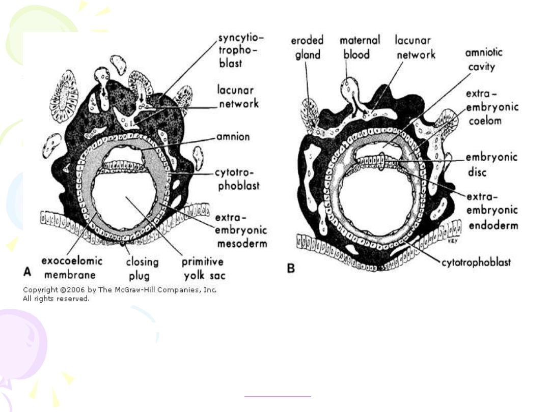
Drawing of sections through implanted blastocysts.
A. At 10 days. B. At 12 days
after fertilization. The stage of development is characterized by
the
intercommunication of the lacunae filled with maternal blood. Note in B that
large
cavities have appeared in the extraembryonic mesoderm, forming the beginning of the
extraembryonic coelom. Also note that extraembryonic endodermal cells have begun to form
on the inside of the primitive yolk sac. (From
CHORIONIC VILLI
• 12th day after fertilization – chorionic villi can first be
distinguished
• Secondary villi – formed by mesenchymal cords
invading the solid trophoblast columns
• Tertiary villi – secondary villi with angiogenesis
• 14th or 15th day after fertilization
• maternal arterial blood enters the intervillous space
• 17th day
• fetal blood vessels are functional
• Placental circulation is established
DECIDUAL SPIRAL ARTERY INVASION
• Extravillous trophoblast
• Interstitial trophoblast
• surround the maternal spiral arteries
• constitute a major portion of the placental bed
• penetrates the decidua and adjacent myometrium
• functions to prepare vessels to facilitate invasion by
the endovascular trophoblast
• Endovascular trophoblast
• penetrates the lumen of the spiral arteries
• Timing of the development of uteroplacental vessels
• 1st wave - before 12th weeks postfertilization
• 2nd wave - 12th-16th weeks postfertilization
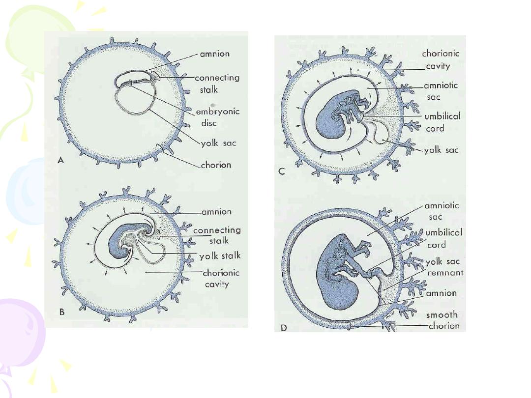
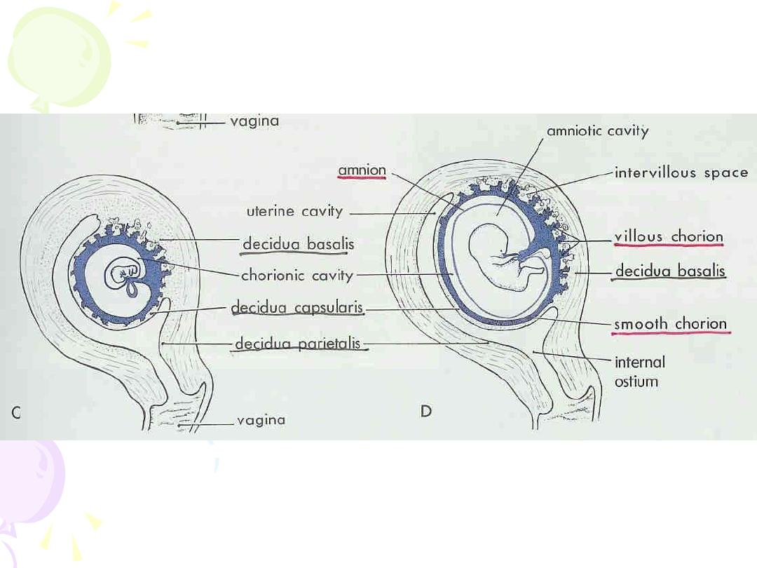
DEVELOPMENT OF CHORION AND DECIDUA
• Chorion frondosum
• so-called leafy chorion
• fetal component of the placenta
• proliferation of the chorionic villi in contact with the
deciduas basalis
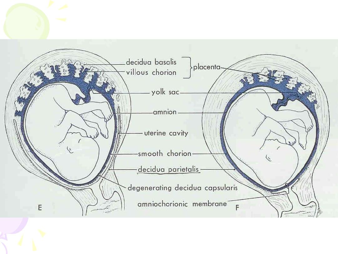
• Chorion Laeve
• smooth chorion
• Avascular fetal membrane that touches the decidua
parietalis
• Composed of cytotrophoblasts and fetal mesodermal
(mesenchymal) cells that survive in a relatively low-
oxygen atmosphere

• Chorion leave and amnion
• separated by exocoelomic cavity until the end of 3rd
month
• form an avascular amniochorion
• important sites of molecular transfer and metabolic
activity
• constitute an important paracrine arm of the fetal-
maternal communication system
• Decidua vera
• area where deciduas Capsularis and deciduas
Parietalis merge

Placental Cotyledons
• Certain villi of the chorion frondosum extend from
chorionic plate to the decidua and serve as anchoring
villi
• Each of the main stem villi(truncal) and their
ramifications (rami) constitute a placental cotyledon
(lobe)
• For each cotyledon, a 1:1:1 ratio of artery to vein to
cotyledon
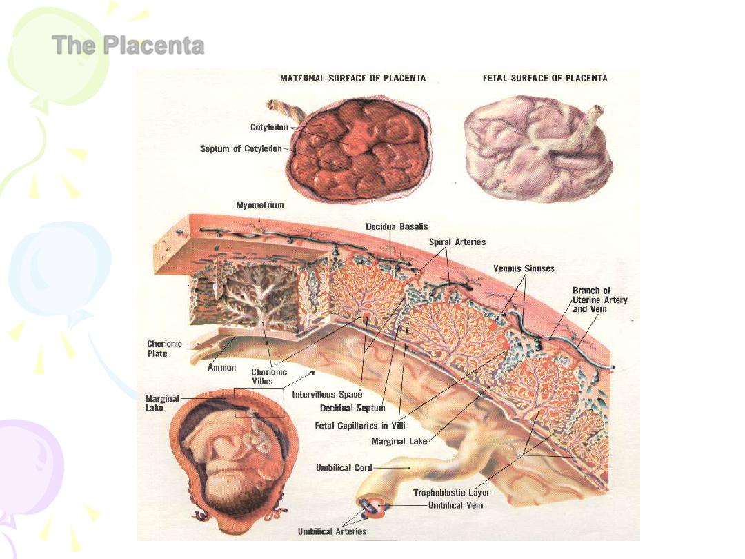
The Placenta
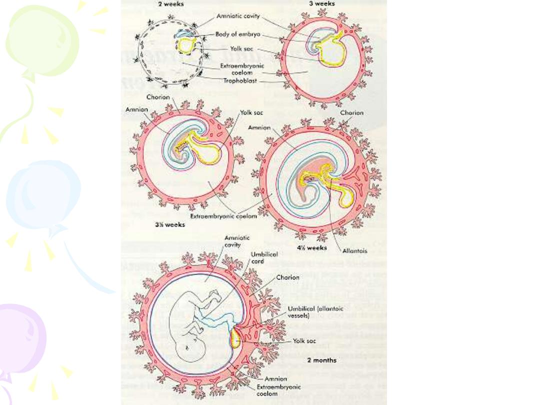
The Amnion
Innermost fetal membrane and is contiguous with amnionic
fluid
Avascular structure
Provide almost all of the tensile strength of the fetal
membranes
• protect against rupture or tearing
FROM CONCEPTION TO BIRTH
Phases of Prenatal Development
• Period of the zygote: conception through
implantation
• Period of the embryo: 3rd-8th week, organ
formation, heart beat
• Period of the fetus: 9th week-birth
THE PERIOD OF THE ZYGOTE
Blastocyst: 60-80 cells
• Embryo – inner layer of blastocyst
• Protective/nourishing tissues – outer layer
Implantation:
• 7-10 days after conception
• Tapping mother’s blood supply through uterine
wall
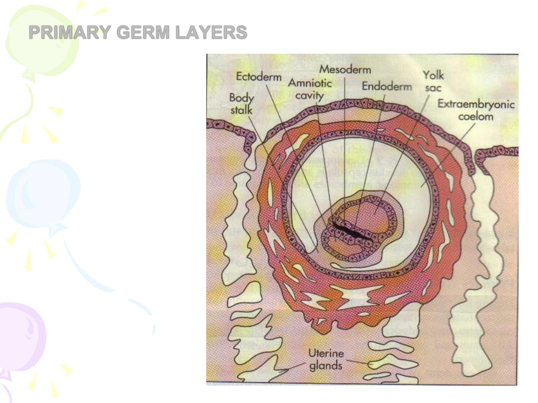
PRIMARY GERM LAYERS
• Ectoderm
• Mesoderm
• Endoderm
THE PERIOD OF THE EMBRYO
Ectoderm (outer layer)
• Nervous system
• Skin
• Hair
Mesoderm (middle layer)
• Muscles
• Bones
• Circulatory system

Endoderm (inner layer)
• Digestive system
• Lungs
• Urinary tract
• Vital organs (pancreas, liver, etc.)

Developmental Milestones
• 3rd week – neural tube
• 4th week – heart beat
• 7th week – a rudimentary skeleton
• 7th-8th weeks – sexual development
• If male, the Y chromosome triggers a
reaction to produce testes, otherwise
ovaries result
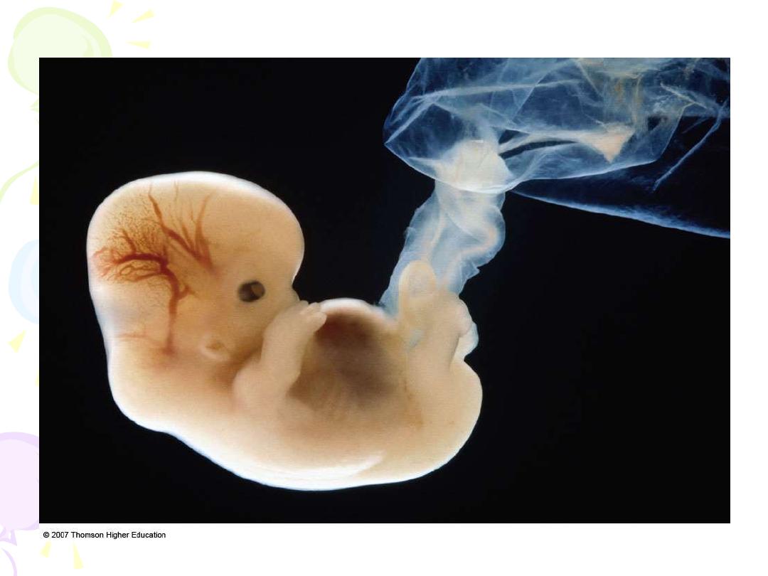
•
Figure 4.3 A human embryo at 40 days.
THE PERIOD OF THE FETUS
Third Month
• Movement – cannot yet be felt by mother
• Digestive system and excretory systems
functioning
• Reproductive system contains immature ova or
sperm cells

Fourth-Sixth Months
• Sucking, swallowing, breathing
• Movements – felt by mother
• Heart beat can be heard with stethoscope
• Sweat glands functioning
• Vernix – protects skin from chapping
• Lanugo – fine hair helps vernix stick to skin
• Visual and auditory senses are functional

Seventh
– Ninth Months
• Age of viability - 22-28 weeks – survival outside
the womb is possible
• Weight is 4 pounds (at end of 7th month)
• 9th month – activity slows, sleep increases
• Birth occurs
GESTATIONAL AGE
Gestational age=Time since last menstrual period (LMP)
EDC, EDD, EDB
266 Days after fertilization
280 Days after onset of LMP
Fertilization
LMP
Days
266
280
Weeks
38
40
Calendar Months
8 3/4
9
Lunar Months
9 1/2
10

Nagele’s Rule
Add 7 days to the first day of the last normal menstrual
period, subtract 3 months and add 1 year.
Example:
1st day of LNMP=December 16 2006
add 7 days = Dec. 23 2006
subtract 3 months = Sept 23 2006
add 1 year =Sept 23 2007, estimated due date (EDD)
TRIMESTERS
Trimester
1st
=
week’s 1-13
2nd
=
week’s 14 - 26
3rd
=
week’s 27 and on

Thank you