The eye
Color Visiondifferent cones are sensitive to different colors of light.
Tricolor Mechanism of Color Detection
All theories of color vision are based on the well known observation that the human eye can detect almost all gradations of colors when only red, green and blue monochromatic lights are appropriately mixed in different combinations.
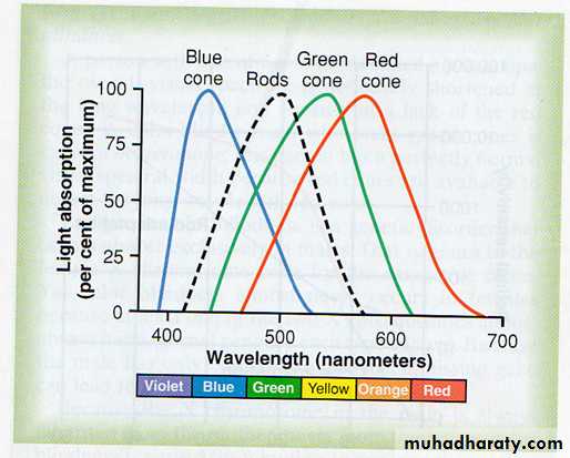
Spectral Sensitivities of the Three Types of Cones.
monochromatic orange light with a wavelength of 580 nanometers stimulates the red cones to a stimulus value of about 99; it stimulates the green cones to a stimulus value of about 42, but the blue cones not at all. Thus, the ratios of stimulation of the three types of cones in this instance are 99:42:0. The nervous system interprets this set of ratios as the sensation of orange. Conversely, a monochromatic blue light with a wave length of 450 nanometers stimulates the red cones to a stimulus value of 0, the green cones to a value of 0, and the blue cones to a value of 97.This set of ratios—0:0:97—is interpreted by the nervous system as blue. Likewise, ratios of 83:83:0 are interpreted as yellow, and 31:67:36 as green.
Perception of White Light:
About equal stimulation of all the red, green, and blue cones gives one the sensation of seeing white. Yet there is no single wavelength of light corresponding to white; instead, white is a combination of all the wavelengths of the spectrum.Yellow color sensation: no wave length for yellow but equal stimulation of red and green cones at the same time.
An object appears red when all wave lengths are absorbed, except red wave length is reflected, by the object
An object appears white when it reflects all wave lengths.
An object appear black when the object absorbs all the wave lengths of light.
Color Blindness
1-Red-Green Color Blindness. When a single group of color receptive cones is missing from the eye, the person is unable to distinguish some colors from others; the person is especially unable to distinguish red from green and is therefore said to have red-green color blindness.
A person with loss of red cones is called a protanope(2%)
A color-blind person who lacks green cones is called a deuteranope(6%)
Red-green color blindness is a genetic disorder that occurs in males. That is, genes in the female X chromosome code for the respective cones.
Yet color blindness almost never occurs in females because at least one of the two X chromosomes has a normal gene . Because the male has only one X chromosome, a missing gene can lead to color blindness. Because the X chromosome in the male is always inherited from the mother, color blindness is passed from mother to son in about 8 per cent, and the mother is said to be a color blindness carrier.
2-Blue Weakness. Only rarely are blue cones missing, although sometimes they are underrepresented, which is a genetically inherited state giving rise to the phenomenon called blue weakness.
Color Test Charts A rapid method for determining color blindness is based on the use of spot charts. These charts are arranged with a confusion of spots of several different colors called Ishihara charts.
Neural circuit of the retina:
1-The photoreceptor them selves: the rods and cones .
2-The horizontal cells : transmit signals horizontally from rods and cones to the bipolar cell . Horizontal cells connect laterally between the synoptic bodies of rods and cones as well as with dendrites of bipolar cells. The output of horizontal cells is always inhibitory. So visual pathway from the area where the light strikes is excitatory while the area on the side is inhibited allowing high visual acuity
3-The bipolar cells: transmitting signals from rods and cones and horizontal cells and synapsing with ganglion cells and Horizontally between bipolar cells, ganglion cells and/or other amacrine cells
Some bipolar cell receive direct excitation from rods and cones.Other receives its signals indirectly through the horizontal cells which cause inhibition .So this provides a second mechanism of lateral inhibition
4-The amacrine cells, which transmit signals from bipolar cells to ganglion cells
5-Ganglion cells: transmit output signals from retina through optic nerve to the brain Transmission of signals in the retinal neurons
Visual pathway:
After impulses leave the :Retina they pass back ward through the optic nerve.All fibers from nasal halves of the retinas cross at the optic chiasma to the opposite side where they join the fibers from the opposite Temporal retinas to form the optic tract then pass to the Lateral geniculate body: (located at the dorsal end of thalamus). In the lateral geniculate body, the fibers from here the geniculo- calocarin- fibers pass through the Optic radiation to the primary visual cortex in the calcarin area of the occipital lobe.
From optic tracts fibers pass to the suprachiasmatic nucleus of hypothalamus for controlling circadian rhythm. Into pretectal nuclei for control of fixation of eyes on an object of importance. Also of activating the pupillary light reflex. Into superior colliculius for controlling bilateral simultaneous movement of the two eyes.
Pupillary Diameter
The major function of the pupil is to increase the amount of light that enters the eye during darkness and to decrease the amount of light that enters the eye in daylight
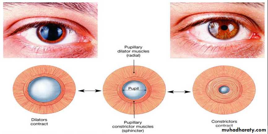
Control of pupillary diameter:
Miosis: decreasing pupillary opening due to parasympathetic stimulation of circular fibers of iris .Mydriasis: pupillary dilatation due to stimulation of the sympathetic nerves excites the radial fibers of the iris.
Accommodation for near object
Reflex pupillary constriction in
Pupillary light reflex
Pupillary light reflex:
Is constriction of pupil under effect of light on retina.The neural pathway: the effect of light on retina impulses pass through optic nerve , optic tract to pretectal nuclei in mid brain. Impulses pass to Edinger-Westphal nucleus through parasympathetic nerves to constrict circular muscle fiber of iris. The other eye also constrict a reflex called consensual light reflex.
pupillary reaction to accommodation
when the eyes fixate on a near object, the signals that cause accommodation of the lens and those that cause convergence of the two eyes cause a mild degree of pupillary constriction at the same time. This is called the pupillary reaction to accommodation.