1
Fifth stage
Radiology
Lec-7
.د
هديل
30/3/2016
Mammogram
Mammography
Mammography is a dedicated radiographic technique for imaging the breast. An x-ray
(radiograph) is a noninvasive medical test that helps physicians diagnose and treat medical
conditions. Imaging with x-rays involves exposing a part of the body to a small dose of ionizing
radiation to produce pictures of the inside of the body.
Types of mammography
In general terms, there are two types of mammography: screening and diagnostic.
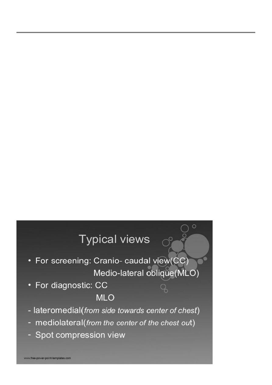
There are numerous mammography views that can broadly be split into two groups
1. Standard views
2. Supplementary views - additional information or problem solving
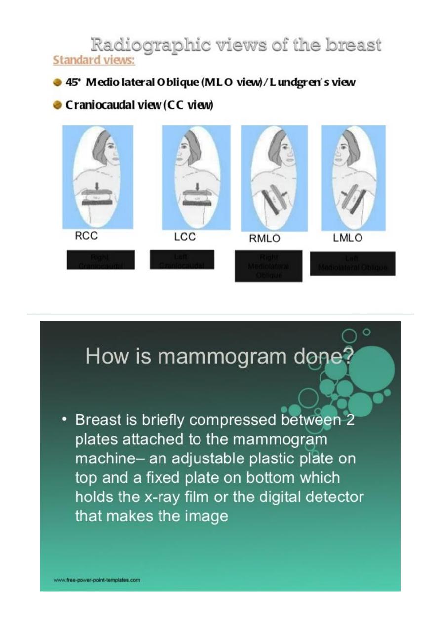
The mediolateral oblique (MLO) view is one of standard mammographic views. It is the most
important projection as it allows to depict most breast tissue.
The craniocaudal view (CC view), is one of the two standard projections in a screening
mammography. It must show the medial part as well the external lateral portion of the breast
as much as possible.
2

3
Screening mammography is the type of mammogram that checks you when you have no
symptoms. It can help reduce the number of deaths from breast cancer among women ages
40 to 70. But it can also have drawbacks. Mammograms can sometimes find something that
looks abnormal but isn't cancer. This leads to further testing and can cause you anxiety.
Sometimes mammograms can miss cancer when it is there. It also exposes you to radiation.
You should talk to your doctor about the benefits and drawbacks of mammograms. Together,
you can decide when to start and how often to have a mammogram.
Mammograms are also recommended for younger women who have symptoms of breast
cancer or who have a high risk of the disease.
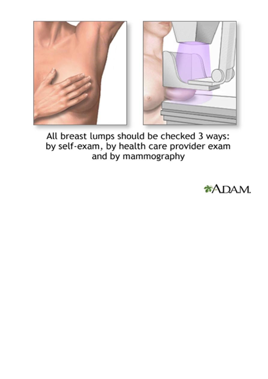
When you have a mammogram, you stand in front of an x-ray machine. The person who takes
the x-rays places your breast between two plastic plates. The plates press your breast and
make it flat. This may be uncomfortable, but it helps get a clear picture. You should get a
written report of your mammogram results within 30 days.
Mammogram facts
Mammograms are images of the breast tissue produced on X-ray film.
Mammograms are the most efficient screening method to detect early breast cancer.
A mammogram only takes a few seconds and may be mildly uncomfortable.
An abnormal mammogram does not necessarily mean that a cancer is present; other
tests, including biopsy, MRI, or ultrasound examination may be performed for further
clarification of an abnormal mammogram.
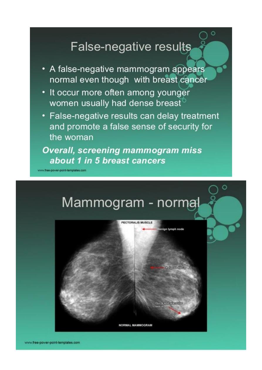
A normal mammogram does not exclude the presence of cancer.
Screening mammography is performed at regular intervals to detect abnormalities.
What are the benefits vs. risks
Benefits
Imaging of the breast improves a physician's ability to detect small tumors. When cancers are
small, the woman has more treatment options.
The use of screening mammography increases the detection of small abnormal tissue
growths confined to the milk ducts in the breast, called ductal carcinoma in situ (DCIS). These
early tumors cannot harm patients if they are removed at this stage and mammography is an
excellent way to detect these tumors. It is also useful for detecting all types of breast cancer,
including invasive ductal and invasive lobular cancer.
No radiation remains in a patient's body after an x-ray examination.
X-rays usually have no side effects in the typical diagnostic range for this exam.
4
Risks
There is always a slight chance of cancer from excessive exposure to radiation. However, the
benefit of an accurate diagnosis far outweighs the risk.
The effective radiation dose for this procedure
Things to consider before asses mammogram film
5
6
7
Finding a Lump
Remember that the majority (about 80%) of breast lumps are not due to cancer. Cysts, benign
tumors, or changes in consistency due to the menstrual cycle can all cause benign breast
lumps. Still, it's important to let your doctor know about any lumps or changes in your breast
that you find. Early detection of breast cancer is associated with high cure rates.
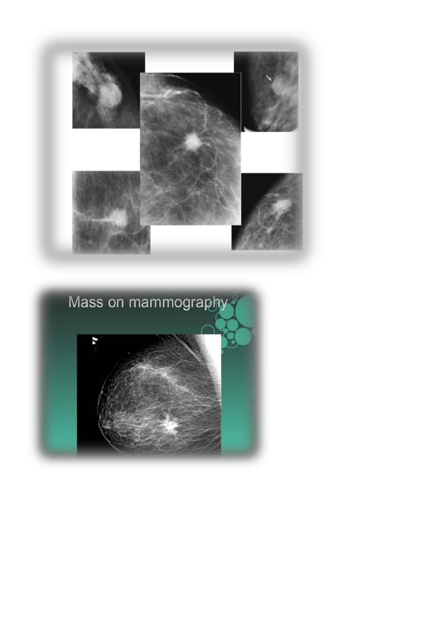
What if my mammogram is abnormal?
Do not panic if you are told that your mammogram is abnormal or that there is a "spot" on
your mammogram. An abnormal mammogram does not mean you have cancer. The
overwhelming majority of abnormal mammograms are caused by benign (harmless)
processes. In some cases, it may just be an area of thicker or denser breast tissue, a cyst, or
a benign lump such as a fibroadenoma. When a mammogram detects a suspicious area, the
patient may be advised to obtain further mammograms of that area, to have an ultrasound
or other imaging.
8
Mammogram deal with the breast mass
Women with a breast lump need to have a mammogram of both breasts. A mammogram
is estimated to be able to detect about 90% of breast cancers. This means that about 10% of
breast cancers are missed by mammography. Therefore, if a woman or her physician feels a
lump and the mammogram is normal, further studies or biopsies are carried out to rule out
cancer. Sometimes, a certain pattern of calcium deposits appears on the mammogram that
makes the doctor suspicious of cancer. In these cases, it is often recommended that a biopsy
be taken that is guided by mammogram images to be sure the correct area is sampled
9
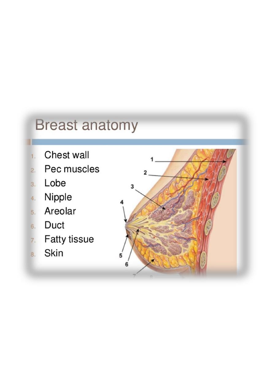
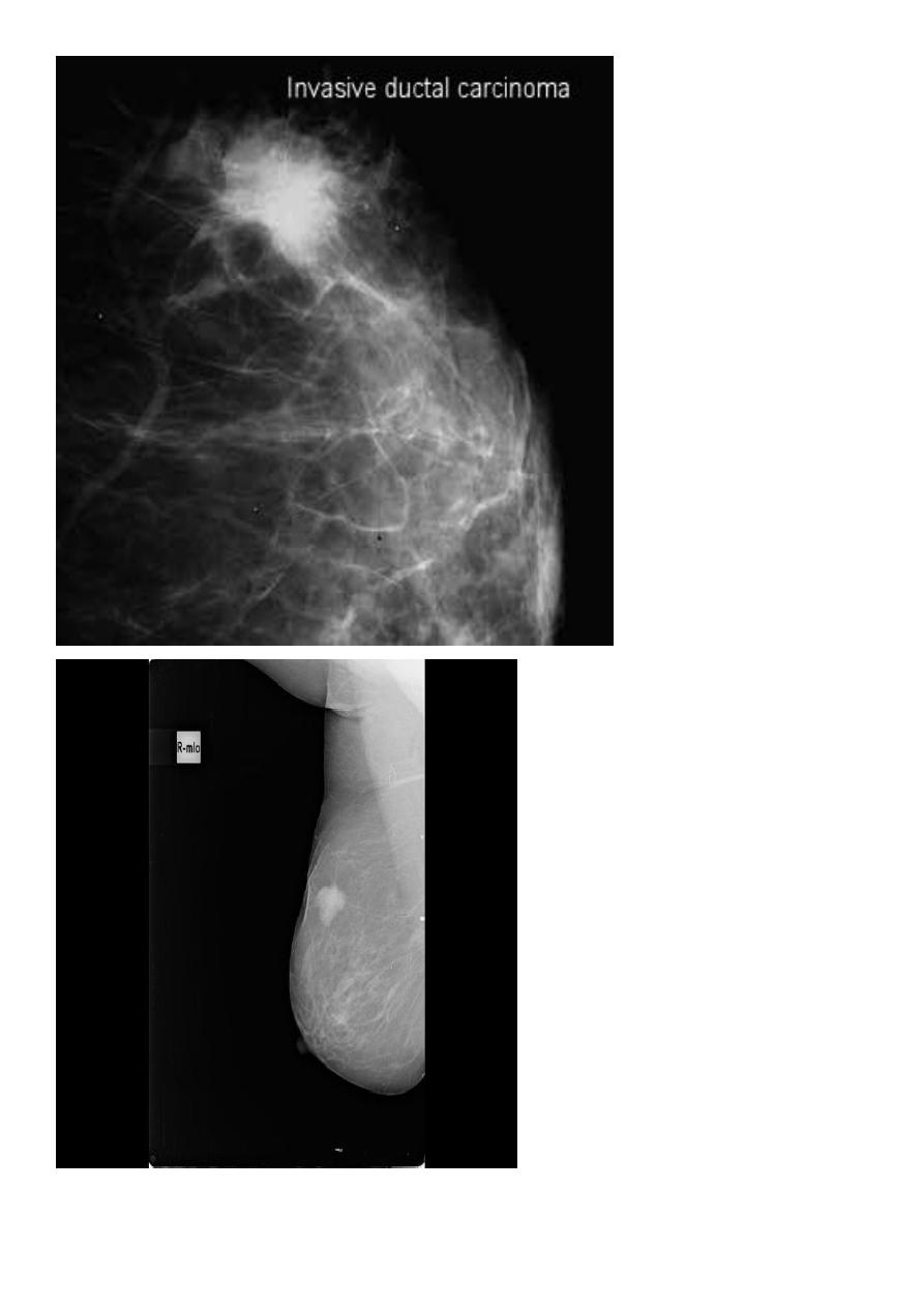
Breast cancer
Breast cancer is cancer that starts in the tissues of the breast. There are two main types of
breast cancer:
Ductal carcinoma starts in the tubes (ducts) that carry milk from the breast to the
nipple. Most breast cancers are of this type.
Lobular carcinoma starts in the parts of the breast, called lobules, which produce milk.
In rare cases, breast cancer can start in other areas of the breast.
When you find a lump U must adjust the following things
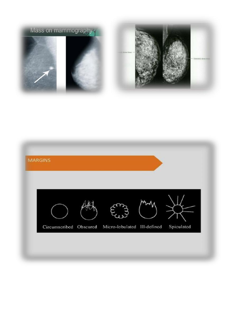
Breast density & who can U detect a mass lesion
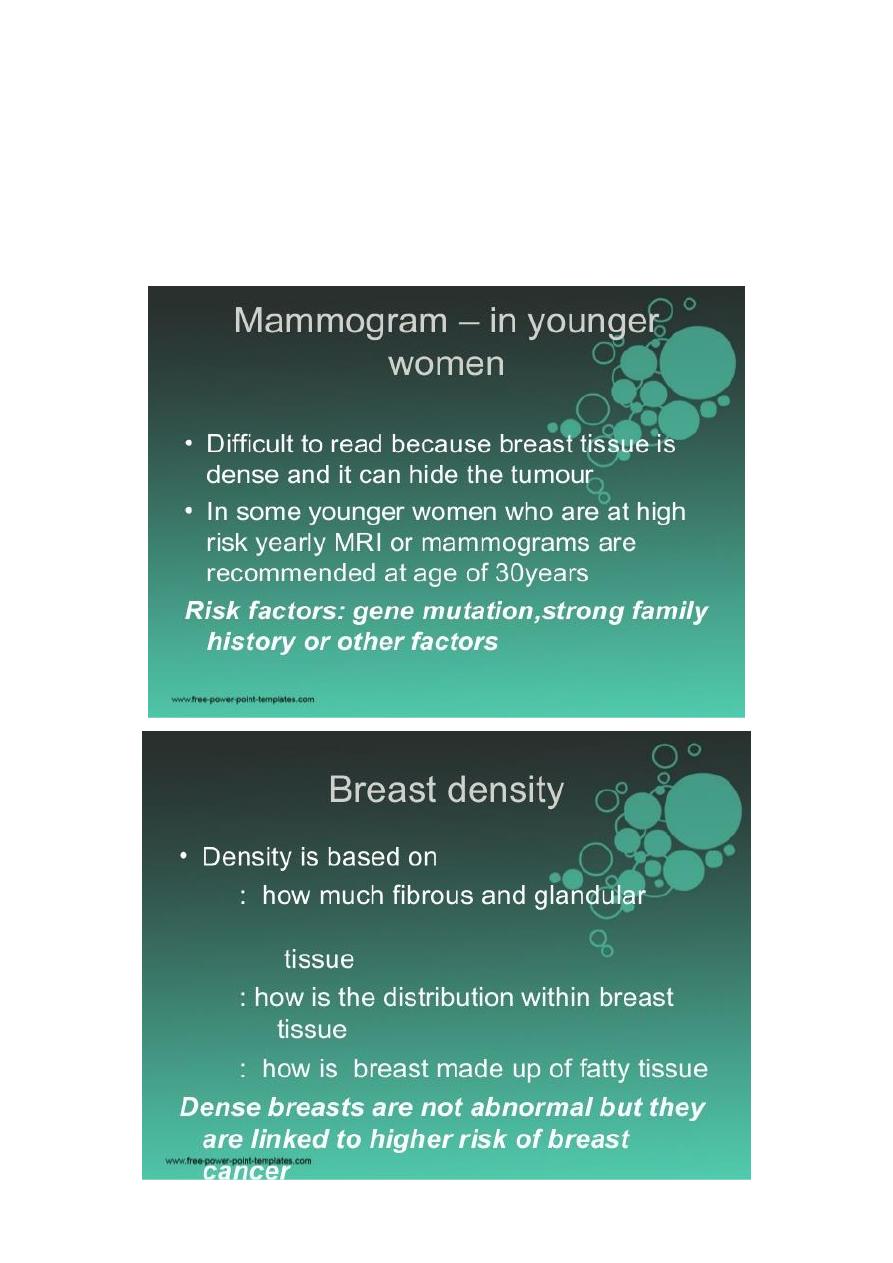
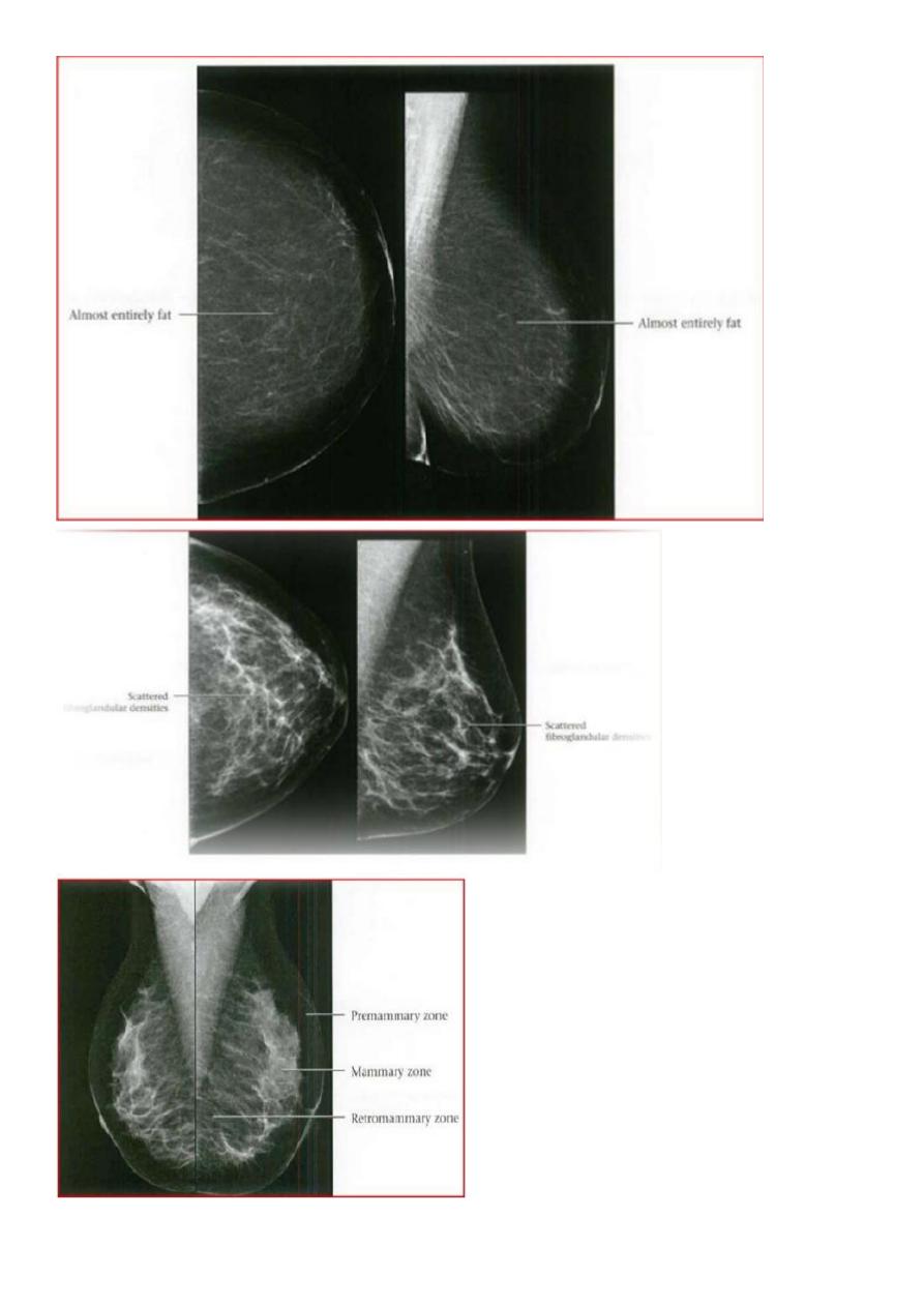
Your mammogram report must take in consideration & assessment the breast density. Breast
density is based on how fibrous and glandular tissue tissues are distributed in your breast,
vs. how much of your breast is made up fatty tissue.
Dense breasts are not abnormal, but they are linked to a higher risk of breast cancer. We
know that dense breast tissue can make it harder to find cancers on a mammogram. Still
experts do not agree what other tests, if any, should be done in addition to mammograms in
women with dense breasts who aren’t in a high-risk group (based on gene mutations, breast
cancer in the family, or other factors
10
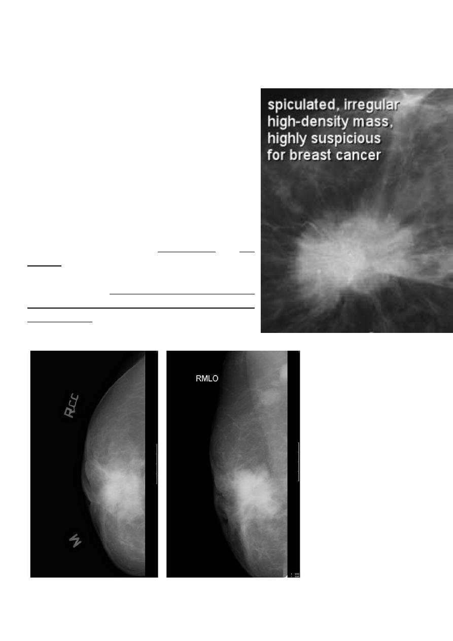
Before u asses the mass look at their margin
11
Infiltrating or invasive lobular carcinoma (ILC)
of the breast is the second most
common type of invasive breast cancer after invasive ductal carcinoma (IDC).
Radiographic features
ILC is more often multicentric and bilateral (10-15%).
Therefore imaging evaluation of the contralateral
breast is crucial. There can be very subtle changes
such as progressive shrinkage or enlargement or
reduced compressibility of the involved breast.
Imaging often underestimates the disease.
Mammography
The sensitivity of mammography for the detection of
ILC reportedly ranges between 55-80% 8. Because of
the limitations of mammography in detecting ILC,
other modalities, such as sonography and MR
imaging, are being used in evaluating clinically
suspicious findings and known cancers to assess the
extent of disease. ILC are more commonly seen on
the craniocaudal (CC), compared to the mediolateral
oblique (MLO).
12
Invasive lobular CA
13
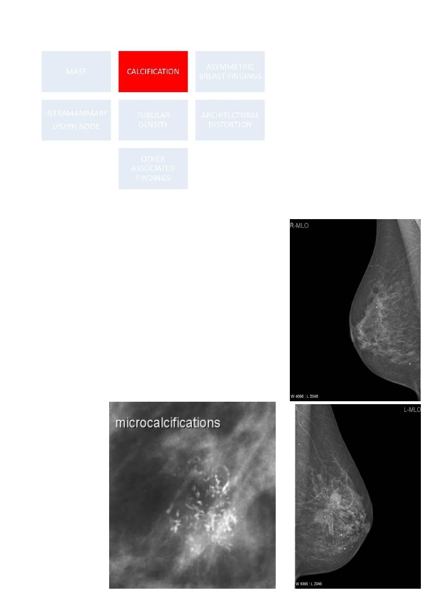
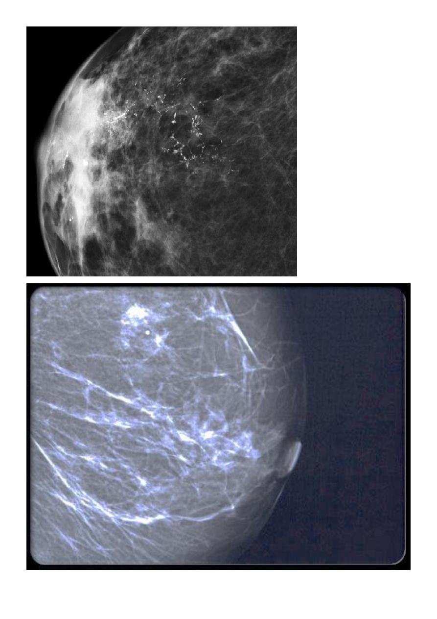
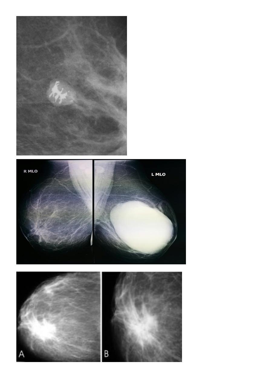
Micro calcifications
Micro calcifications are tiny specks of calcium in the breast.
Micro calcifications seen on a mammogram are of more
concern than macrocalcifications, but they do not always
mean that cancer is present. The shape and layout of
microcalcifications help the radiologist judge how likely it is
that cancer is present.
In most cases, the presence of microcalcifications does not
mean a biopsy is needed. But if the microcalcifications have a
suspicious look and pattern, a biopsy will be recommended.
(During a biopsy, the doctor removes a small piece of the
suspicious area to be looked at under a microscope. A biopsy
is the only way to
tell if cancer is
really present.)
14
15

16
After all description who can U asses the mass lesion ????
A mass it is calcification & margin
>>>>>> A mass will detected
>>>>>> if it is with or without calcifications, change seen on a mammogram.
>>>>>> it is Margin
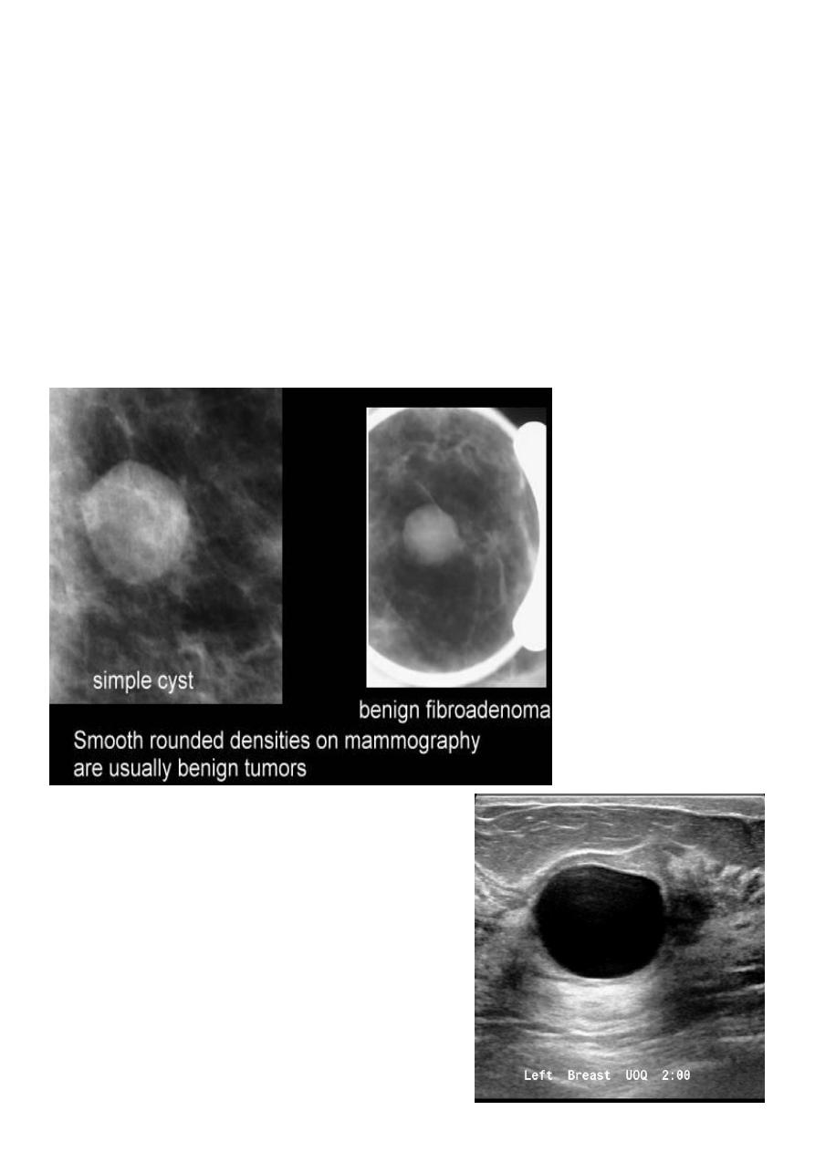
Masses are areas that look abnormal in mammogram that can be many things, not only CA
including cysts (non-cancerous, fluid-filled sacs) and non-cancerous solid tumors (such as
fibro adenomas), but must aware they sometimes may be a sign of cancer
Cysts can be simple fluid-filled sacs (known as simple cysts) or can be partially solid (known
as complex cystic and solid masses). Simple cysts are benign (not cancer) and don’t need to
be biopsied. If a mass is not a simple cyst, it is of more concern and might need to be
biopsied to be sure it isn’t cancer
17
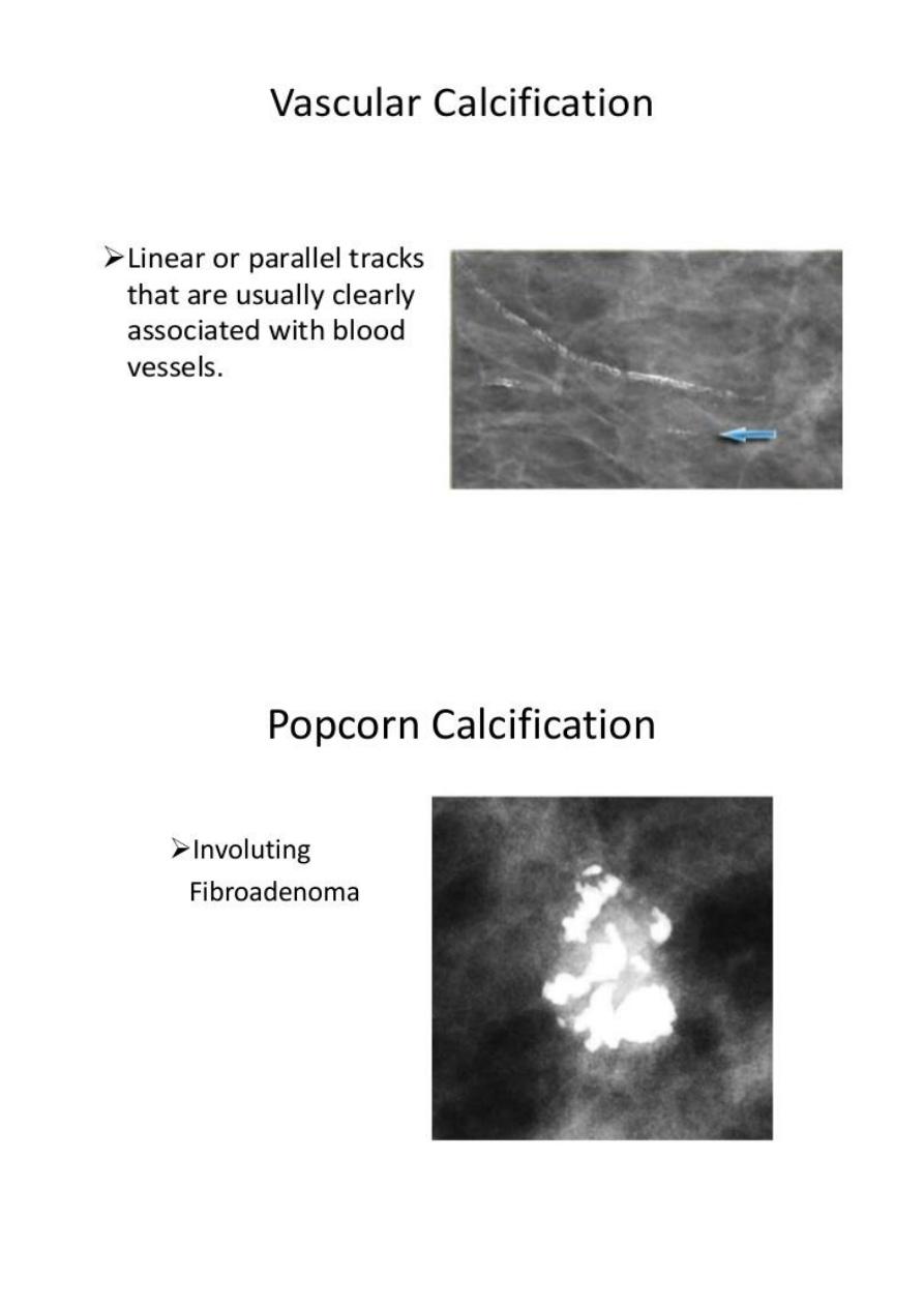
Solid benign (non-cystic lesion) fibro adenoma
Fibroadenoma is a common benign breast lesion and results from excess proliferation of
connective tissue. Fibroadenomas characteristically contain both stromal and epithelial
cells.
Mammography
Fibroadenomas have a spectrum of features from the well circumscribed discrete oval mass
hypo- or isodense to the breast glandular tissue, to a mass with macrolobulation or partially
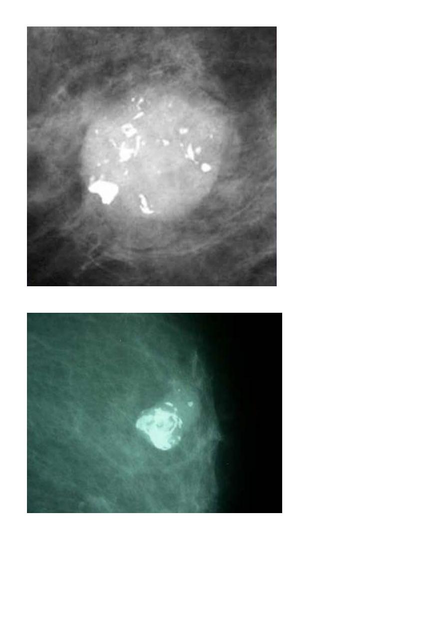
obscured margin. Involuting fibroadenomas in older, typically postmenopausal patients
may contain calcification, often producing the classic, coarse popcorn calcification
appearance.
CYST
18
FIBROADENOMA
19
CA BREAST
20
?????????