
د.رندعبداللطيف
Histology
Organs of special senses
1
THE EAR (Vestibulocochlear Apparatus)
The ear contains a series of receptors specialized for hearing
&equilibrium. The ear is divided into three parts, the external ear, the
middle ear (or tympanic cavity), and the internal ear (or labyrinth)
where the vibrations are transformed to specific nerve impulses which pass
to the central nervous system.
Fenestra Vestibuli (oval window):
Is an oval opening on the medial wall of the tympanic cavity; it is closed
by the footplate, or base, of the stapes. On the medial side of the window is
the perilymph of the scala vestibuli of the internal ear.
Fenestra Cochleae (round window):
Is a circular opening on the medial wall of the tympanic cavity below
and behind the fenestra vestibuli. It is closed by an elastic membrane
called the secondary tympanic membrane. On the medial side of this
window is the perilymph of the blind end of the scala tympani.
When the pressure of the perilymph rises as a result of medial
movement of the base of the stapes, the secondary tympanic membrane
stretches laterally and bulges into the tympanic cavity to accommodate
the increased pressure.
INTERNAL EAR (LABYRINTH):
Is situated in the petrous part of the temporal bone and consists of an
interconnected series of bony-walled chambers and passages containing
similarly shaped membranous sacs and canals. They are known as:
(1) The bony labyrinth, comprising a series of cavities within the bone.
(2) The membranous labyrinth, comprising a series of membranous sacs
and ducts contained within the bony labyrinth.

د.رندعبداللطيف
Histology
Organs of special senses
2
1- Bony Labyrinth:
Consists of three parts; the vestibule, the semicircular canals, and the
cochlea. The bony walls of the vestibule and semicircular canals are
lined with several layers of flattened connective tissue cells that form
the mesothelium. From this layer, thin trabeculae, consisting of fine
fibrils and fibroblasts, extend to the outer walls of the utricle, saccule
and semicircular ducts and support these parts of the membranous
labyrinth. Blood vessels are also found in this connective tissue.
The bony labyrinth contain a tissue fluid very similar to cerebrospinal
fluid, the perilymph, in which is suspended the membranous labyrinth.
Vestibule
Is the central part of the bony labyrinth and lies posterior to the cochlea
and anterior to the semicircular canals. In its lateral wall are the fenestra
vestibuli and the fenestra cochleae. Lodged within the vestibule are the
saccule and utricle of the membranous labyrinth.
Semicircular Canals:
The three semicircular canals the superior (anterior), posterior, &
lateral open into the posterior part of the vestibule. Each canal has a
swelling at one end called the ampulla. Each canal open into the
vestibule. Lodged within the canals are the semicircular ducts. The
superior semicircular canal is positioned vertically and anteriorly and
lies at right angles to the posterior semicircular canal. The lateral canal
is set in a horizontal position.
Cochlea
This structure is a diverticulum of the saccule. It is highly specialized as
a sound receptor. The cochlea resembles a snail’s shell and contains the
cochlear duct. It opens into the anterior part of the vestibule. about
35mm in total length, makes two-and-one-half spiral turns around a
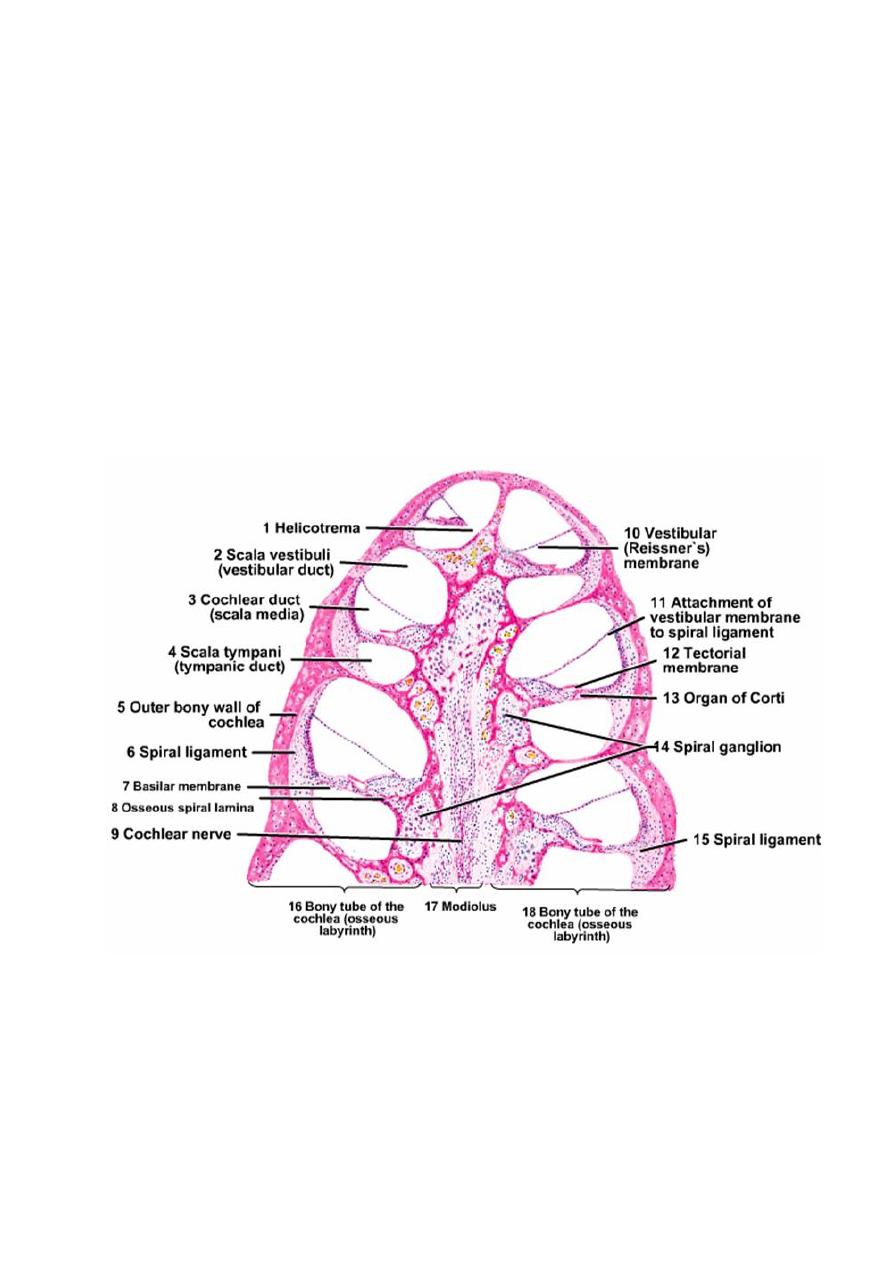
د.رندعبداللطيف
Histology
Organs of special senses
3
spongy bon known as the modiolus. Each successive turn is of
decreasing radius, so that the whole structure is conical. The apex faces
anterolaterally, and the base posteromedially.
The modiolus has spaces containing blood vessels and the cell bodies
and processes of the acoustic branch of the eighth cranial nerve (spiral
ganglion). Extending laterally from the modiolus is a thin bony ridge,
the osseous spiral lamina. The modiolus has a broad base that is
situated at the bottom of the internal acoustic meatus.

د.رندعبداللطيف
Histology
Organs of special senses
4
2- Membranous Labyrinth
These are membranous sacs and canals lodged within the bony
labyrinth, filled with endolymph and surrounded by perilymph.
It consists of the utricle and saccule, which are lodged in the bony
vestibule; the three semicircular ducts; which are lodged in the
semicircular canals, and the duct of the cochlea, which lies within the
bony cochlea. All these structures freely communicate with one another
The membranous labyrinth connected to the periosteum of the osseous
labyrinth by thin strands of connective tissue containing blood vessels.
Flowing between the connective tissue strands is the perilymph.
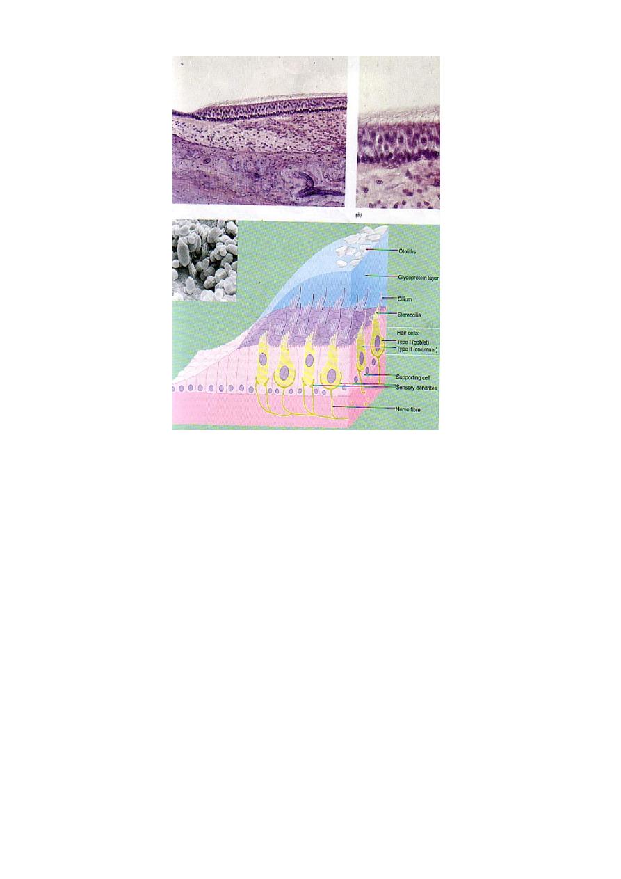
Utricle and Saccule:
The utricle and saccule are two dilated regions of the membranous
labyrinth lying within the vestibule of the inner ear and are joined by
the short utriculosaccular duct, which leads into the slender
endolymphatic duct.
Both the utricle and the saccule are filled with endolymph, (a fluid with
high K
+
and low Na
+
content) and are suspended in the perilymph of the
vestibule.
Internally, the utricle and saccule are composed of a thin sheath of
connective tissue lined with simple squamous epithelium but in each
there is a small region of highly specialized epithelium called the
macula, containing differentiated neuroepithelial cells that are
innervated by branches of the vestibular nerve.
Maculae in both locations have the same basic histologic structure and
consist of a thickening of the wall and possess two types of receptor
cells (hair cells), some supporting cells, and the afferent and efferent
nerve endings.
These receptors are sensitive to the orientation of the head in relation to
gravity or other acceleration forces. The maculae of the utricle and
saccule are disposed perpendicularly to one another. The macula

د.رندعبداللطيف
Histology
Organs of special senses
5
consists of a thickened area of neuroepithelium resting on a basal
lamina and it consist of three types:
•
(1) Type I hair cells, (2) Type II hair cells, (3) supporting cells.
Type I hair cells are flask-shaped; and stains poorly; their nuclei
tending to lie lower level than those of Type II hair cells which are
cylindrical.
Each hair cell possesses on its free surface 40-80 elongated highly
specialized microvilli, or stereocilia, and a single kinocilium; the latter
has the microtubular structure of a cilium but is longer than the
microvilli.
The microvilli and the kinocilium constitute the so-called hairs
embedded in a thick gelatinous glycoprotein membrane called the
otolithic membrane (probably secreted by the supporting cells; this is
lost during histological preparation). Within the membrane are
numerous crystalline bodies called otoliths, composed of calcium
carbonate and associated with protein.
The type I & II hair cells are invested by a meshwork of dendritic
processes of afferent sensory neurons. Both cell types have efferent
nerve endings that are probably inhibitory.
The supporting cells are tall and columnar, lie on the basal lamina
between the hair cells, each possesses small microvilli on its free
surface and basally located nucleus. These cells may assist in the
nutrition of the hair cells or modify the composition of the endolymph.
د.رندعبداللطيف
Histology
Organs of special senses
6
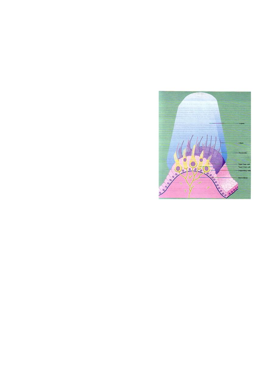
Semicircular Ducts:
Three semicircular canals arise from the vestibule of the inner ear, each
containing a membranous semicircular duct which opens at both ends
into the utricle.
At one end of each duct is a dilated portion, the ampulla, which
contains a receptor organ, called the crista ampullaris Which is an
elongated epithelial structure situated on a ridge of supporting tissue
arising from the membranous wall of the ampulla and oriented at right
angles to the direction of flow of endolymph in the semicircular duct.
Structurally, the crista is composed of Type I and Type II hair cells
supported by a single layer of columnar supporting cells which is
continuous with the simple cuboidal epithelium lining the rest of the
membranous labyrinth.
Like those of the maculae, the hair cells of the cristae have numerous
stereocilia and a single kinocilium. The stereocilia and the kinocilia of
د.رندعبداللطيف
Histology
Organs of special senses
7
the hair cells are embedded in a conical form of gelatinous glycoprotein
called cupula. In contrast to the macula, the cupula does not contain
otolithic crystals.
Movement of the endolymph in the semicircular ducts, associated with
an angular movement of the head, causes the cupula to move and pull
on the hairs of the hair cells. These cells
are stimulated and in turn stimulate the
nerve endings of the vestibular division
of the eighth cranial nerve.
Duct of the Cochlea (Scala media):
The duct of the cochlea is a spirally arranged tube lying within the bony
cochlea.
It is triangular in cross section.
The sides of the triangular cross section of the duct are made up of the
following structures: (1) the basilar membrane (forming the floor of
the triangular space) (2) the stria vascularis (forming the lateral wall)
and (3) the vestibular membrane (forming the roof the space).
Walls of the Cochlear Duct:
The basilar membrane consists of a thin sheet of fibrous tissue
(collagenous and some elastic fibers). It is stretched between the
osseous spiral lamina of the modiolus and the spiral ligament laterally.
The membrane is thinnest at the base of the cochlea and becomes
progressively thicker as it spirals towards the apex.

د.رندعبداللطيف
Histology
Organs of special senses
8
The highly specialized epithelium that lies on the upper surface of the
basilar membrane forms the spiral organ of Corti; the undersurface
exposed to the scala tympani, is lined by simple epithelium (flattened
mesothelial cells).
Stria vascularis : It forms the outer wall of the cochlear duct. The lining
epithelial cells are stratified cuboidal with many epithelial and
subepithelial rich plexus of capillaries, a rare example of vascularized
epithelium in the human body. It is believed to form endolymph.
Vestibular membrane (Reissner’s membrane): It forms the third wall of
the cochlear duct. It composed of two layers of flattened epithelial cells
separated by a basal lamina. The cells possess numerous microvilli and
may be involved in fluid transport.
Examination of a cross section of the cochlea shows that the basilar and
vestibular membranes divide it into three distinct portions: a scala
vestibuli above (filled with perilymph), a scala tympani below (filled
with perilymph), and the cochlear duct sometimes called scala media
(filled with endolymph).
The perilymph within the scala vestibuli is separated from the tympanic
cavity by the base of the stapes and the annular ligament at the fenestra
vestibuli (oval window).
The perilymph in the scala tympani is separated from the tympanic
cavity by the secondary tympanic membrane at the fenestra cochleae
(round window)
The helicotrema is the point at the apex of the cochlea where the
scala vestibuli and the scala tympani become continuous.
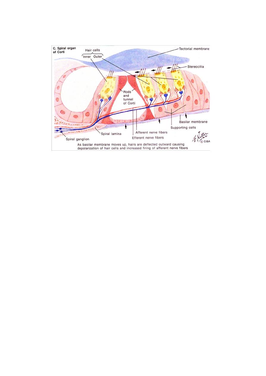
Spiral Organ Of Corti :
The spiral organ of Corti contains the sensory receptor cells for hearing
and is situated on the cochlear duct surface of the basilar membrane.
The organ of Corti consists of two types of cells, sensory (hair) cells
and support cells of several different types, including the inner and

د.رندعبداللطيف
Histology
Organs of special senses
9
outer pillar cells and inner and outer phalangeal cells. The bases of
the cells are wide, and their cell bodies incline toward each other so that
the upper extremities meet to enclose the tunnel of Corti.
The tunnel of Corti is a triangular-shaped canal at the centre of the
organ, bounded on each side by a single row of tall columnar cells
called pillar cells.
On the inner aspect of the inner row of pillar cells is a single row of
flask-shaped cells (inner sensory cells) or (hair cells).
Beyond the outer row of pillar cells there are three to five rows of outer
phalangeal cells which support the same number of rows of outer
sensory (hair) cells.
The hair cells are similar in structure to the Type Ι hair cells of the
maculae and ampullae, having many long stereocilia, but they lack
kinocilia.
From the layer of border cells which cover the spiral limbus, there
extends a flap-like mass of glycosaminoglycans called the tectorial
membrane overlying the sensory cells and within which the tips of the
stereocilia are embedded.
The support cells are tall columnar cells, their function appears to be to
support the hair cells and the nerve endings near the bases of the hair
cells. Afferent and efferent nerve endings are present between the inner
and outer hair cells. The efferent nerve fibers reach the brain via the
cochlear division of the eighth cranial nerve.
Spiral Ganglion
The spiral ganglion is a spiral-shaped mass of nerve cell bodies lying in
a canal at the extremity of the osseous spiral lamina of the modiolus.
The efferent nerve fibers consists of bipolar neurons whose central
processes converge to form the cochlear division of the eighth cranial
nerve. The vestibular division of the eighth cranial nerve, which
receives sensory information from the maculae of the utricle and
د.رندعبداللطيف
Histology
Organs of special senses
11
saccule and from the ampullae of the semicircular canals, has a
vestibular ganglion situated in the internal auditory meatus.