
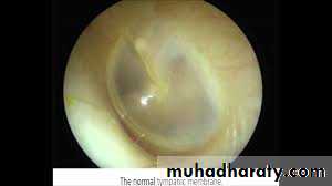
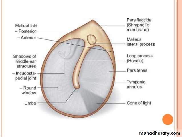
Tympanic Membrane
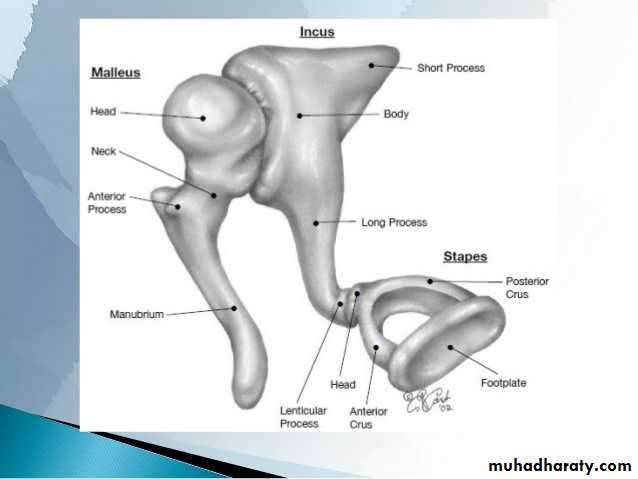
The tympanic membrane is a thin membrane that separates the external ear from the middle ear. It acts to transmit sound waves from air in the external auditory canal to the ossicles of the middle ear. The malleus is the first bone in the ossicular chain that eventually sees the sound wave transmitted to the oval window of the cochlea.
Innervation
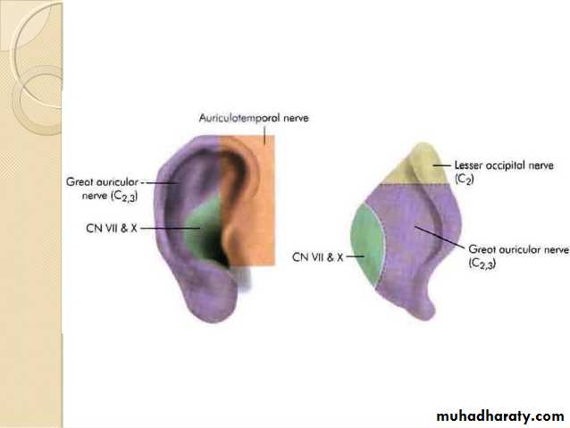
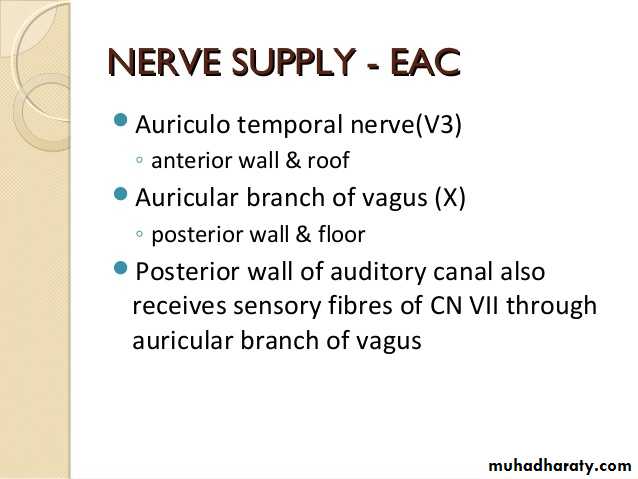
The membrane has two distinct nerve supplies based on the different embryological origins of the internal and external surfaces.
external surface
• predominantly the auriculotemporal nerve (CN V3)
• auricular branch of the vagus nerve (CN X)
internal surface: glossopharyngeal nerve (CN IX)
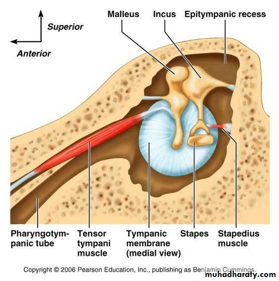
1. Stapedius muscleIs the smallest of the skeletal muscles in the human body.
Arises from the pyramidal eminence, and its tendon emerges from the eminence.Inserts on the neck of the stapes.
Is innervated by a branch of the facial nerve.
Pulls the head of the stapes posteriorly, thereby tilting the base of the stapes.
Prevents (or reduces) excessive oscillation of the stapes and thus protects the inner ear from injury from a loud noise.
Its paralysis results in hyperacusis.
2. Tensor tympani muscleArises from the cartilaginous portion of the auditory tube.
Inserts on the handle (manubrium) of the malleus.
Is innervated by the mandibular branch of the trigeminal nerve.
Draws the tympanic membrane medially and tightens it (in response to loud noises), thereby increasing the tension and reducing the vibration of the tympanic membrane.
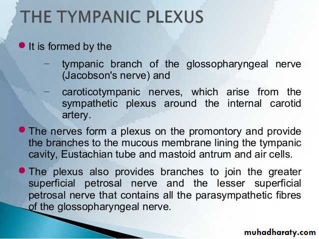
Sensory nerve and blood supply to the middle ear1. Is innervated by the tympanic branch of the glossopharyngeal nerve, which forms the tympanic plexus with caroticotympanic nerves from the internal carotid plexus of sympathetic fibers. The tympanic nerve continues beyond the plexus as the lesser petrosal nerve, which transmits preganglionic parasympathetic fibers to the otic ganglion.
2.Receives blood from the stylomastoid branch of the posterior auricular artery and the anterior tympanic branch of the maxillary artery.
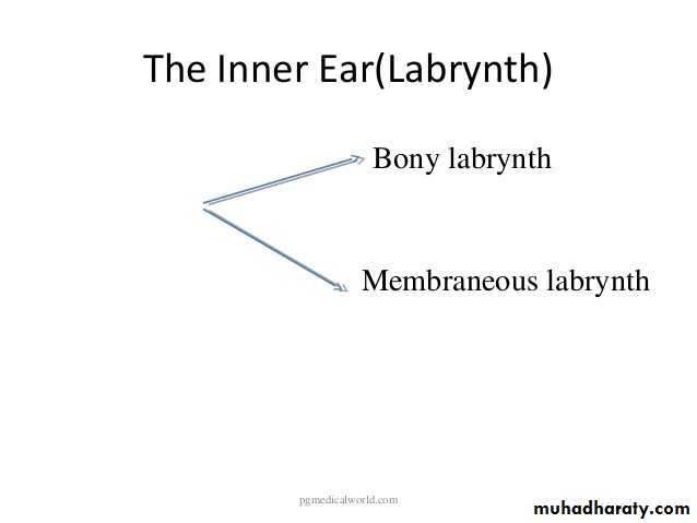
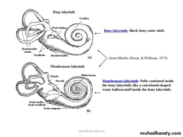
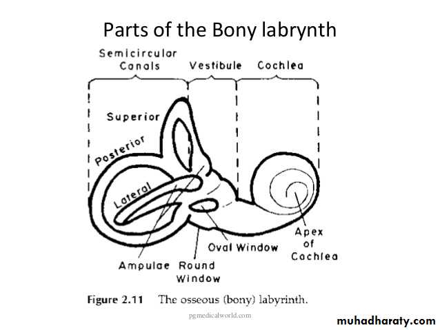
Vasculature of the inner ear
The bony labyrinth and membranous labyrinth have different arterial supplies. The bony labyrinth receives three arteries, which also supply the surrounding temporal bone:• Anterior tympanic branch (from maxillary artery).
• Petrosal branch (from middle meningeal artery).
• Stylomastoid branch (from posterior auricular artery).
The membranous labyrinth is supplied by the labyrinthine artery, a branch the basilar artery). It divides into three branches:
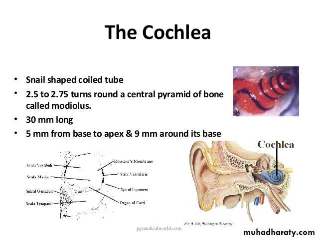
Cochlear branch – supplies the cochlear duct.
Vestibular branches (x2) – supply the vestibular apparatus.
Venous drainage of the inner ear is through the labyrinthine vein, which empties into the sigmoid sinus or inferior petrosal sinus.
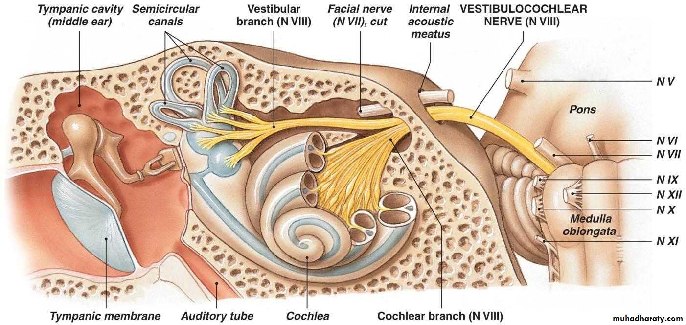
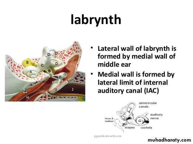
Innervation
The inner ear is innervated by the vestibulocochlear nerve (CN VIII). It enters the inner ear via the internal acoustic meatus, where it divides into the vestibular nerve (responsible for balance) and the cochlear nerve (responsible for hearing)The facial nerve, CN VII, also passes through the inner ear, but does not innervate any of the structures present.