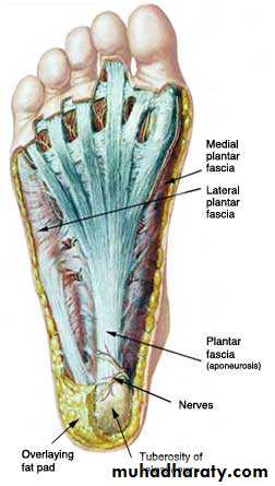
Sole of the foot
Deep fascia (planter fascia)Planter aponeurosis
compartments of the sole
first layer ( most superficial layers) contains 3 muscles: ,abductor hallucis ,flexor digitorum brevis (FDB) ,bductor digiti minimi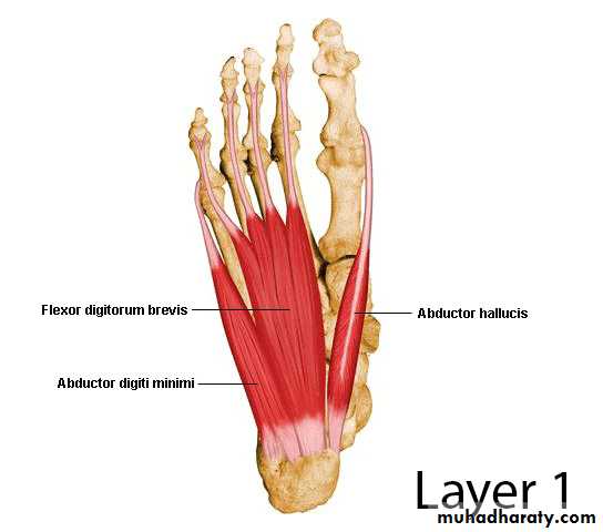
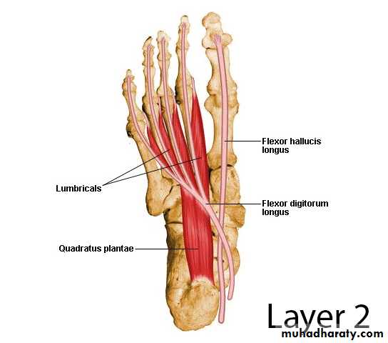
2nd layer contains 2 Muscles and 2 tendonand Neurovascular structures medial and lateral plantar arteries
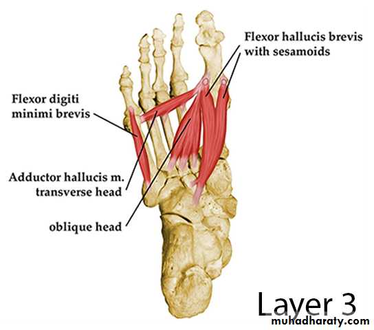
3- 3rd layer contains 3 muscles
4th layer contains 2 muscles and 2 Tendons
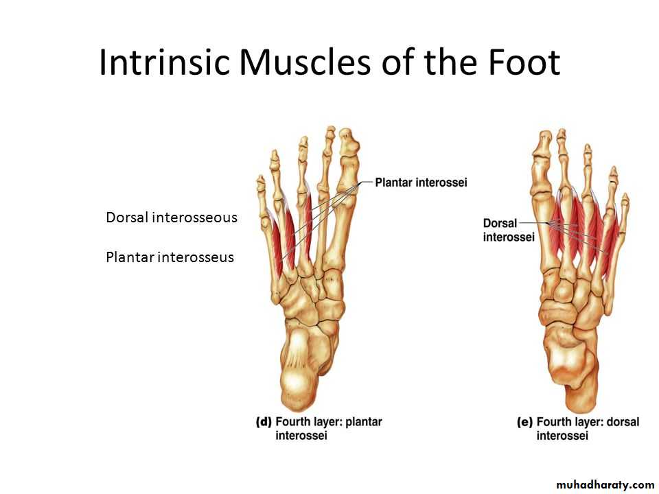
4th layer
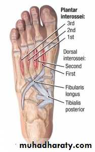
contains 2 muscles and 2 Tendons
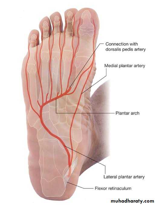
Arteries of the sole of the footThe medial planter artery
Lateral planter arteryThe planter arch :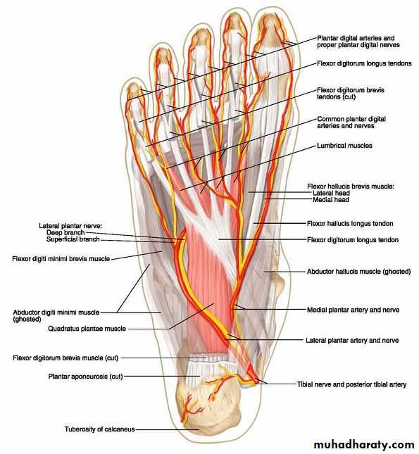
The planter arch : It is formed from the lateral planter artery, the arch completed medially by its union with the deep planter branch of the dorsalis pedis . The arch gives:four planter metatarsal arteries run between the metatarsal bones, each artery divided into pairs of proper digital arteries
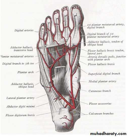
Arteries of the sole of the foot
The medial planter arteryLateral planter arteryThe planter arch :
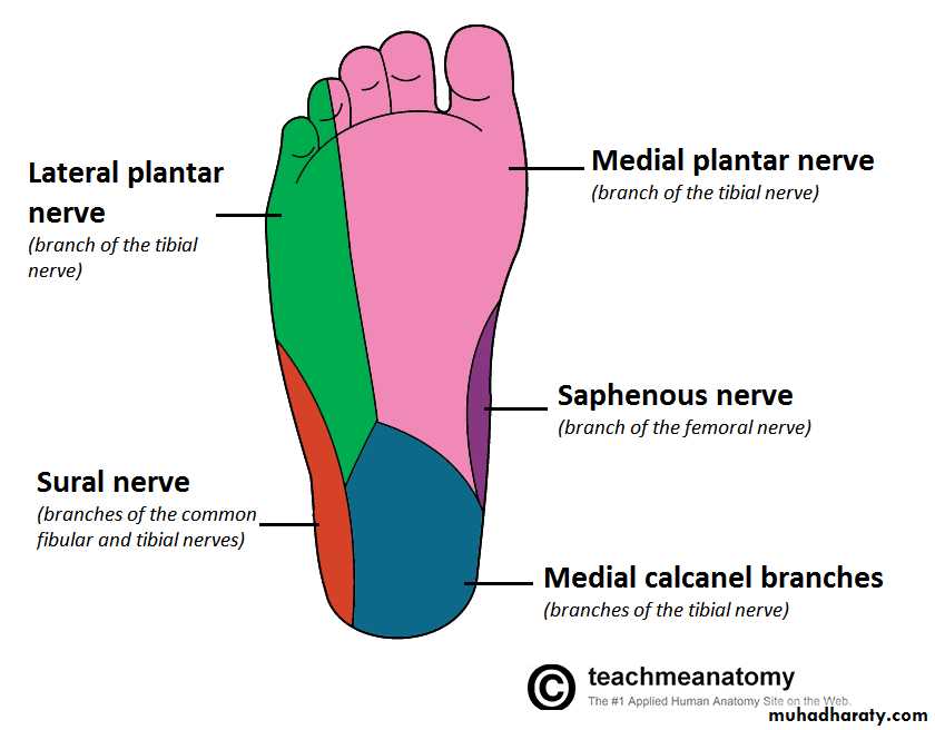
The medial and lateral planter nerve
medial planter .n gives:muscular branches
2- articular branches
3- planter cutaneous
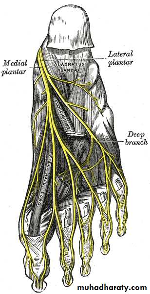
Lateral planter nerve
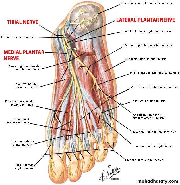
The smaller terminal branches of the tibial nerve passes forward and laterally.
branches
muscular branches
articular branches.
Superficial branch (Cutaneous) supply the skin of the lateral 1/3 of sole and lateral 1 and half of lateral digit
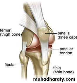
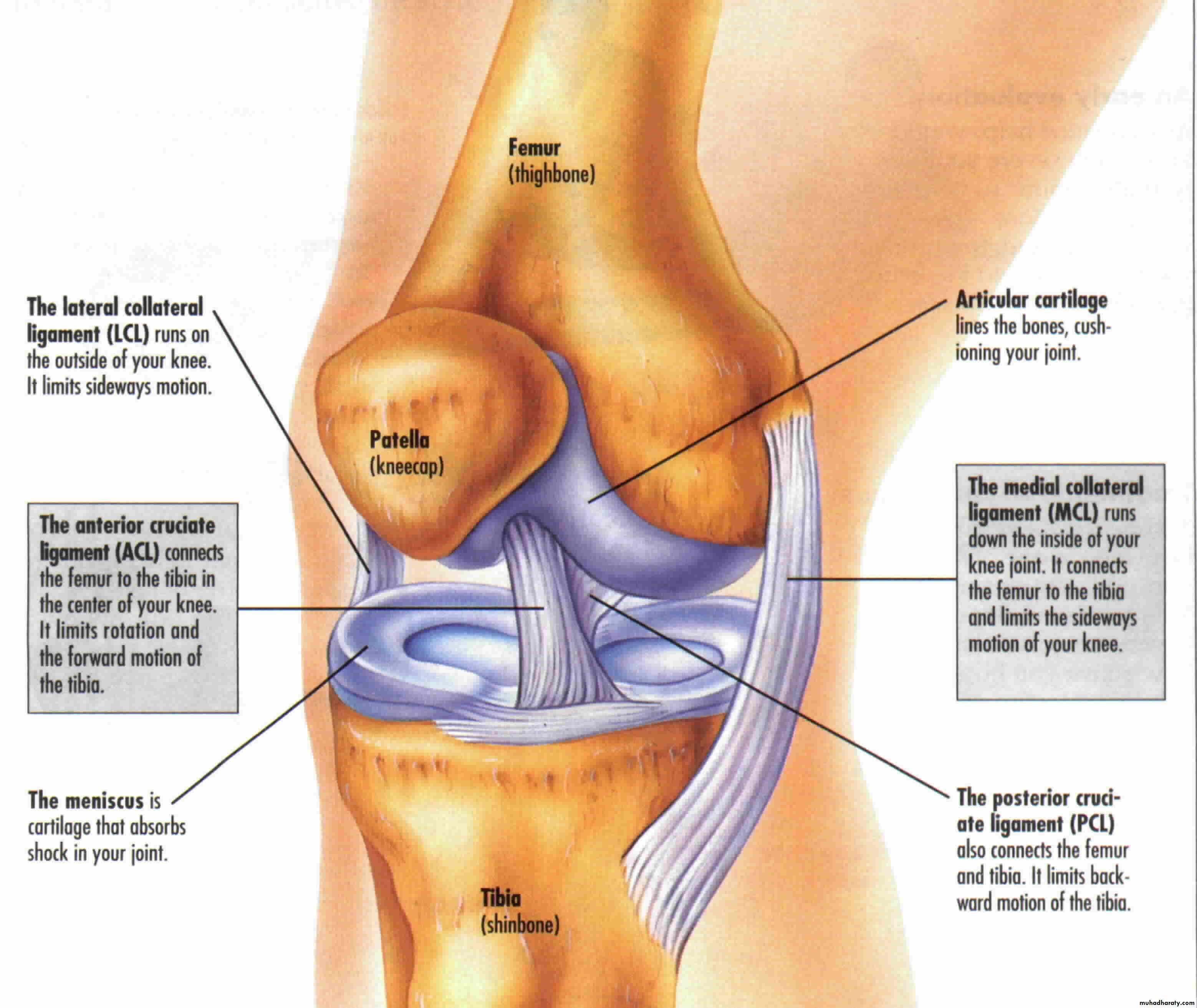
The knee joint
It is a synovial joint of the hinge type, it is unstable joint but this overcome by certain mechanism:1- expansion of the upper end of the tibia and lower end of the femur.
2- Presence of the strong collateral ligament and tendons.
3- Strong capsule.
4- Presence of the intra-articular ligaments.
The capsule is strengthened by number of ligaments
include:1- lateral and medial patellar retinacula
2- Iliotibial tract.
3- The ligamentum patellae which is a continuation of the quadriceps femoris tendon run on the patella to reach the tibial tuberosity.
4- Oblique popliteal ligament it is the posterior reinforcement of the capsule of the joint and it is extension from the tendon of the semimembrenosus m.
5- Arcuate popliteal ligament arise from the back of the head of the fibula and runs medially over the popliteus m.
6- Collateral ligament they are tibial and fibular collateral ligaments. They are very strong ligaments.
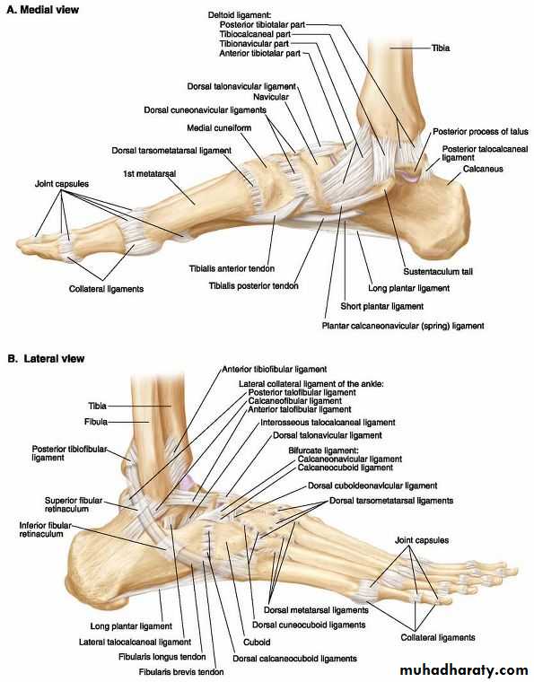
Ankle joint
This is a hinge type of joint between the trochlea of the talus with the distal end of the tibia and medial malleolus medially and the lateral surface of the body of the talus with the lateral malleolus laterally