
Color Atlas of Veterinary
Histopathology
By
Hafidh I. AL- Sadi
BVMS, University of Mosul - Iraq
MS. Pathology, Davis University - USA
Ph.D. Pathology, Davis University - USA
Saevan Saad Al-Mahmood
BVMS, University of Mosul - Iraq
M.Sc. Pathology, University of Mosul - Iraq
Ph.D. Pathology, University of Mosul - Iraq
First edition - 2010
Systemic Pathology (10) : The Respiratory System
54
Systemic Pathology (10)
The Respiratory System
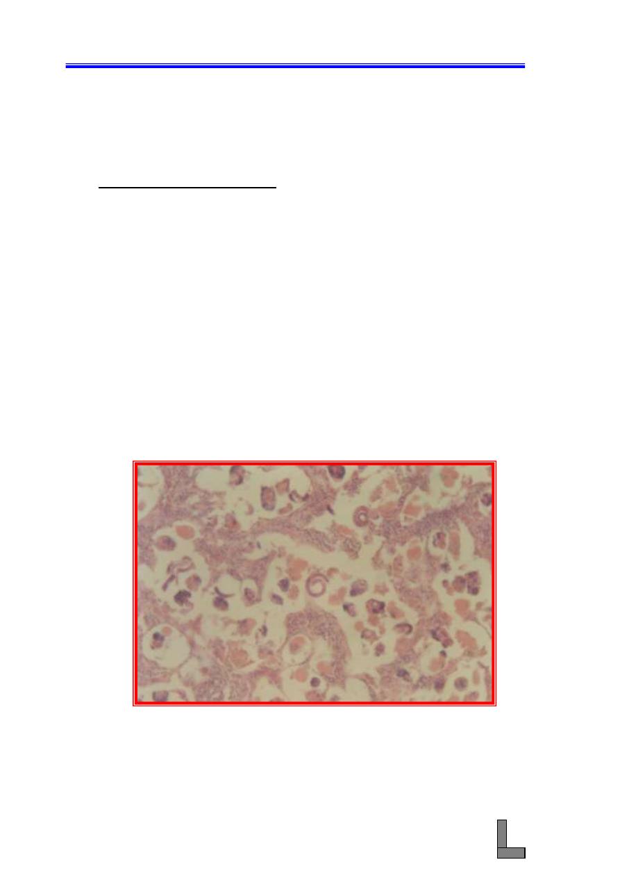
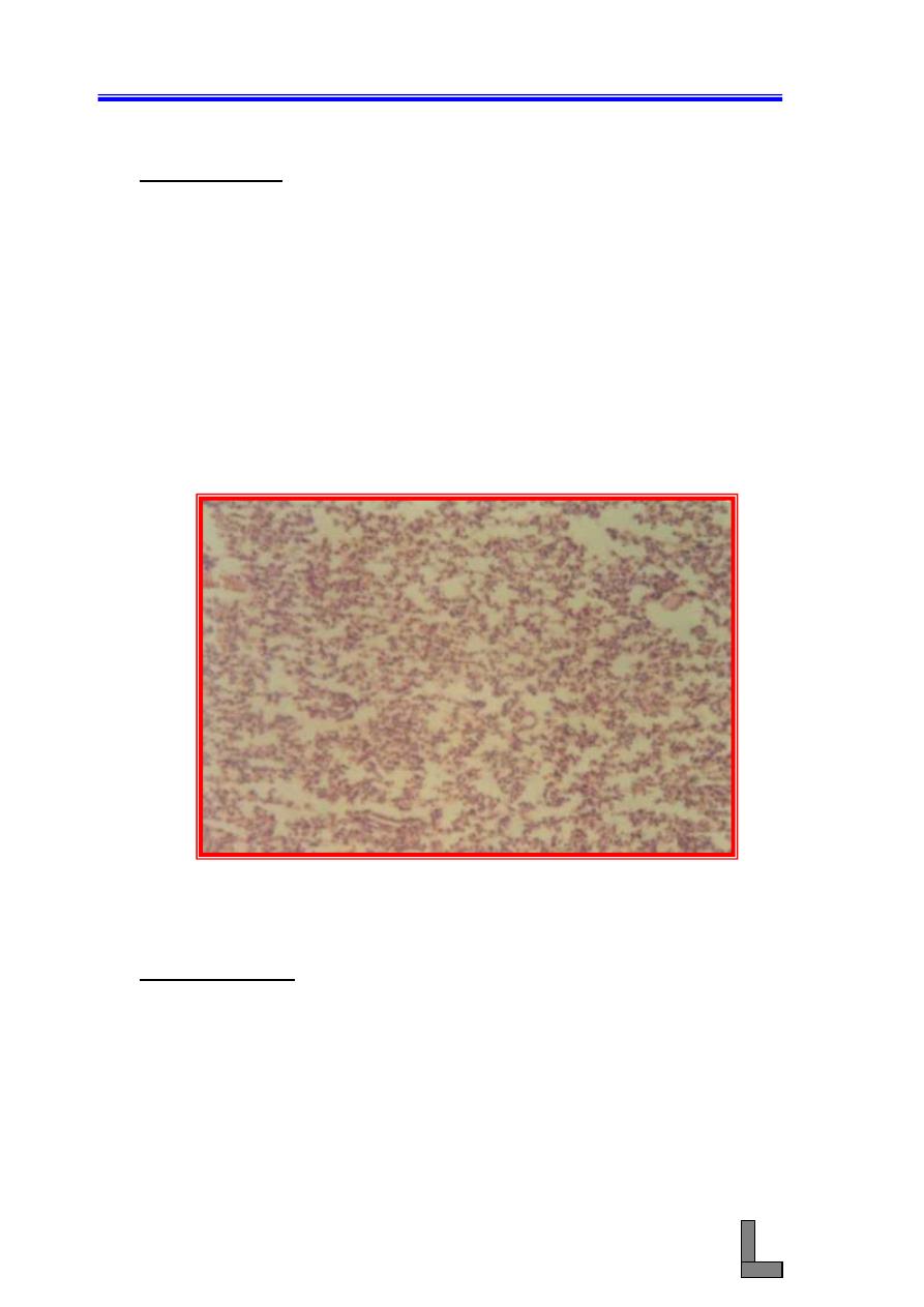
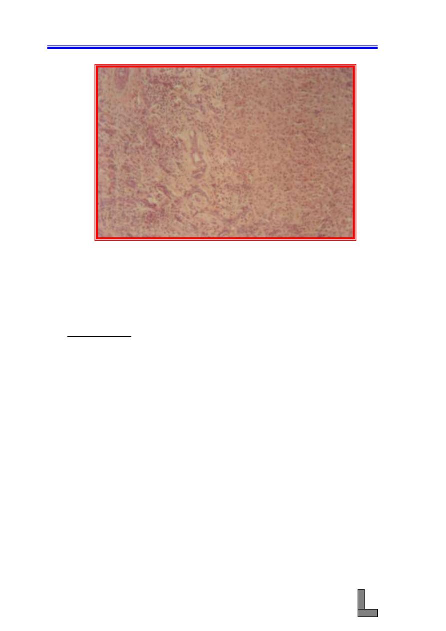
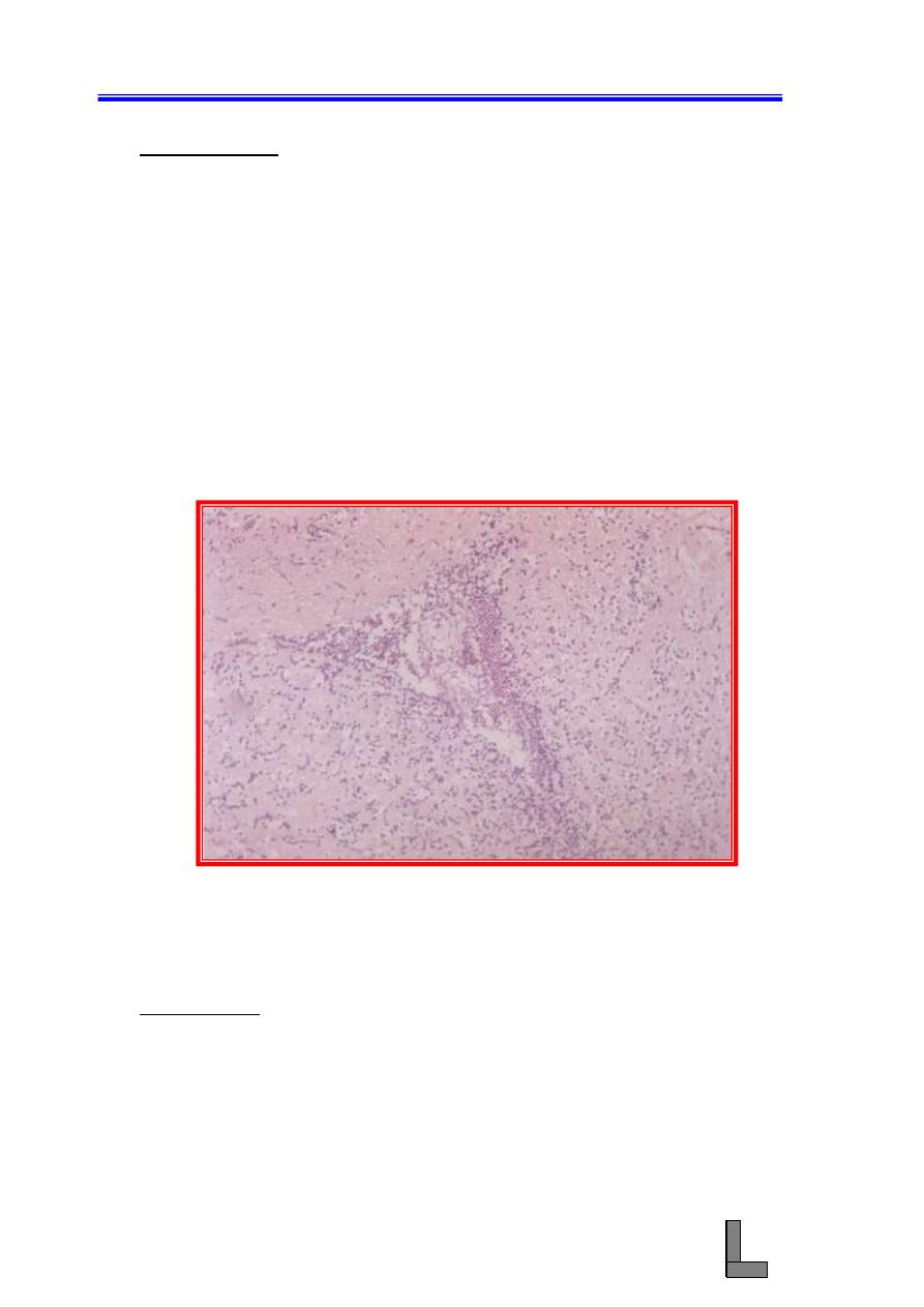
(1) Verminous pneumonia: Also called pulmonary nematodiasis,
lungworm
disease,
Dictyocauliasis,
Dictyocaulosis,
and
Metastrongyloidosis.
Important
parasite
species
include
Dictyocaulus viviparous (cattle, deer, buffalo, camels), D. filaria
(sheep and goats) and D. arnifieldi (horses and donkeys),
Protostrongylus rufescens (sheep and goats), Mullerius capillaris
(sheep and goats).
(A) Note the larvae in the alveoli inciting inflammatory cellular infiltrate
chiefly composed of eosinophils, which fill the alveoli, alveolar
septae, and terminal bronchioles.
(B) Heavy infection is often associated with extensive pulmonary edema
and interstitial emphysema.
(C) The larvae break through the capillary and alveolar walls, they cause
hemorrhage and necrosis.
(D) Remember that when the number of larvae is large, the lesion could
be seen grossly as foci of consolidation randomly distributed
throughout the lungs.
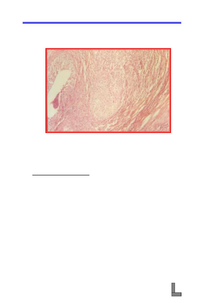
Fig. 41-
Photomicrograph of a lung showing verminous pneumonia.
Note the presence of numerous developmental stages of
lungworms in the alveoli and bronchioles. Emphysema,
edema, and inflammatory exudate could also be seen. H&E
stain. X 100.
Systemic Pathology (10) : The Respiratory System
55
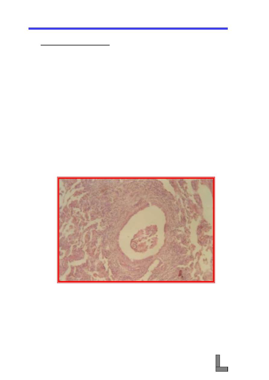
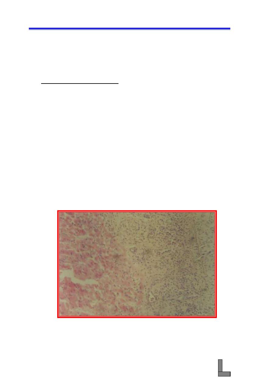
(2) Parasitic bronchitis: See the pervious slide: Adult pulmonary
nematodes reside in the bronchi, where eggs are deposited, some of
which may hatch in the airways. Eggs and / or larvae are coughed
up, swallowed, and passed in the feces, they then develop to
infective 3
rd
stage larvae in the soil. Ingested larvae penetrate the
intestinal mucosa and migrate via lymphatic to mesenteric lymph
nodes, where they develop to 4
th
stage larvae. These reach the lungs
by way of lymphatics and pulmonary arteries. They enter the
pulmonary alveoli, and bronchioles and bronchi, where they reach to
sexual maturity.
(A) The bronchial epithelium is hyperplastic, and eosinophils and
lymphocytes infiltrate the wall and peribronchial tissues.
(B) Eosinophils and mucous plug the bronchial lumens, which when
occluded, lead to atelectasis or consolidation of the related
alveoli.
(C) Adults feed on mucus and cellular detritus, deposit ova; these are
coughed up as embryonated eggs or hatched larvae, or they may
lodge in alveoli, initiating a foreign body reaction.
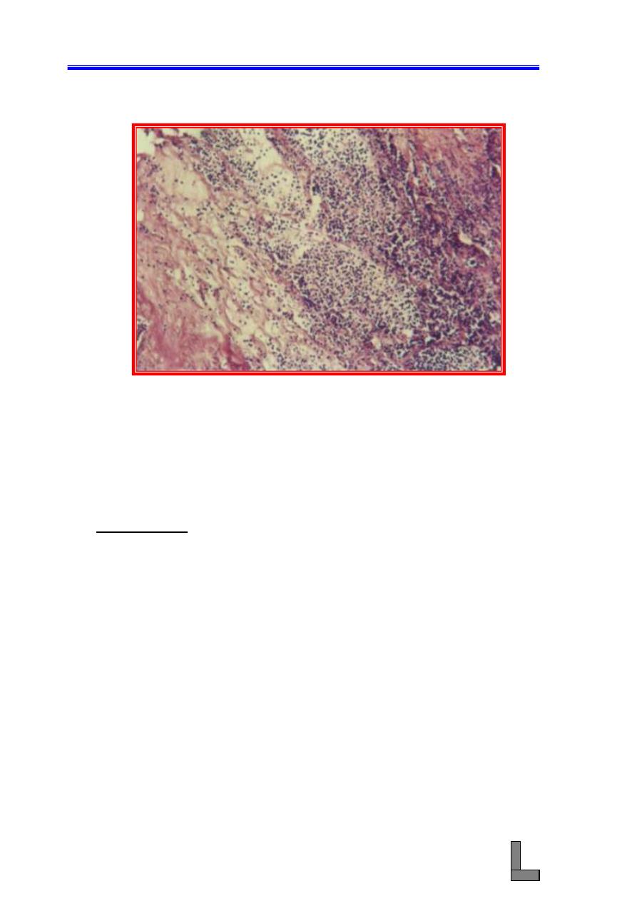
Fig. 42-
Photomicrograph of a lung showing parasitic bronchiolitis.
Developmental stages of lungworms, mucus, and debris
could be seen in the bronchiole (arrow). Thickening of the
bronchiolar wall due to fibrosis and the accumulation of
inflammatory cells could be seen. H&E stain. X 100.
Systemic Pathology (10) : The Respiratory System
56
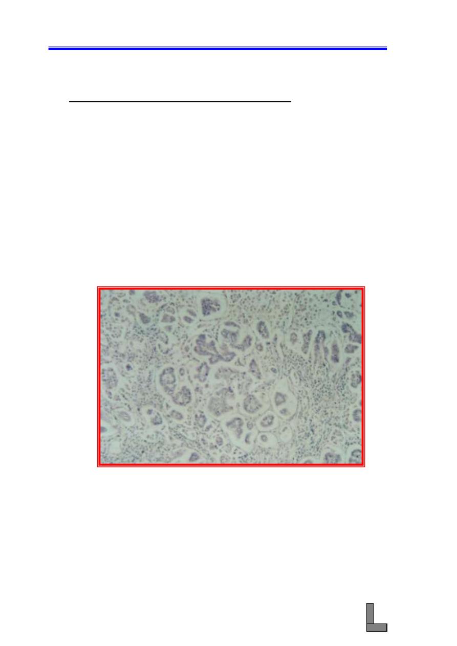
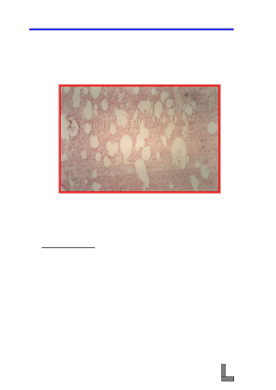
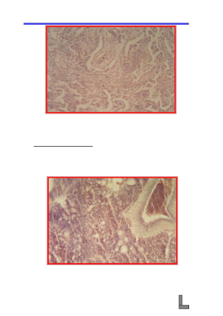
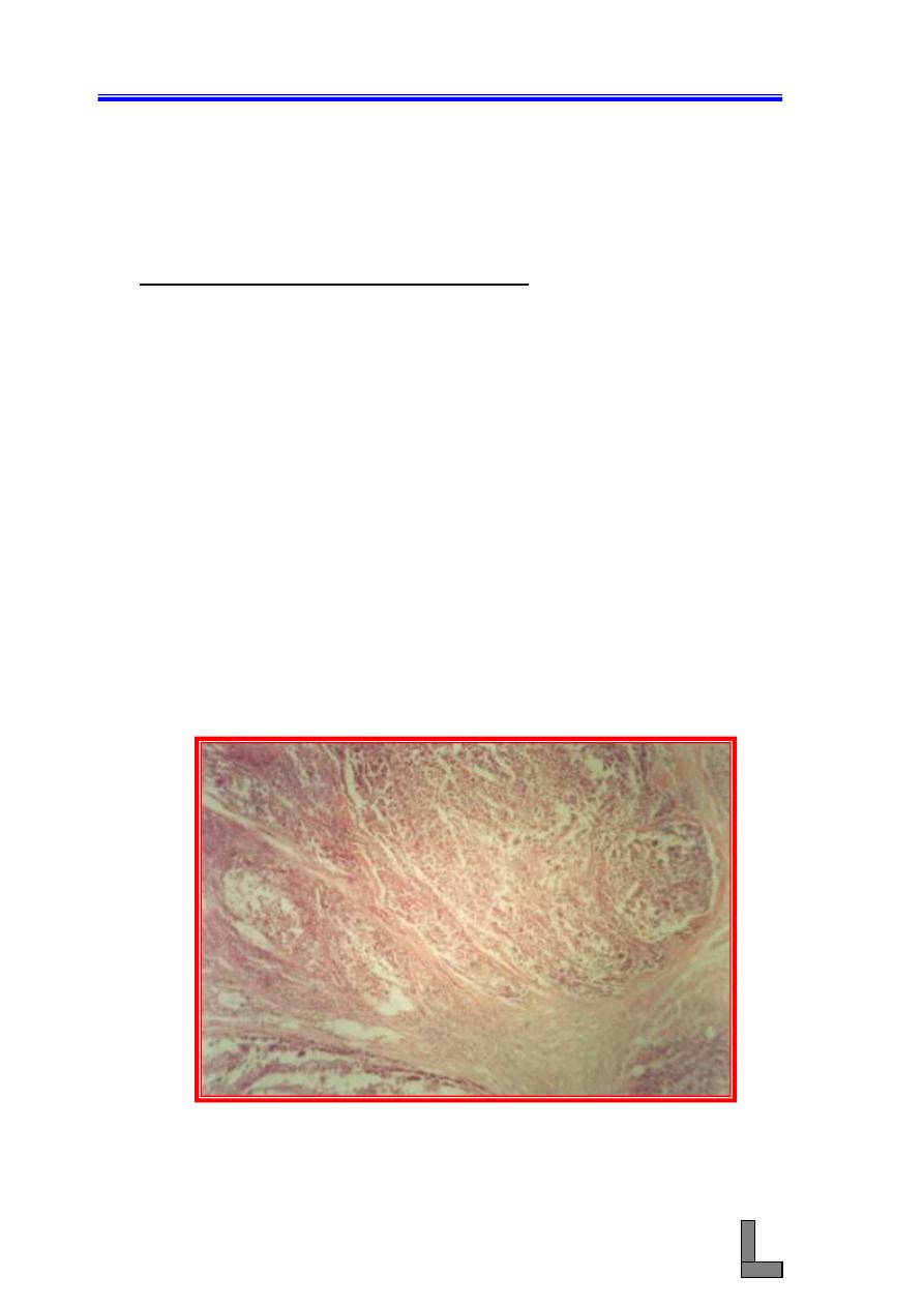
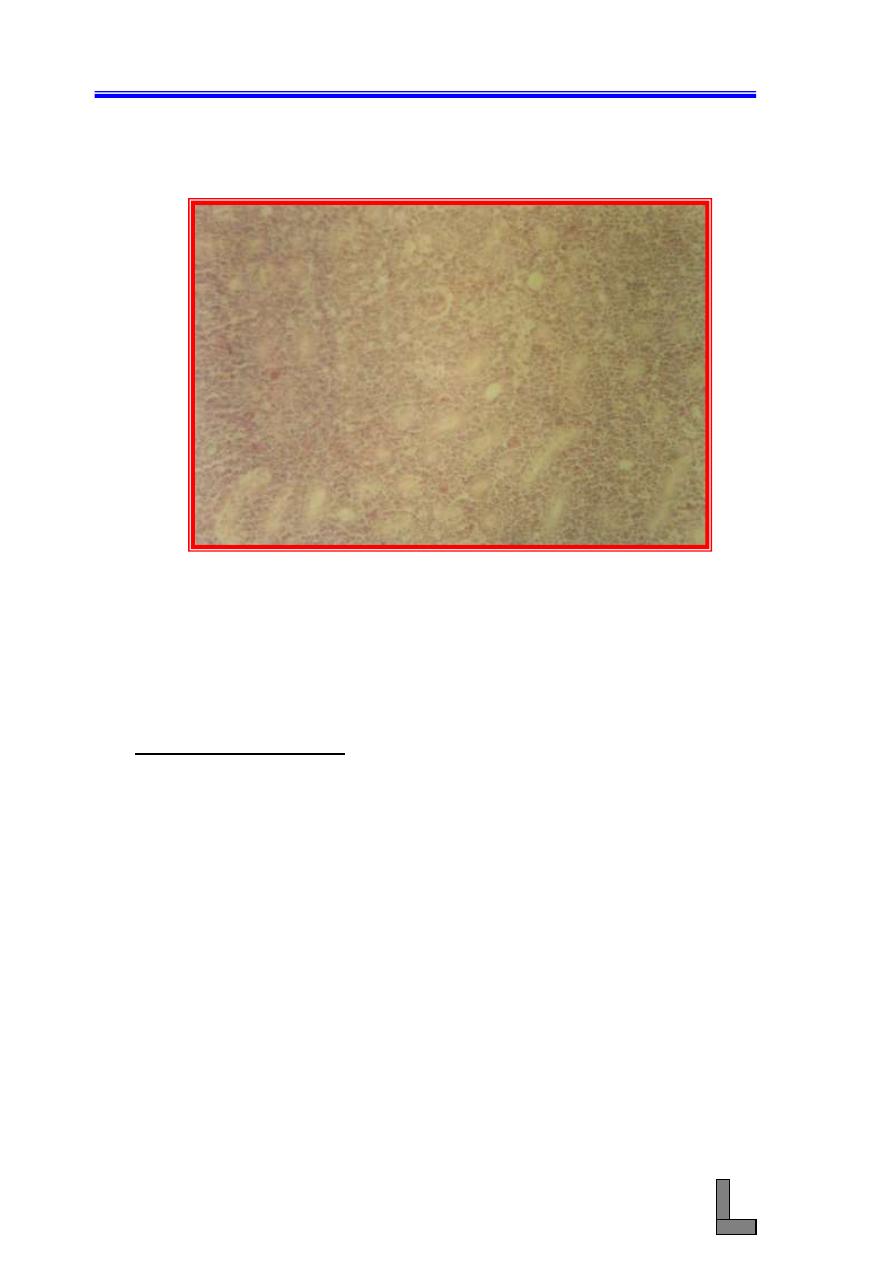
(3) Sheep Pulmonary Adenomatosis (SPA): Also called ovine
pulmonary adenoma, pulmonary carcinoma of sheep, and jaagsiekte.
(A) Note multiple foci of neoplastic alveolar type II cells in acinar
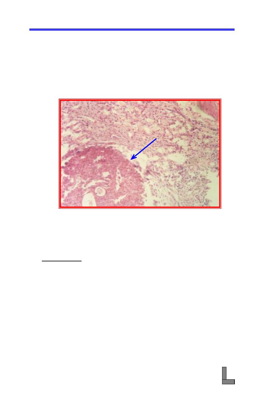
and papillary patterns. The result is a pronounced thickening of
the alveolar walls and their interstices and partial obliteration of
the alveolar spaces by small adenocarcinomas.
(B) Lymphocytes accompany the reticuloendothelial cells, and
fibroblasts appear in the later stage (fibrosis).
(C) Mononuclear cells spill over into the alveoli, and accompanied
by a few neutrophils, appear as an exudate in some of the
bronchi.
(D) The peribronchiolar lymph nodes are hyperplastic (markedly
enlarged).
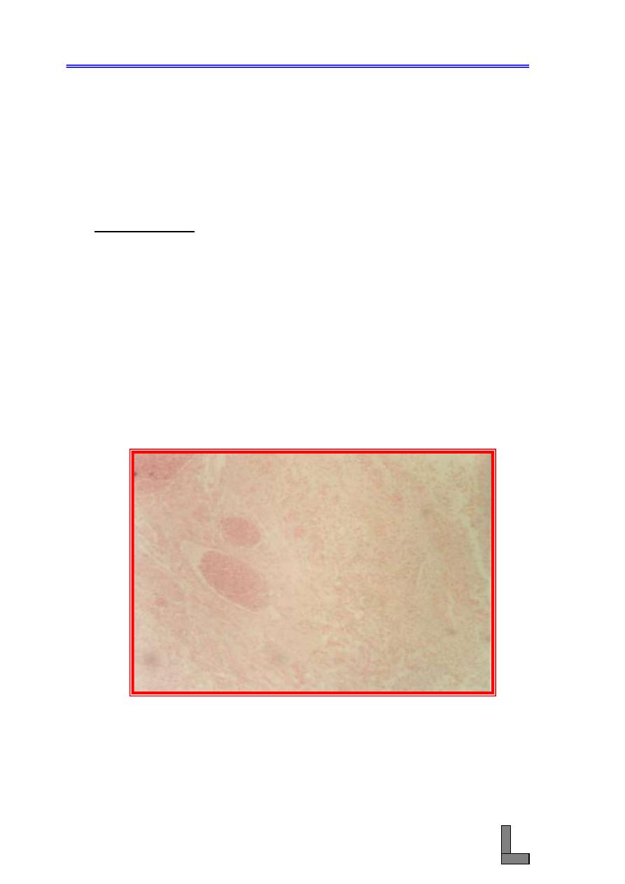
Fig. 43- Photomicrograph of a lung affected with sheep pulmonary
adenomatosis. Note the acinar and papillary patterns of
proliferation of the neoplastic alveolar type II cells. H&E
stain. X 100.
Systemic Pathology (10) : The Respiratory System
57
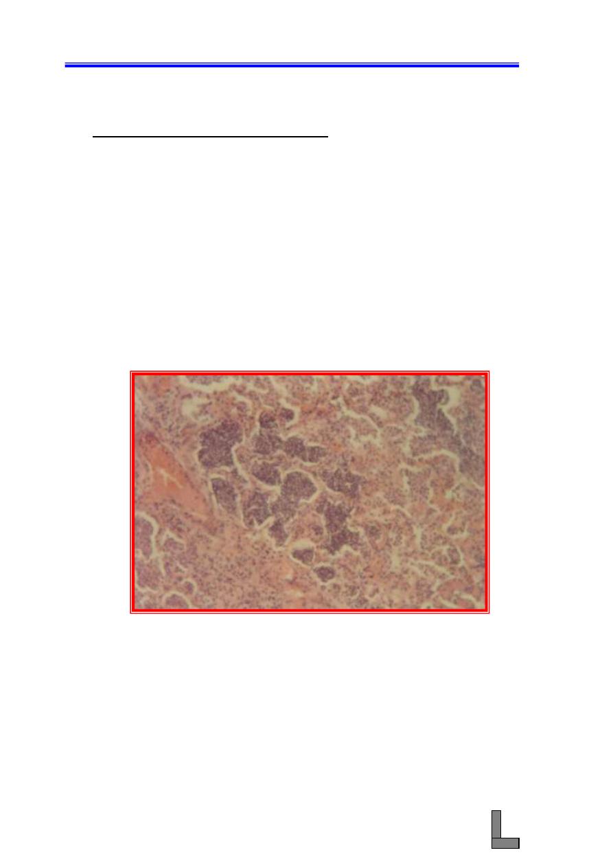
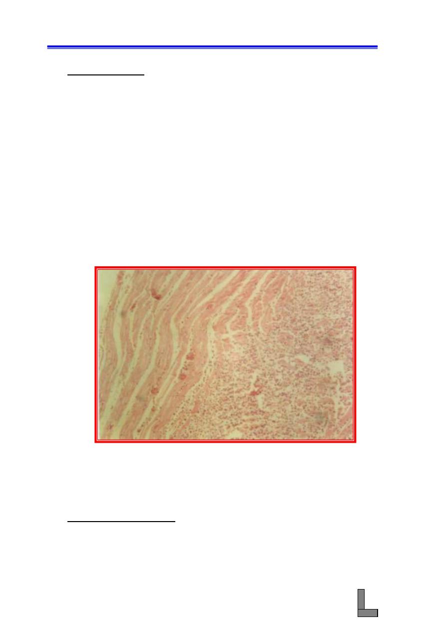
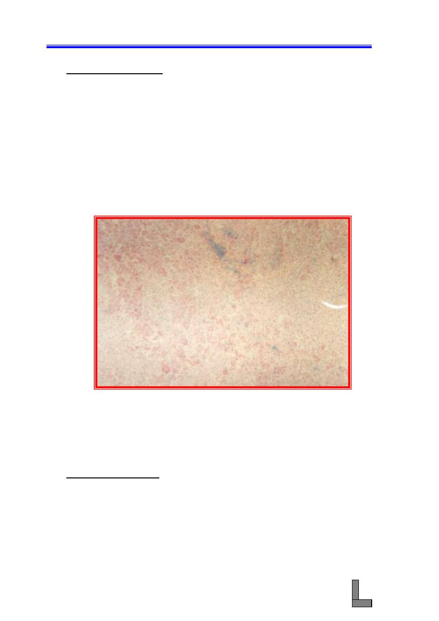
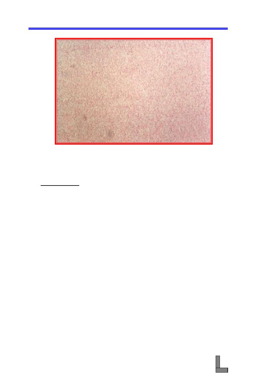
(4) Suppurative bronchopneumonia: When the infectious particles
enters through the bronchial passages the pneumonia is called
bronchial pneumonia or bronchopneumonia. In some cases alveolitis
results from extension of the inflammatory exudates from the
bronchi (called bronchitis) and bronchioles (called bronchiolitis).
(A) Note extensive infiltration of inflammatory cells (mainly
neutrophils) in the walls and lumens of the bronchi and
bronchioles.
(B) The alveoli are filled with neutrophilic exudate.
(C) Other changes indicative of acute inflammation such as
hyperemia of blood vessels in the walls of bronchioles and
alveoli could also be seen.
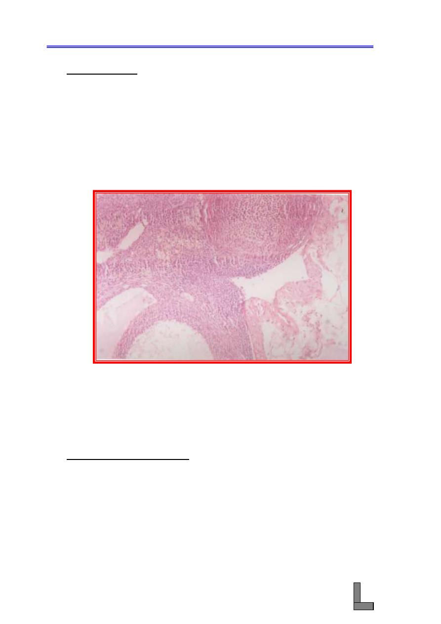
Fig. 44- Photomicrograph of a lung affected with suppurative
bronchopneumonia. Note that the alveoli (and the
bronchioles) are filled with neutrophilic exudate. H&E stain.
X 100.
Systemic Pathology (10) : The Respiratory System
58
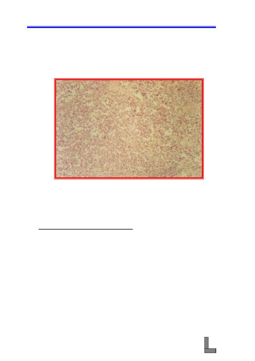
(5) Atelectasis: This term means failure of the alveoli to open or to
remain open; in other words, the empty alveoli are collapsed and do
not contain air. Atelectic portion is dull red in color and has the
consistency of hepatic tissue. Since the atelectic alveoli do not
contain air, the atelectic portion sinks in water.
(A) The alveoli are compressed into scarcely recognizable slits, all
laying parallel in a direction determined by adjacent pressures.
(B) Note that the cells are those of normal lung tissue.
(C) Well – filled capillaries could be seen, but the total blood
content of the atelectic tissue is less than normal.
(D) The bronchioles are collapsed as far as their structure permits.
Fig. 45- Photomicrograph of a lung showing atelectasis. Note collapse of
the alveoli and terminal bronchioles. H&E stain. X 100.
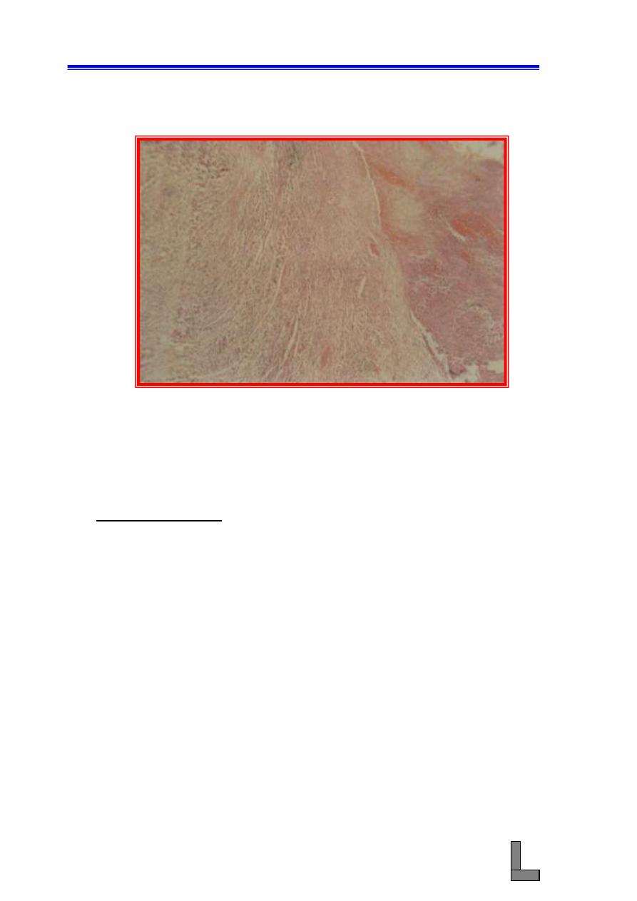
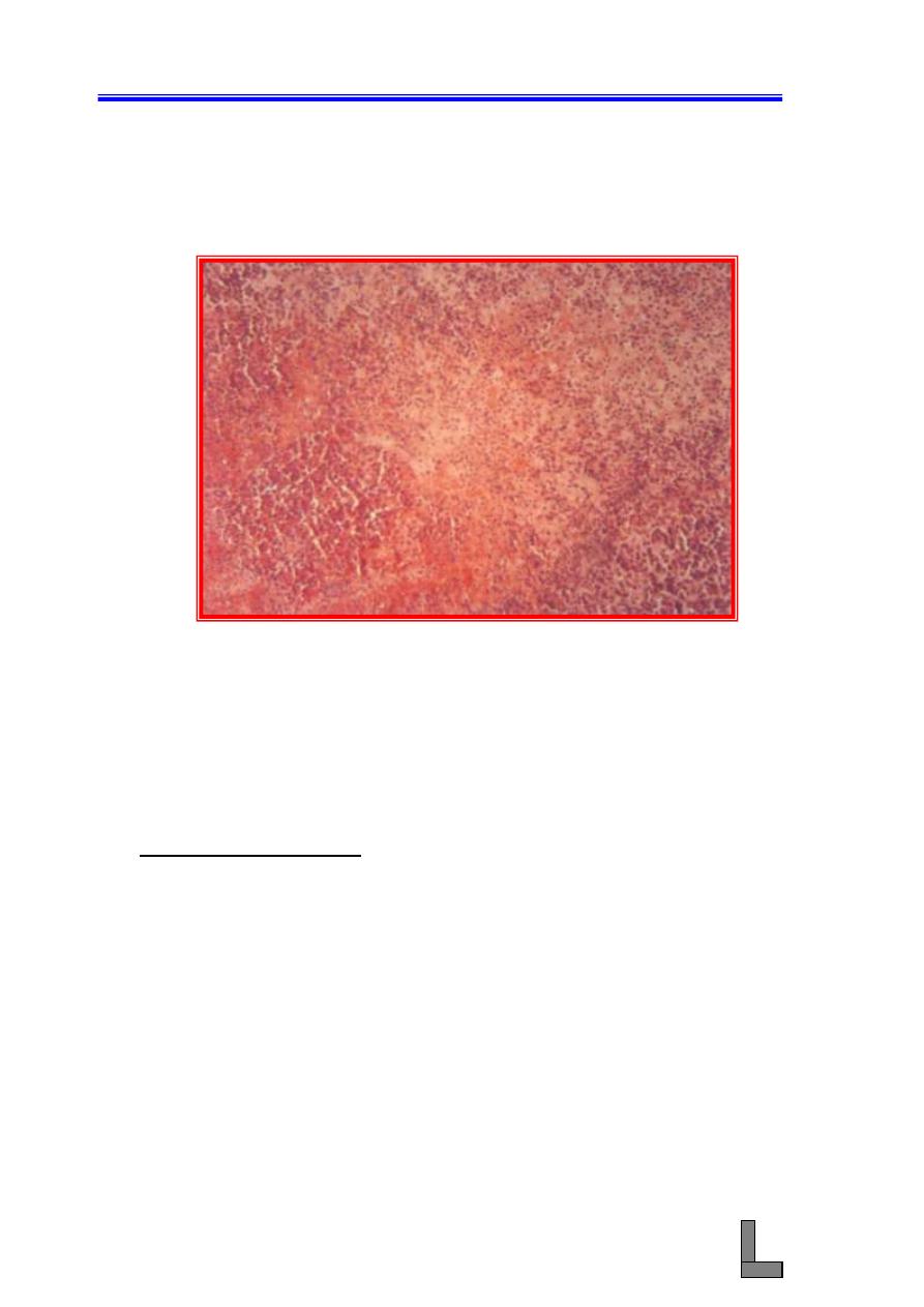
(6) Emphysema: Means the presence of air in the tissues and there are
two kinds, alveolar and interstitial. Alveolar emphysema is more
common and is called pulmonary emphysema.
(A) Many alveoli are too large and many have wide openings into
each other or into a common space due to rupture of alveolar
walls.
(B) The blunt ends of alveolar walls which have been broken often
persist and become thickened and hyperplastic rounded knobs.
Systemic Pathology (10) : The Respiratory System
59
(C) Some of the walls are slightly thickened while others are
stretched and very thin.
(D) Blood – filled capillaries are scarce and the smooth muscle of
the alveolar ducts is often hyperplastic.
Fig. 46- Photomicrograph of a lung affected with emphysema. Many of
the alveoli are too large and open into each other. H&E stain.
X 40.
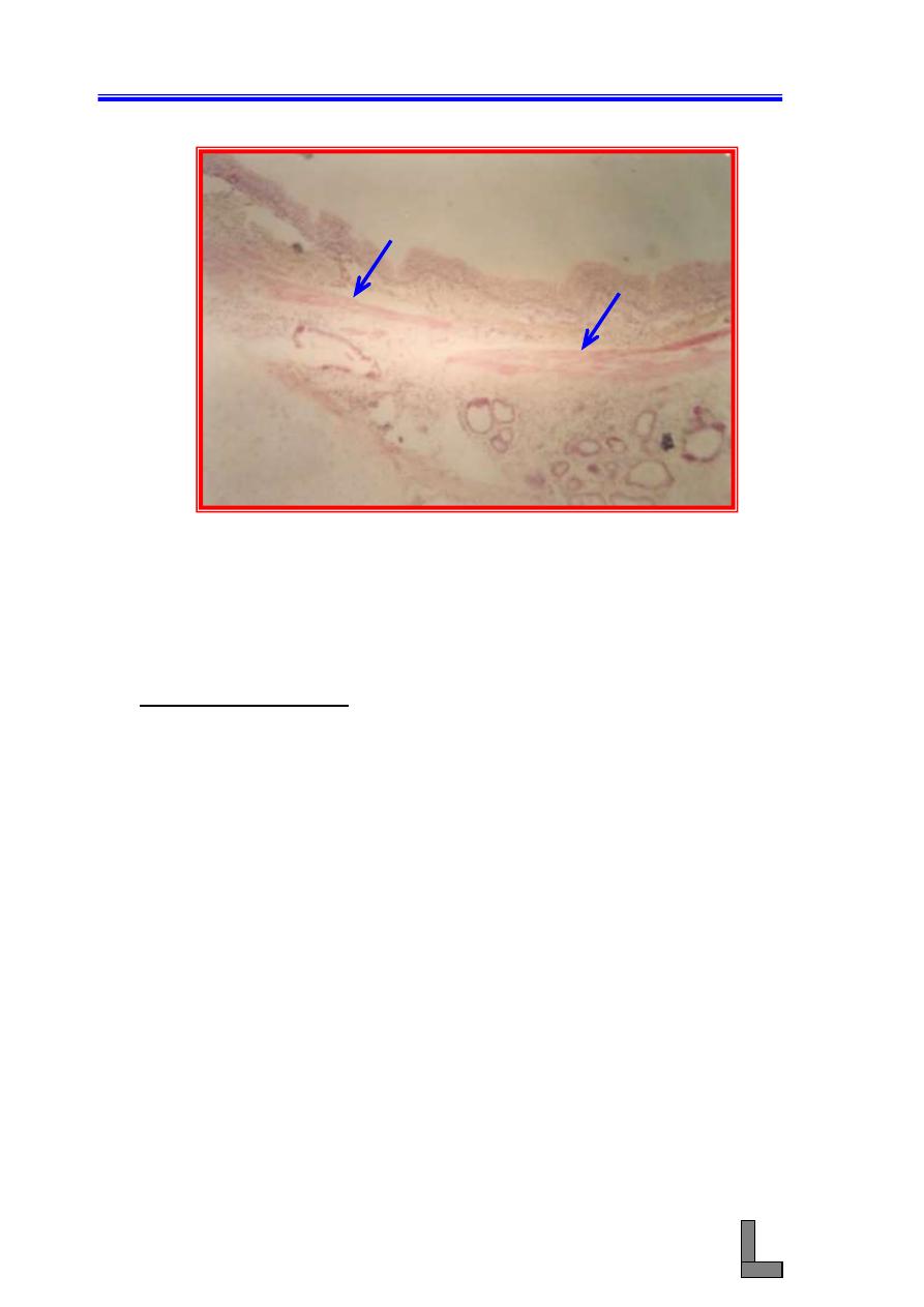
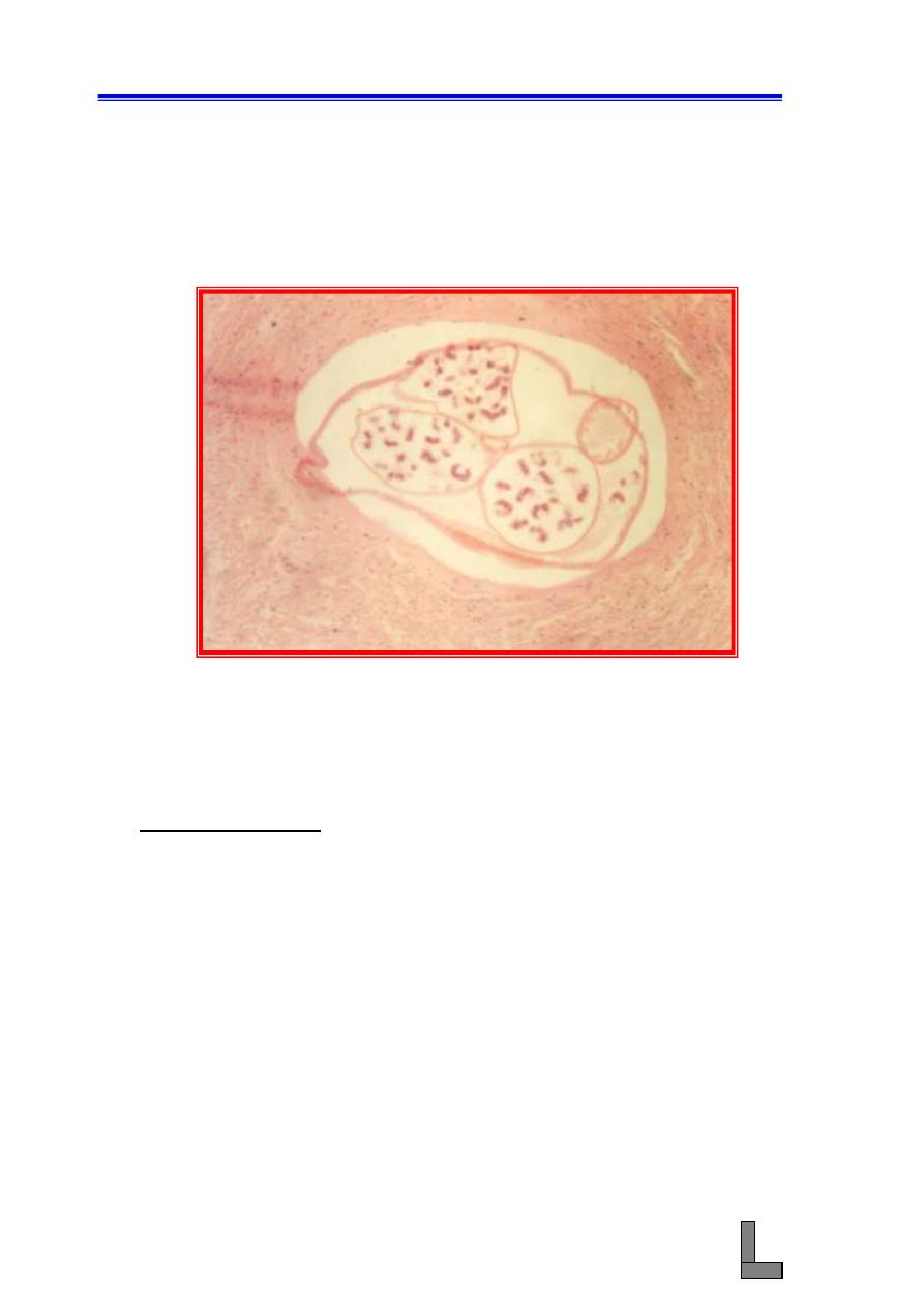
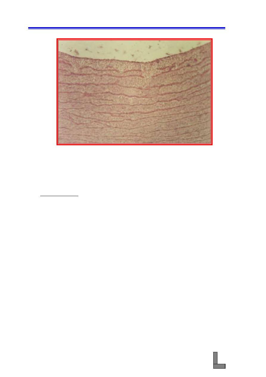
(7) Bronchiectasis: Is one of the most devastating sequel that follows
chronic bronchitis. It consists of pathologic and permanent dilation
of a bronchus as a result of the accumulation of exudates in the
lumen and partial rupture of bronchial walls. Destruction of walls
occurs, in part, when proteolytic enzymes relapsed from phagocytic
cells during chronic inflammation degrade and weaken the smooth
muscle and cartilage that help to maintain normal bronchial
diameter.
(A) The presence of purulent exudate in the dilated bronchus.
(B) Remnants of the bronchial wall surround the exudate.
(C) Differentiate this lesion from pulmonary abscess.
Systemic Pathology (10) : The Respiratory System
60
Fig. 47- Photomicrograph of a lung affected with bronchiectasis. Note the
very large lumen (
L
) and the infiltration of inflammatory
cells in the wall of the bronchiole. Hyperplasia of smooth
muscle could also be noted (
arrows
). H&E stain. X 40.
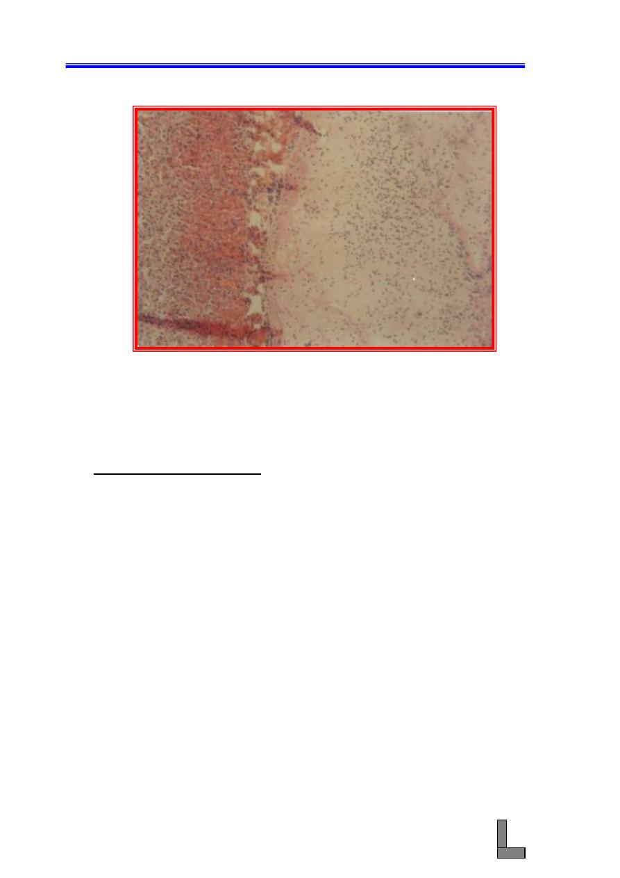
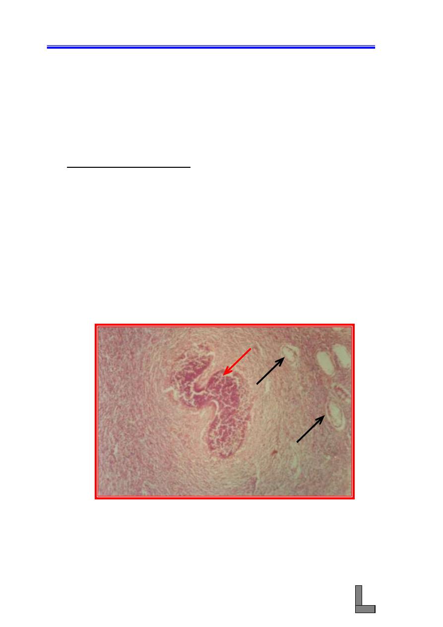
(8) Pleuropneumonia: Pleuritis (pleurisy) means inflammation of the
visceral or parietal pleura. It can occur as part of pneumonia,
particularly in fibrinous bronchopneumonia, and here the condition
is called pleuropneumonia. Bovine and ovine pneumonic
pasteurellosis and bovine pleuropneumonia are good examples of
pleuritis associated with fibrinous bronchopneumonia.
(A) Note the presence of a serofibrinous exudate in the pleura
leading to increased thickness of the pleura.
(B) Widely distributed hyperemia of blood vessels and bacterial
colonies could be seen.
(C) The alveoli are filled with a serofibrinous exudate containing
polymorphonuclear cells and large numbers of erythrocytes.
(D) Inflammatory exudate could also be seen in the bronchi and
bronchioles.
L
Systemic Pathology (10) : The Respiratory System
61
Fig. 48- Photomicrograph of a lung affected with pleuropneumonia. Note
the accumulation of serofibrinous exudate in the pleura (1)
and hyperemia and inflammation of the lung tissue (2). H&E
stain. X 100.
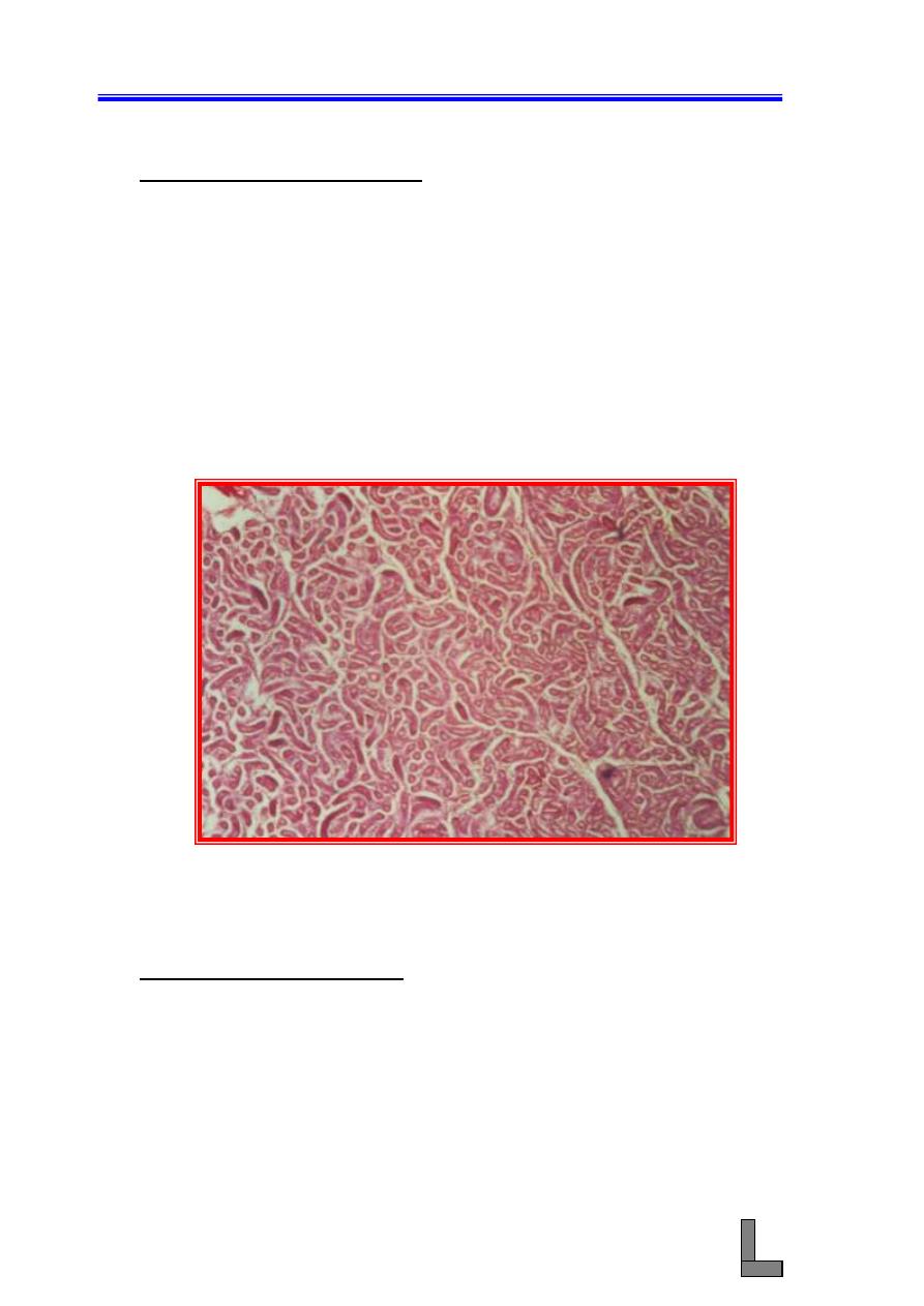
(9) Interstitial Pneumonia: In this type of pneumonia, the inflammatory
process takes place primarily in any of the three layers of the
alveolar walls (endothelium, basement membrane, and alveolar
epithelium) and the contiguous bronchiolar interstitium.
(A) Note the thickened bronchiolar walls due to accumulation of
inflammatory cell infiltrate in the mucosa and its extension
peribronchially into the alveolar interstitium. This change is
called bronchiointerstitial pneumonia.
(B) Although the lesions in interstitial pneumonia are centered in the
alveolar walls and interstitium, a mixture of desquamated
epithelial cells, macrophages, and mononuclear cells are usually
present in the lumen of bronchioles and alveoli.
(C) Hyperplasia of smooth muscle in airways or pulmonary
vasculature, and microscopic granulomas could be seen.
1
2
Systemic Pathology (10) : The Respiratory System
62
Fig. 49- Photomicrograph of a lung affected with interstitial pneumonia.
Note the increased thickness of the alveolar interstitium due
to infiltration of mononuclear cells. X 100.
(10) Mycotic Pneumonia: The most common causes of granulomatous
pneumonia in animals include systemic fungal diseases such as
Cryptococcosis (Cryptococcus neoformans), Coccidioidomycosis
(Coccidioides immitis), Histoplasmosis (Histoplasma capsulatum),
and blastomycosis (Blastomyces dermatitidis).
Fig. 50- Photomicrograph of an avian lung showing mycotic pneumonia.
Note severe inflammation of the pulmonary tissue. A
thrombosed blood vessel could be seen. H&E stain. X 100.

Systemic Pathology (10) : The Respiratory System
63
References:
Brogden KA, Lehmkul HD, Cutlip RC. Pasteurella haemolytica complicated
respiratory infections in sheep and goats. Vet Res 1998; 29 :
233-254.
Carrman S, Rosendal S, Huber L, Gyles C, Mckee S, Willoughby RA, Dubovi
E, Thorsen J, Lein D. Infectious agents in acute respiratory
disease in horses in Ontario. J Vet Diag Invest 1997; 9:17-23.
De La Concha
– Bermejillo A. Maedi – Visna and ovine progressive
pneumonia. Vet clin North Am Food Anim Pract 1997; 13:13-33.
Dungworth DL. The respiratory system. In : Jubb KVF, Kennedy PC, Palmer
N, eds. Pathology of domestic animals. 4th ed. New York :
Academic Press, 1993: 539.
Hondalus MK. Pathogenesis and virulence of Rhodococcus equi. Vet
Microbiol 1997; 56:257-268.
Lopez A. Respiratory system, thoracic cavity, and pleura. In: McGavin MD,
Carlton WW, Zachary JF. Eds. 3rd ed. Thomson's special
veterinary pathology. St. Louis : Mosby, 2001 : 125 - 195.
Moulton JE. Tumors of the respiratory system. In: Moulton JE, ed. Tumors in
domestic animals. 3rd ed. Berkeley: University of California
Press, 1990:308.
Systemic Pathology (11) : The Digestive System
64
Systemic Pathology (11)
The Digestive System
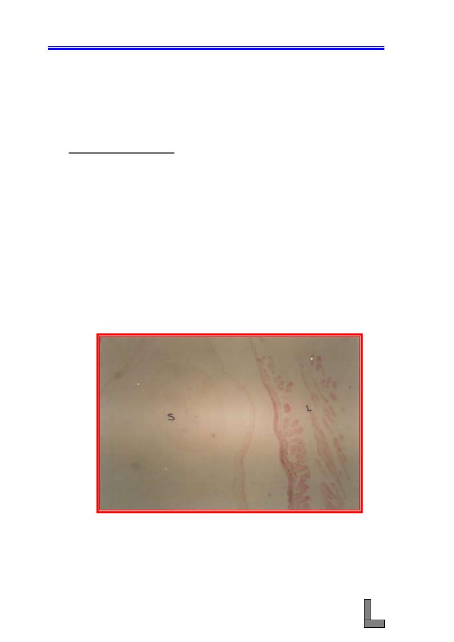
(1) Parasitic Glossitis: Among parasites that are seen in the tongue of
ruminants are the tapeworms (Cysticercus bovis, Cysticercus ovis),
protozoa (Sarcocystic spp), and round worms (Gongylonema spp).
Cysticercus bovis, the larva of Taenia saginata occurs in muscle,
liver, heart, lungs, diaphragm, lymph nodes, and other parts. On the
other hand, Cysticercus ovis, the larva of Taenia ovis occurs in the
heart, voluntary muscles, diaphragm, esophagus, and rarely the lung.
(A) Note the presence of multiple larvae beneath the lingual mucosa
and in between the bundles of muscle fibers of the tongue.
(B) Minimal tissue response occurred against the presence of the
larva. This response is in the form of thin inflammatory layer
with connective tissue response.
(C) Note atrophy of some of the bundles of muscle fibers close to
the larva (pressure atrophy).
(D) Sarcocystic spp could also be seen in the muscle fibers.
Fig. 51- Photomicrograph of a tongue affected with both cysticercosis
and sarcocystosis (parasitic glossitis). Note the presence of
the cysticercus larva (
S
) in the lingual tissue (
L
). H&E stain.
X 40.
S
L
Systemic Pathology (11) : The Digestive System
65
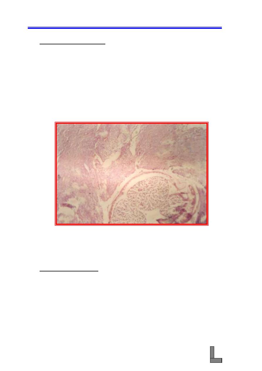
(2) Parasitic esophagitis: Infection with the spiruroid worm, Spirocerca
lupi (also called the esophageal worm) is particularly common in
dogs in many parts of the world. The adult worms are usually found
in nodules in the wall of the esophagus, aorta, stomach, and other
organs of the dog, fox, wolf, and cat. Grossly, one or more nodules
are seen in the luminal surface, elevating the epithelium with the
presence of the worms in these nodules. Microscopically:
(A) Sections of adult Spirocerca lupi worms could be seen in the
wall of the esophagus.
(B) The worms are surrounded by thick metaplastic connective
tissue.
Fig. 52- Photomicrograph of the wall of the esophagus of a dog infected
with Spirocerca lupi . A cross section of an adult worm (
W
)
could be seen in a fibrous tissue nodule (
F
). H&E stain.
X 40.
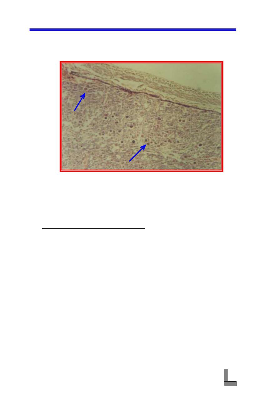
(3) Parasitic Enteritis: Coccidia are host – and tissue – specific protozoa
and obligate intracellular pathogens. They cause lesions that could
be proliferative or hemorrhagic. In epithelial cells of the villi or
crypts, the organism undergo one or more asexual reproductive
cycles, with the resulting sporozoites producing schizonts that
contain from few to thousands of merozoites. The latter enter new
cells and repeat the cell cycle. Microscopically:
(A) Section of the intestine showing vascular and cellular changes
indicative of inflammation (enteritis).
F
F
F
W
Systemic Pathology (11) : The Digestive System
66
(B) Numerous stages of the life cycle of Eimeria spp could be seen
in the mucosa and submucosa.
Fig. 53- Photomicrograph of the small intestine of a laboratory animal
affected with intestinal coccidiosis. Note the presence of
various developmental stages of the parasite (Eimeria spp.)
(
arrows
) and the infiltration of inflammatory cells in the
intestinal mucosa. H&E stain. X 100.
(4) End - Stage liver or Cirrhosis: This is characterized by loss of
normal hepatic architecture due to nodular regeneration of
parenchyma, fibrosis, and often biliary duct hyperplasia. The
condition could be defined as a diffuse process characterized by
fibrosis and conversion of normal liver architecture into structurally
abnormal lobule. It could also be defined as the total absence of any
normal lobular architecture. Among causes of this condition are
toxic plants (hepatotoxins) and anticonvulsant drugs such as
Primidone. Microscopically:
(A) The architecture of the liver is altered by loss of hepatic
parenchyma, condensation of reticulin frame work, and
formation of tracts of fibrous connective tissue.
(B) Regeneration of hepatic tissue between fibrous bands leads to the
formation of variably sized regenerative nodules.
(C) Diffuse infiltration of inflammatory cells could be seen in both
the hepatic parenchyma (hepatitis) and the fibrous tissue.
Systemic Pathology (11) : The Digestive System
67
Fig. 54- Photomicrograph of a liver exhibiting cirrhosis. Lack of normal
lobular architecture of the liver (L) and the formation of
fibrous tissue tract (T) could be seen. Heavy infiltration of
inflammatory cells could be seen in both the liver tissue and
the fibrous tract. H&E stain. X 100.
References:
Bowman DD. Georgi's parasitology for veterinarians. 7th ed. Philadelphia:
Saunders, 1999.
Gelberg HB. Alimentary system. In : McGavin MD, Carlton WW, Zachary JF.
Eds. 3rd ed. Thomson's special veterinary pathology. St. Louis :
Mosby, 2001 : 1 - 124.
Kelly WR. The liver and billiary system. In: Jubb KVF, Kennedy PC, Palmer N.
eds. Pathology of domestic animals. 4th ed. New York:
Academic Press, 1985:239.
Moon HW. Intestine. In : Cheville NF, ed. Cell pathology. 2nd ed. Ames, IA:
Iowa state University Press, 1983:503-529.
T
L
Systemic Pathology (12) : Cardiovascular System
68
Systemic Pathology (12)
Cardiovascular System
(1) Fibrinous Pericarditis:- This is the most common type of
pericardial inflammation in animals, is usually the result of
hematogenously spread infection. Specific diseases with this lesion
include pasteurellosis, blackleg, coliform septicemias, and sporadic
bovine encephalomyelitis in cattle; streptococcal infections in horse;
and psittacosis in birds.
Grossly, both the visceral and parietal pericardial surfaces
are covered by variable amounts of yellow fibrin deposits that can
result in adherence between parietal and visceral layers. When the
pericardial sac is opened at necropsy, these attachments are torn
away (so – called bread – and – butter heart). Microscopically:
(A) Note the presence of an eosinophilic layer of fibrin with
admixed neutrophils over the congested pericardium.
(B) Bacterial colonies could also be seen in the pericardium.
(C) Fibrous organization of the exudates could be visualized
and this leads to adhesion between the pericardial surfaces.
(D) Atrophy and edema of myocardial fibers could be seen.
Fig. 55- Photomicrograph of a heart showing fibrinous pericarditits. An
extensive fibrinous exudate (
F
) could be seen closely
attached to the myocardium (
M
). H&E stain. X 40.
M
F
Systemic Pathology (12) : Cardiovascular System
69
(2) Myocarditis:- Most commonly results from hematogenous spread of
infections to the myocardium and occurs in various systemic
diseases. Infrequently, myocarditis is the primary lesion in affected
animals and responsible for their death. Myocarditis could be
suppurative, necrotizing, hemorrhagic, lymphocytic, or eosinophilic.
In the suppurative type, the inflammation results from localization of
pyogenic bacteria in the myocardium. These often originate from the
vegetations of vegetative valvular endocarditis on the mitral and
aortic valves.
(A) Note the presence of foci consisting of bacterial colonies
neutrophils, and necrotic myocytes (abscesses) in the
myocardium.
(B) A tract could be seen in the myocardium indicating that the
inflammation may be traumatic.
(C) Zenker’s necrosis of myocytes could be seen in the viscinity
of the tract.
Fig. 56- Photomicrograph of a heart exhibiting myocarditis. Note the
heavy infliltration of inflammatory cells between the
bundles. The exudate has replaced some of the muscle fibers.
H&E stain. X 100.
(3) Parasitic arteritis:- Arteritis is a prominent feature of several
parasitic diseases. Examples include dirofilariasis (Dirofilaria
immitis), Spirocercosis (Spirocerca lupi), Onchocerciasis, filariasis,
and angiostrongylosis.
Systemic Pathology (12) : Cardiovascular System
70
(A) Note thickening of wall of the artery (aorta) due to extensive
infiltration of inflammatory cells and proliferation of
fibroblasts.
(B) Remember that live or dead parasites (or larvae) could be
seen in the vascular lesion.
Fig. 57- Photomicrograph of a blood vessel (aorta) showing parasitic
aortitis (parasitic arteritis).Note a cross section of the adult
worm in a fibrous nodule in the wall of the aorta. H&E stain.
X 100.
(4) Acute arteritis: Arteritis occurs as a feature of many infections and
immune-mediated diseases. In inflamed vessel, leukocytes will be
present within and surrounding the walls, and damage to the vessel
wall will be evident as fibrin deposits or necrotic endothelial and
smooth muscle cells. These changes are accompanied by thrombosis
which can result in ischemic injury or infarction in the circulatory
field.
(A) Note the presence of extensive infiltration of leukocytes in
almost all of the layers of the vessel wall.
(B) Engorgement of blood vessel within the vessel wall as well
as hemorrhages could also be seen.
Systemic Pathology (12) : Cardiovascular System
71
Fig. 58- Photomicrograph of a blood vessel showing acute arteritis. Note
the heavy infiltration of inflammatory cells in the wall of the
blood vessel (
BV
) and the presence of blood clot and tissue
debris (
TD
). H&E stain. X 100.
(5) Atherosclerosis: This is a very important vascular disease in human
beings, and it occurs only infrequently in animals. The principal
alteration is accumulation of deposits (atheroma) of lipid, fibrous
tissue, and calcium in vessel walls, which eventually results in
luminal narrowing.
(A) There is increased thickness of the arterial wall due to
accumulation of lipid globules in the cytoplasm of smooth
muscle cells and macrophages (foam cells) in the media
and intima.
(B) There is fibrous tissue accumulation in the arterial wall.
(C) Infiltration of inflammatory cells (mainly macrophages)
could be seen in the affected part of the arterial wall.
BV
TD
Systemic Pathology (12) : Cardiovascular System
72
Fig. 59- Photomicrograph of the wall of a blood vessel (artery) affected
with atherosclerosis. Note the accumulation of lipid globules
in the smooth muscle cells and macrophages (foam cells) in
the media and intima. H&E stain. X 100.
References:
Jones JET. Experimental streptococcal endocarditis in the pig: The
development of lesions 3 to 14 days after inoculation. J Comp
Pathol 1981; 91:51-62.
Jones TC; Hunt RD; King NW. Veterinary Pathology. 6th ed., Philadelphia :
Lippincott Williams & Wilkins, 1997 .
Van Vleet JF, Ferrans VJ. Myocardial disease of animals. Am J Pathol 1986;
124:98-178.
Van Vleet JF, Ferrans VJ. Cardiovascular system. 3rd ed. Tomson's special
Veterinary pathology. St. Louis: Mosby, 2001: 197-2
Systemic Pathology (13) : Urinary System
73
Systemic Pathology (13)
Urinary System
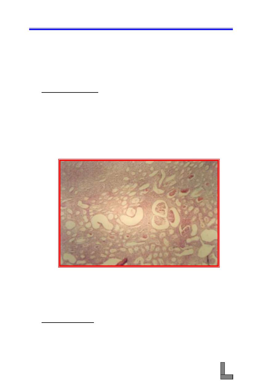
(1) Hydronephrosis: This refers to dilatation of the renal pelvis due to
obstruction of urinary outflow and is associated with increased
pelvic pressure, dilatation of the pelvis, and progressive renal
parenchymal atrophy.
(A) Note dilatation of the renal tubules and degeneration of the
tubular epithelial cells.
(B) Hyaline casts could be seen in the lumens of the tubules.
(C) Note also infiltration of inflammatory cells in the renal
parenchyma.
Fig. 60- Photomicrograph of a kidney affected with hydronephrosis. Note
dilatation of the renal tubules, degeneration of the tubular
epithelium, the presence of hyaline casts, and the infiltration
of inflammatory cells in the renal interstitium. H&E stain. X
40.
(2) Cystic kidney: Polycystic kidneys have many cysts that involve
numerous nephrons such that the kidney can have a (Swiss cheese)
appearance when incised. As cysts enlarge, they compress the
adjacent parenchyma. When kidneys are polycystic, renal function
can be impaired.
Systemic Pathology (13) : Urinary System
74
(A) Multiple cysts could be seen in the kidney.
(B) The cyst wall is lined by flattened epithelial cells.
(C) Note the presence of hyaline casts in the lumens of the
tubules.
(D) Numerous inflammatory cells could be seen in the walls of
the cyst.
Fig. 61- Photomicrograph of a kidney affected with cystic kidney. Note
the presence of multiple cysts (
C
) that are lined by flat cells.
The cysts compress the surrounding tissue. Hyaline cast
(
arrow
) and the infiltration of inflammatory cells into the
renal parenchyma could be seen. H&E stain. X 40.
(3) Focal Interstitial Nephritis: Aggregates of inflammatory cells can
be present in the renal interstitium in various systemic infectious
diseases of domestic animals. When the interstitial inflammation
appears to be the primary lesion and there is no evidence of embolic
nephritis or pyelonephritis, this lesion has traditionally been called
interstitial nephritis. This inflammatory response can be acute,
subacute, or chronic, and is traditionally associated with a
lymphoplasmacytic infiltrate, however, other types of leukocytes can
also be present.
(A) Note the accumulation of monocytes, macrophages,
lymphocytes, and plasma cells.
(B) Escherichia coli septicemia in claves can result in
interstitial nephritis (so- called white spotted kidney).
C
C
C
Systemic Pathology (13) : Urinary System
75
Fig. 62- Photomicrograph of a kidney affected with focal interstitial
nephritis. Note the focal accumulation of inflammatory
mononuclear cells in the renal interstitium (
F
). H&E stain. X
100.
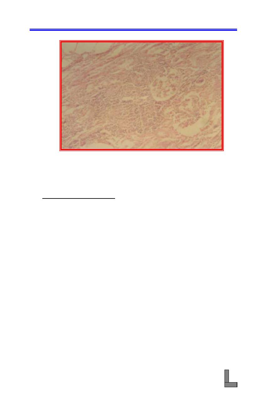
(4) Acute Pyelonephritis: This means inflammation of both the renal
pelvis and renal parenchyma and is an excellent example of
supprative tubulointerstitial disease. Infection may be ascending
(most common) via the ureters to the kidney and establish in the
pelvis and inner medulla, or descending (from the kidneys) wherein
bacterial infection of the kidneys occurs via the hematogenous route,
i.e. embolic nephritis.
(A) The transitional epithelium is usually focally or diffusely
necrotic and desquamated. Necrotic debris, fibrin,
neutrophils, and bacterial colonies can be adherent to the
denuded surface.
(B) Medullary tubules are markedly dilated, and their lumina
contain neutrophils and bacterial colonies.
(C) The tubular epithelium is necrotic and an intense
neutrophilic infiltrate, present in the renal interstitium,
can be accompanied by marked interstitial hemorrhages
and edema.
F
Systemic Pathology (13) : Urinary System
76
Fig. 63-Photomicrograph of a kidney affected with acute pyelonephritis.
Note the dilated renal tubules (
blue
arrows
) and heavy
infiltration of the renal interstitium with inflammatory cells
(
red arrows
). H&E stain. X 40.
(5) Embolic Nephritis (Suppurative Glomerulitis): This is the result of
a bacteremia, in which bacteria randomly lodge in glomeruli and to a
lesser extent in interstitial capillaries and cause the formation of
multiple foci of inflammation thoughout the renal cortex.
(A) Glomerular capillaries have numerous bacterial colonies.
(B) Necrosis and extensive infiltration with neutrophils often
obliterate the glomeruli.
(C) Glomerular or interstitial hemorrhage can also occur.
(D) With time, the neutrophilic infiltrates will progressively have
increasing numbers of lymphocytes, plasma cells, and
macrophages.

Systemic Pathology (13) : Urinary System
77
Fig. 64- Photomicrograph of a kidney showing embolic nephritis
(suppurative glomerulitis). Note the infiltration of the
glomeruli
with
inflammatory
cells
(
red
arrow
),
hemorrhages in the interstitium, and the presence of a plug
of tissue debris and bacterial colonies in one of the tubules
(black arrow). H&E stain. X 100.
References:
Catran RS, Rennke H, Kumar V. The Kidney and its collecting system. In:
Kumar V, Cotran RS, Robbins SL. Eds. 7th ed. Robbins basic
pathology. Philadelphia: Saunders; 2003 : 509
– 542.
Confer AW, Panciera RJ. The urinary system. In: McGavin MD, Carlton WW,
Zachary JF. Eds. 3rd ed. Thomson's special veterinary
pathology. St. Louis : Mosby, 2001 : 235 - 277.
Rebhun WC, Dill SG, Perdrizet JA, Hatfield CE. Pyelonephritis in cows: 15
cases (1982
– 1986). J Am Vet Med Assoc; 1989; 194 : 953 –
955.
Systemic Pathology (14) : Skin and Appendages
78
Systemic Pathology (14)
Skin and Appendages
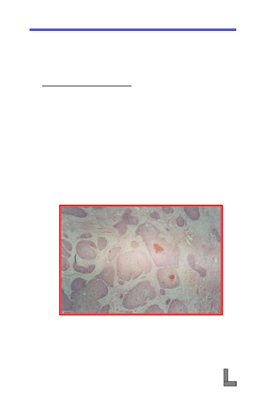
(1) Squamous Cell Carcinoma:- This is a malignant tumor of squamous
epithelial cells and is common in all domestic animals. The tumor
appears grossly as papillary growths of varying size, many of which
have a cauliflower-like appearance. Ulceration and hemorrhages of
the surface of the tumor are common. Another form of the tumor
(the erosive form) is characterized by the appearance of shallow,
crusted ulcer which, if allowed to develop, can become deep and
craterlike.
(A) The tumor consists of irregular masses or cords of epidermal
cells that proliferate downward and invade the dermis and
subcutis.
(B) This tumor is well – differentiated since large numbers of "horn
pearls" (cancer pearls) are present and they consists of
concentric layers of sequamous cells showing gradually
increasing keratinization toward the centers.
(C) Mitotic figures are abundant and many of them are atypical.
Fig. 65- Photomicrograph of a skin affected with squamous cell
carcinoma. Neoplastic cells could be seen invading the
dermis in the form of finger-like projections or nests. Some
of the nests contain keratin. H&E stain. X 100.
Systemic Pathology (14) : Skin and Appendages
79
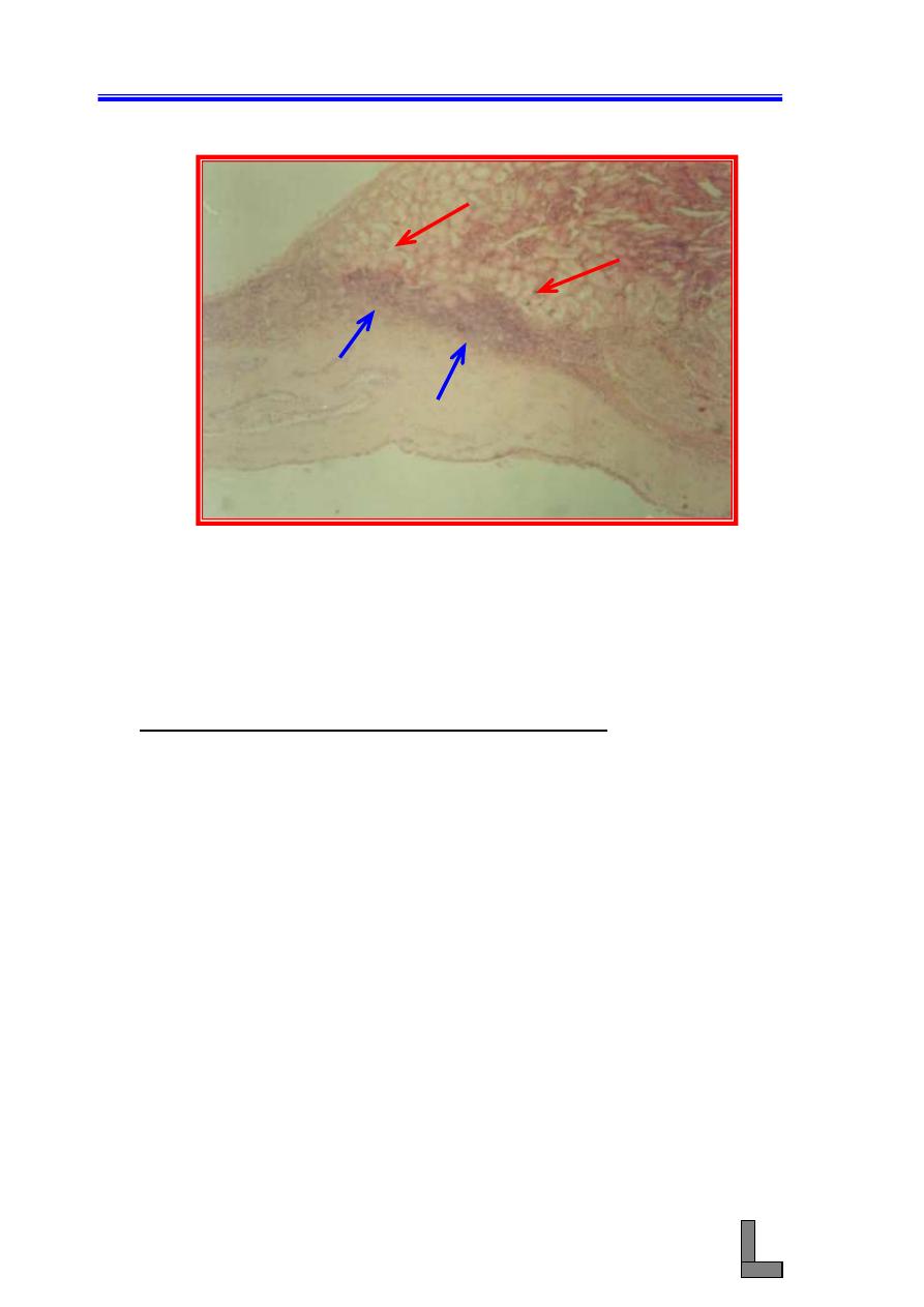
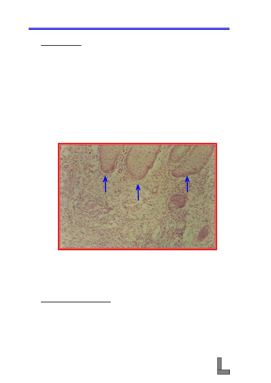
(2) Dermatitis: Dermatitis is an inflammation of the skin, and it could be
serous, papular, suppurative, necrotic, or parasitic. Other types of
dermatitis are also exist. Among the non-infectious causes of
dermatitis are solar energy, photosensitization, chemical causes,
immunological causes, temperature extremes, bites, and others. The
common infectious causes include poxvirus, herpesviruses,
rickettsial infections, bacteria, fungi, parasites, metabolic disorders,
and nutritional causes.
(A) This is a section of the skin stained with hematoxylin and eosin.
Note ulceration of the skin (necrosis and sloughing of the
epidermis).
(B) Note the increased thickness of the dermis due to the infiltration
of inflammatory cells and proliferation of local fibrocytes.
Fig. 66- Photomicrograph of a skin showing dermatitis. note the heavy
diffuse infiltration of inflammatory cells into the dermis (
D
).
Small portions of the epidermis could also be seen (
arrows
).
H&E stain. X 100.
(3) Parasitic Dermatitis: Ectoparasites that cause cutaneous lesions
include mites and ticks (have eight legs), and lice, and flies, (have
six legs). Endoparasites that cause lesions include nematodes,
trematodes, and protozoa. Cutaneous lesions that result from
parasitic infections depends on the parasite number, location, feeding
habits, and host immune response.
D
Systemic Pathology (14) : Skin and Appendages
80
(A) This is a section of the skin stained with hematoxylin and eosin.
Note the presence of many parasitic nodules in the epidermis
and dermis.
(B) Each nodule consists of a section in the parasite surrounded by
many mixed – type inflammatory cells (eosinophils, mast cells,
neutrophils, lymphocytes and macrophages).
(C) Hyperemia and hemorrhages could also be seen.
Fig. 67- Photomicrograph of a skin affected with parastitic dermatitis.
Note heavy infiltration of the dermis with mixed type
inflammatory cells and the presence of a parasitic nodule
(
arrow
). H&E stain. X 100.
References:
Gross TL, Ihrke PJ, Walder EJ. Veterinary dermatopathology: a macroscopic
and microscopic evaluation of canine and feline skin disease. St.
Louis : Mosby
– Year Book, 1992.
Hargis AM, Ginn PE. Integumentary system. In: McGavin MD, Carlton WW,
Zachary JF. Eds. 3rd ed. Thomson's special veterinary
pathology. St. Louis : Mosby, 2001 : 537-599.
Stannard AA, Pulley LT. Tumors of the skin and soft tissues. In: Moulton JE,
ed. Tumors in domestic animals. 3rd ed. Berkeley: University of
California Press, 1990: 23-87.
Yager JA, Scott DW. The skin and appendages. In: Jubb KVF, Kennedy PC,
Palmer N, eds. Pathology of domestic animals. 4th ed. New
York : Academic Press, 1993: 531-738.
Systemic Pathology (15) : The Musculo-Skeletal System
81
Systemic Pathology (15)
The Musculo – Skeletal System
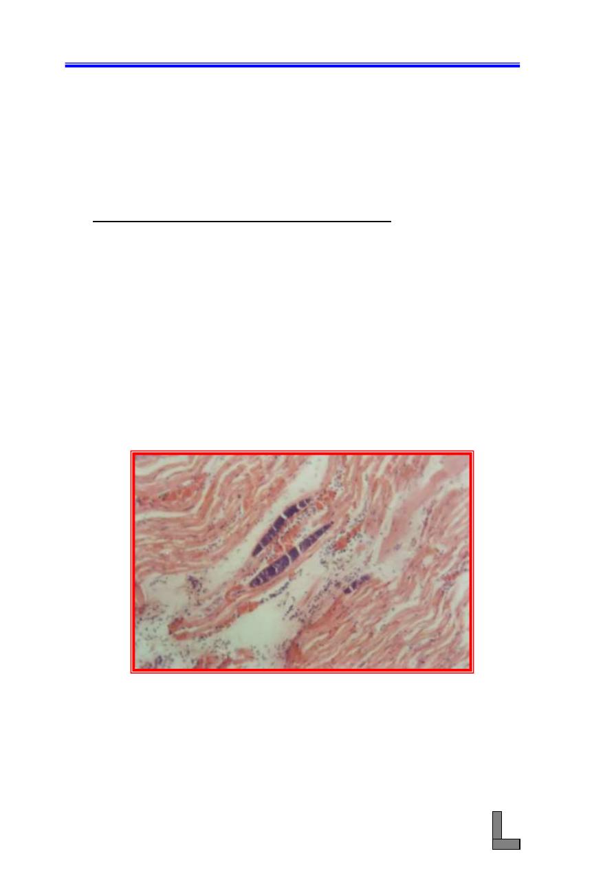
(1) Parasitic Myositides (Sarcocystis Infection):Parasitic infections of
the skeletal muscle of domestic animals include sarcocystis sp. (in
cattle, sheep, goats), Neosporum caninum (in dogs), and Trichinella
spiralis (in pigs). Sarcocystis spp occur as intracytoplasmic protozoal
cysts. Because they are intracellular, they are protected from the host's
defense mechanisms and thus, there is no inflammatory response or
reaction by the myocyte. Eosinophilic myositis may occur in cattle as
a manifestation of Sarcoystis infection and this may involve
hypersensitivity.
(A) Rounded and elongated parasitic cysts could be seen in myofibers
without the occurrence of inflammation.
(B) Atrophic muscle fibers are separated by an exudate consisting
chiefly of eosinophils.
Fig. 68- Photomicrograph of a skeletal muscle showing parasitic
myositides (Sarcocystis infection). Elongated parasitic cysts
could be seen in the myofibers. A mild inflammatory
exudates could also be seen between the bundles of muscle
fibers. H&E stain. X 100.
Systemic Pathology (15) : The Musculo-Skeletal System
82
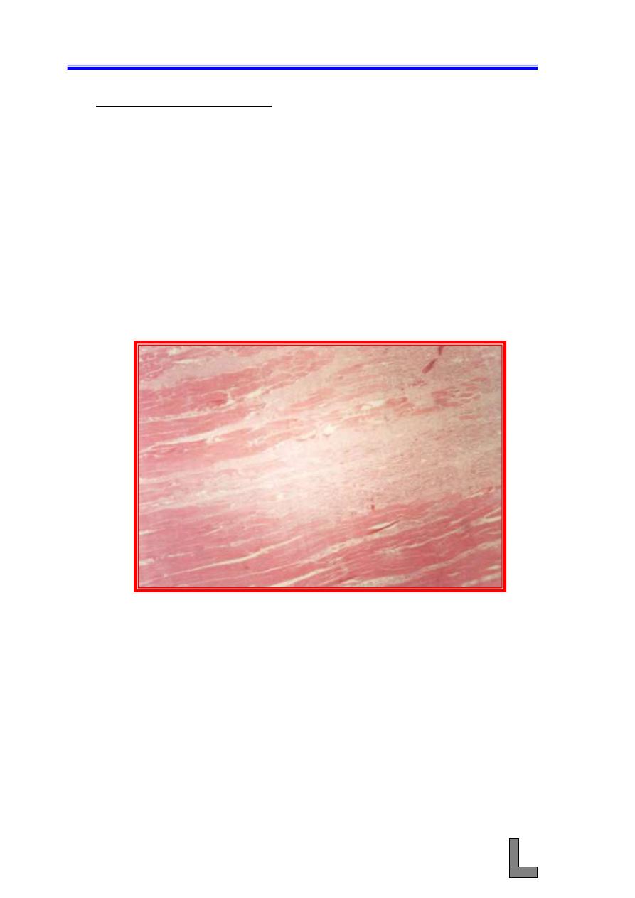
(2) Nutritional Myopathy: This condition also called white muscle
disease and it is common in calves, lambs,, and horses. It is due to
vitamin E and selenium deficiency. Grossly, numerous gray and white
streaks which are areas of segmental necrosis and calcification could
be seen. Necrotic portions of the fiber may become floccular or
granular as that portion of the myofiber starts to fragment. The
adjacent histologically normal portion of the myofiber can separate
from the necrotic segement, forming so – called retraction caps.
Occasionally, necrotic segments of myofibers are mineralized.
(A) This is a section in skeletal muscle stained with hematoxylin and
eosin. Segmental necrosis of the myofibers could be seen.
(B) Calcification of the necrotic myfibers could also be seen.
Fig. 69- Photomicrograph of a skeletal muscle of a case of nutritional
myopathy. Note necrosis, disappearance, and replacement of
bundles of muscle fibers by fibrous tissue. Some atrophied
bundles of muscle fibers could also be seen. H&E stain. X
100.
Systemic Pathology (15) : The Musculo-Skeletal System
83
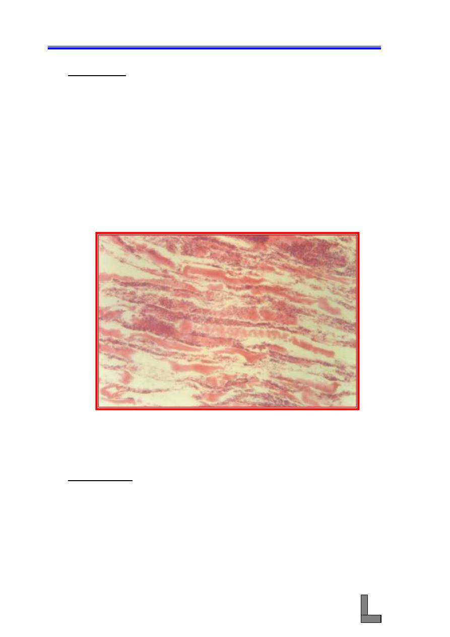
(3) Myositis: This is inflammation of muscle (plural: myositides) and is
caused by a wide array of agents (bacteria , viruses, protozoa,
helminths, and immuno-mediated mechanisms). In true myositis the
inflammatory cells are directly responsible for initiating and
maintaining myofiber injury. The types of inflammation could be
suppurative, serohemorrhagic, or granulomatous.
(A) The necrotic segment is infiltrated by macrophages which " clean
– up" the cellular debris.
(B) Severe acute necrotizing myopathy is accompanied by a certain
degree of infiltrating lymphocytes, plasma cells, neutrophils, and
even eosinophils.
(C) Cytokines released from damaged muscle fibers recruit a variety
of inflammatory cells.
Fig. 70- Photomicrograph of a skeletal muscle showing myositis. Note
the presence of accumulation of inflammatory cells between
the bundles of muscle fibers. H&E stain. 40X.
References:
Carpenter JL, Schmidt CM, Moore FM, Albert DM, Abrams KL, Elner VM.
Canine bilateral extraocular polymyositis. Vet Pathol 1989; 26 :
510 - 512.
Hulland TJ. Muscle and tendon. In : Jubb KVF, Kennedy PC, Palmer N, eds.
Pathology of domestic animals. 4th ed. New York : Academic
press, 1993 : 183
– 265.
McGavin MD, Carlton WW, Zachary JF. Eds. 3rd ed. Thomson's special
veterinary pathology. St. Louis : Mosby, 2001 : 474 - 477.
Systemic Pathology (16) : The hemic and lymphatic System
84
Systemic Pathology (16)
The Hemic and Lymphatic Systems
(1) Chronic Focal Suppurative Splenitis: chronic splenitis occurs in
many infectious diseases, and leads either to a diffuse enlargement
or irregular enlargement (nodular) depending on the causative
organism. Examples of infectious diseases causing chronic splenitis
include brucellosis, tuberculosis, toxoplasmosis, histoplasmosis,
tularemia,
caseous
lymphadenitis,
pseudotuberculosis,
and
leishmaniasis. Lymphoid hyperplasia occurs in many of these
diseases and contributes to hypersplenism.
(A) this is a section in the spleen stained with hematoxylin and
eosin. Note the presence of multiple foci of inflammation in the
splenic parenchyma.
(B) Each focus consists of a central area of necrosis (liquefactive
necrosis)
surrounded
by
inflammatory
cells
(mainly
neutrophils) and then mononuclear cells (lymphocytes, plasma
cells, and macrophages) and finally by a thick connective tissue
capsule.
(C) Hemorrhages and deposition of hemosiderin pigment could also
be seen.
Fig. 71- Photomicrograph of a spleen showing chronic focal suppurative
splenitis. Multiple foci of suppurative inflammation could be
seen. H&E stain. X 40.
Systemic Pathology (16) : The hemic and lymphatic System
85
(2) Leukosis (liver): Avian leucosis complex is an economically
important disease in the domestic fowl. Two main forms of the
disease are known and they include lymphatic leukosis and
myelocytic leukosis. Lymphocytic leukosis occurs in one of 5 forms
: neural, visceral, ocular, osteopetrotic and skin forms. The visceral
form of the disease is characterized by a diffuse or localized
infiltration of lymphoid cells in any of the visceral organs.
(A) This is a section in the liver stained with hematoxylin and eosin.
Note the presence of densely packed collections of lymphoid
cells having the appearances of immature lymphocytes.
(B) Large areas within the liver have been replaced by the neoplastic
cells.
Fig. 72- Photomicrograph of a liver of a chicken affected with leucosis.
Densely packed collections of neoplastic lymphoblasts could
be seen in the hepatic parenchyma. Only remnants of
hepatocytes could be found (arrows). H&E stain. X 100.
(3) Lymphosarcoma: This type of neoplasms is also called lymphoma or
malignant lymphoma and it is a common neoplasm in animals. The
tumor arises from lymphocytes which are present in lymphoid tissue
anywhere in the body. Many cytological forms of lymphosarcoma
are known. Microscopically, the neoplastic cells cause disappearance
of the normal architecture of the involved lymphoid organ.
(A) This is a section of lymph node stained with hematoxylin and
eosin. Note disappearance of the normal architecture of the
Systemic Pathology (16) : The hemic and lymphatic System
86
lymph node due to the presence of numerous pleomorphic
neoplastic cells and mitotic figures.
(B) The tumor cells have replaced the normal lymphoid tissue of the
node.
Fig . 73- Photomicrograph of a lymph node affected with lympho
sarcoma. Note that large number of neoplastic
lympoblasts obscuring the normal architecture of the
lymph node. H&E stain. X 100.
(4) Acute Serous Lymphadenitis: The most common causes of
lymphadenitis are infectious agents or their products. In most
examples, there is an associated reactive hyperplasia. Although there
are many specific diseases which affect lymph nodes, many
examples of lymhadenitis are nonspecific. Lymhadenitis occurs in
nodes draining primary inflammatory disease of other organs or
tissues. In this type of lymphadenitis, hyperplasia and edema with
distension of the lymph sinuses are seen. The distended sinuses
contain a variable number of neutrophils and macrophages.
Eosinophils are seen in large numbers in lymph nodes draining
chronic skin disease and hypersensitivity lesions.
(A) this is a section of lymph node stained with hematoxylin and
eosin. Note the vascular and cellular changes typical of
inflammation.
(B) A serous exudate stained pink could be seen particularly
underneath the capsule.
Systemic Pathology (16) : The hemic and lymphatic System
87
(C) The exudate consists mainly of serous fluid. Polymorphonuclear
leukocytes could also be seen.
Fig. 74- Photomicrograph of a lymph node affected with acute serous
lymphadenitis. A pinked colored serous exudates and
numerous
polymorphonnuclear
leukocytes
could
be
visualized between the lymph follicles and under the
capsule. H&E stain. X 100.
(5) Leukosis in kidneys: See the description given to slide No. 2
(A) Large numbers of neoplastic lymphoblasts could be seen
infiltrating the renal interstitium.
(B) The tumor cells have replaced the normal renal tissue and caused
distortion of the normal architecture of the kidney.
Systemic Pathology (16) : The hemic and lymphatic System
88
Fig. 75- Photomicrograph of a kidney affected with leucosis. Nnumerous
neoplastic lymphoblasts have infiltrated the renal
interstitium. Note distortion of the normal architecture of the
kidney. H&E stain. X100.
(6) Leukosis in Spleen : See the description given to slide No. 2
(A) This is a section in spleen stained with hematoxylin and eosin.
Note the numerous pleomorphic lymphoblasts infiltrating the
spleen.
(B) Mitotic figures and lymphoid hyperplasia could also seen.
Systemic Pathology (16) : The hemic and lymphatic System
89
Fig. 76- Photomicrograph of a spleen affected with leucosis. Note diffuse
infiltration of neoplastic lymphoblasts in the splenic
parenchyma. H&E stain. X 100.
References:
Jones TC; Hunt RD; King NW. Veterinary Pathology. 6th ed. Philadelphia :
Lippincott Williams & Wilkins ; 1997 : 1009
– 1042.
McGavin MD ; Carlton WW; Zachary JF. Thomson's special veterinary
pathology. 3rd ed. St. Louis : Mosby ; 2001 : 325
– 379.
Systemic Pathology (17) : Male Reproductive System
90
Systemic Pathology (17)
Male Reproductive System
(1) Granulimatous orchitis: Orchitis means inflammation of the testicle
and in bulls Brucella abortus is probably the most important
example of granulomatous orchitis in domestic animals. This type of
orchitis includes the formation of multiple, possibly coalescing,
inflammatory nodules displacing and replacing the testicular
parenchyma. In most of the cases bacteria are seen microscopically
clustered in the cytoplasm of macrophages in the areas of
pyogranulomatus inflammation.
(A) Note the presence of several infectious granulomas, each
consists of a necrosis center with calcification, surrounded by
inflammatory cells (mononuclear cells) and giant cells.
(B) The whole lesion is surrounded by a thick connective tissue
capsule.
Fig. 77- Photomicrograph of a testis affected with granulomatous
orchitis. Note degeneration and necrosis of the seminiferous
tubules (black arrows) and one tubule is containing pus and
surrounded by fibrous tissue (
red arrow
). H&E stain. X
100.
Systemic Pathology (17) : Male Reproductive System
91
(2) Testicular Degeneration: This condition can be unilateral or
bilateral, depending on whether the cause is local or systemic.
Among the common causes of this condition are fever, local heat,
obstruction of flow of spermatozoa, torsion or severe crushing of the
spermatic cord, nutritional deficiency, hormonal aberrations, toxins,
and irradiation.
(A) The seminiferous tubules are small and have thickened basement
membrane, decreased numbers of seminiferous epithelial cells,
vacuolated Sertoli cells, intratubular giant cells, and interstitial
fibrosis.
(B) Impaction of sperms may or may not be seen as it depends on
whether obstruction of the seminiferous tubules present or not.
Fig. 78- Photomicrograph of a testis affected with degeneration and
atrophy. Note the small size of the seminiferous tubules and
their irregular shape. H&E stain. X 40.
(3) Spermatic granuloma: Epididymitis can be focal, multifocal,
diffuse, unilateral or bilateral. In non-infectious epididymitis,
spermatozoa that escape from the lumen of the epididymis incite a
foreign body response. Congenital and acquired obstructions of the
epididymal tubule is the main cause of escape of spermatozoa.
(A) Sperms that have escaped from the lumen of the duct of the
epididymis are surrounded by inflammatory cells particularly
phagocytic cells, including multinucleated giant cells.
(B) The whole lesion is surrounded by fibrous tissue.
Systemic Pathology (17) : Male Reproductive System
92
Fig. 79- Photomicrograph of a testis affected with spermatic granuloma.
Note the granulomatous reaction (
G
) and the thick fibrous
tissue surrounding the granuloma (F).H&E stain. X 100.
(4) Suppurative Orchitis: Among the most common organisms that
cause infectious orchitis in domestic animals are Actinomyces
pyogenes, Bluetongue virus, Brucella abortus, lumpy skin disease
virus, Mycobacterium tunberculosis, and Nocardia asteroides in
bulls, Brucella melitensis in bucks, Brucella canis, canine distemper
virus, Escherichia coli, Proteus vulgaris, and Pseudomonas
pseudomallei in dogs, Brucella ovis, Chlamydia psittaci,
Corynebacterium ovis, and sheep pox virus in rams, equine infectius
anemia virus, equine viral arteritis virus, Salmonella abortus equi,
and Strongylus edentatus in stallions. It starts as inflammation in the
seminiferous tubules, which then spills over into the interstitium.
(A) Note that many of the seminiferous tubules have undergone
atrophy and coagulative necrosis.
(B) Neutrophilic exudate could be seen in the lumen of the tubules as
well as in the interstitial connective tissue.
F
G
F
Systemic Pathology (17) : Male Reproductive System
93
Fig. 80 -
Photomicrograph of the testis of a donkey affected with
suppurative orchitis. Note extensive destruction of the
testicular tissue, disappearance of the seminiferous tubules,
and the presence of edema and heavy infiltration of
neutrophilic exudate. H&E stain. X 100.
References:
Humphery JD; Ladds PW. Pathology of the bovine testis and epididmyis. Vet
Bull 1975; 45: 787-795.
McEntee K. Pathology of the testis of the bull and stallion. Proc Soc
Theriogenol 1979; 80-91.
McGavin MD; Carlton WW; Zachary JF. Thomson's special veterinary
pathology. 3 rd. ed. St. Louis : Mosby; 2001 : 635-652.
Systemic Pathology (18) : The Female Reproductive tract
94
Systemic Pathology (18)
The female Reproductive Tract
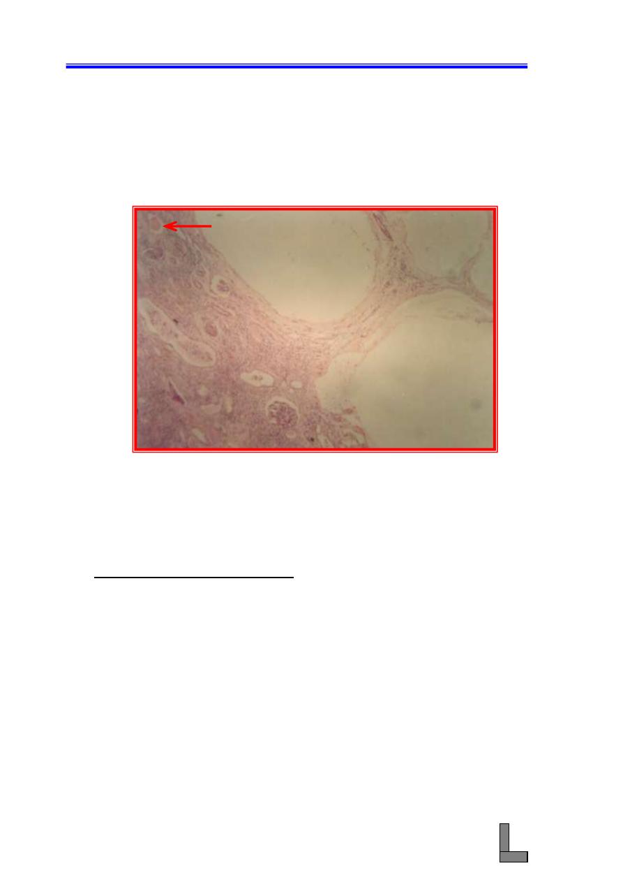
(1) Salpingitis:- This means inflammation of the uterine tubes and it
results from spread of infection from the uterus to the uterine tubes,
and it is usually bilateral. The inflammation ranges from mild to
severe and from acute to chronic, and even mild inflammation can
reduce fertility.
(A) This is a section of the uterine tube stained with hematoxylin and
eosin. Note that the lamina propria of the folds of the uterine
tube is hypercellular and is infiltrated by inflammatory cells
(mainly mononuclear cells).
(B) The lumen of the tube contains inflammatory exudate that
includes many neutrophils.
(C) Hyperemia of blood vessels in the wall of the uterine tube culd
also be seen.
Fig. 81-
Photomicrograph of a uterine tube (oviduct) showing
salpingitis. Note hyperemia and the infiltration of
inflammatory cells in the wall of the tube. H&E stain. X 100.
Systemic Pathology (18) : The Female Reproductive tract
95
(2) Oophoritis:- This condition means inflammation of the ovary and it
is quite rare in domestic animals. Viral infection such as infectious
bovine rhiontracheitis and bovine virus diarrhea are accompanied with
oophoritis when these viral diseases are induced experimentally.
Necrotizing oophoritis is the most common type of oophoritis.
(A) The lesions in the corpus luteum range from focal necrosis and
infiltration of mononuclear cells to diffuse hemoorhage and
necrosis.
(B) Affected ovaries also have necrotic follicles and diffuse
mononuclear cell infiltrates in the stroma.
Fig. 82-
Photomicrograph of an ovary affected with oophoritis. Portion
of a secondary follicle and corpus luteum could be seen.
mononuclear cells could be seen in the ovarian interstitium.
H&E stain. X 100.
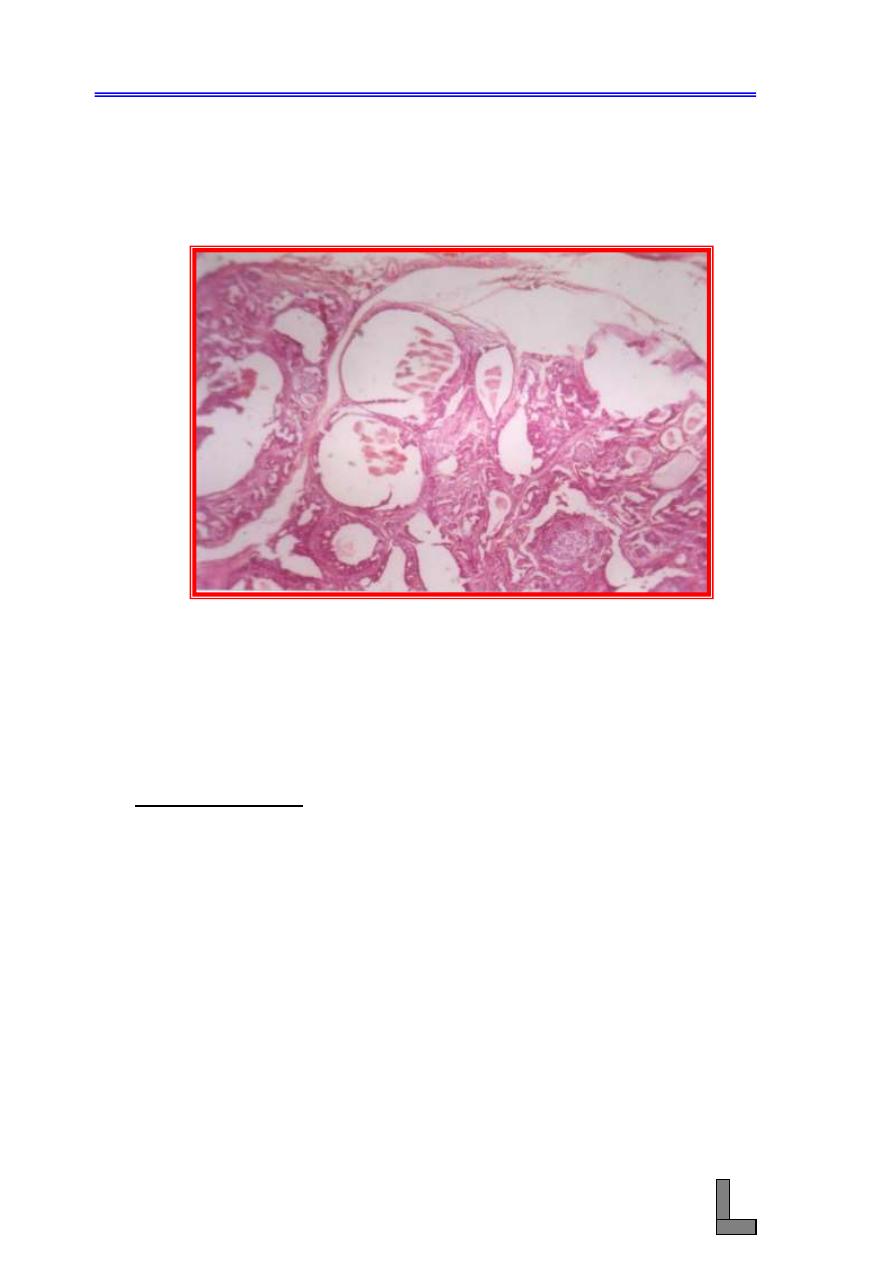
(3) Papillary Cystadenoma: Adenoma in its pure state is uncommon and
is usually a part of mixed tumor. In its pure state, the tumor is usually
occurs as papillary adenoma with dilated ducts and arising in multiple
sites within the gland. The tumor develops within the lobules from the
alveoli (and / or intralobular ducts system), in the interlobular ducts, or
in the teat sinus.
(A) This is a section of the mammary gland stained with hematoxylin
and eosin. Note conversion of the glandular alveoli to cystic
structures.
(B) Cystic spaces containing papillary ingrowths are seen.
Systemic Pathology (18) : The Female Reproductive tract
96
(C) Greatly dilated ducts containing papillomatus growths could also
seen.
Fig. 83-
Photomicrograph of a mammary gland affected with
cystadenoma. Note cystic dilatation of some of the acini and
papillary projections of the lining epithelium of some other
acini.. H&E stain. X 100.
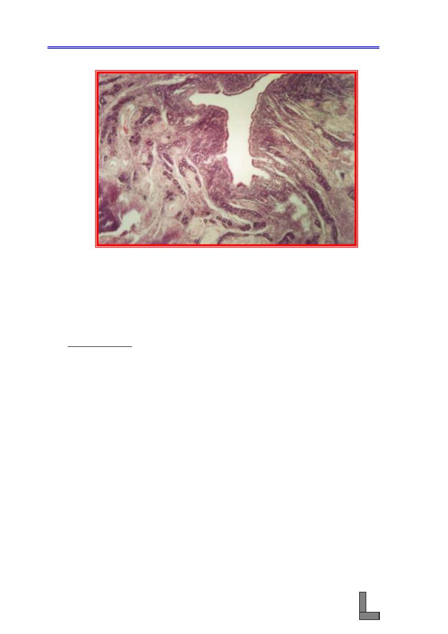
(4) Endometritis: This is inflammation that is limited to the
endometrium. All cases of uterine infections beign as endometritis.
Uterine infections are related to introduction of semen contaminated
with bacteria or to metabolic disturbances that favor an environment
for bacterial organisms in pregnancy, parturition, or postpartum
involution.
(A) A few to many neutrophils are found in the stroma and in the
glands.
(B) A rang of surface changes from desquamation of a few surface
epithelial cells to severe necrosis of the endometrium.
(C) Moderate endometrial fibrosis could also be seen.
Systemic Pathology (18) : The Female Reproductive tract
97
Fig. 84-
Photomicrograph of a uterus affected with endometritis. Note
the heavy infiltration of mononuclear cells ( particularly
lymphocytes) in the endometrium H&E stain. X 100.
References:
Gelberg HB; McEntee K. Pathology of the canine and feline uterine tube. Vet
Pathol 1986; 23:770-775.
Herenda D. An abattoir survey of reproductive organ abnormalities in beef
heifers. Can Vet J 1987; 28: 33-37.
McGavin Md; Carlton WW; Zachary JF. Thomoson's special Veterinary
Pathology. 3 rd. ed. St. Louis: Mosby; 2001: 601-652.
Systemic Pathology (19) : The Endocrine System
98
Systemic Pathology (19)
The Endocrine System
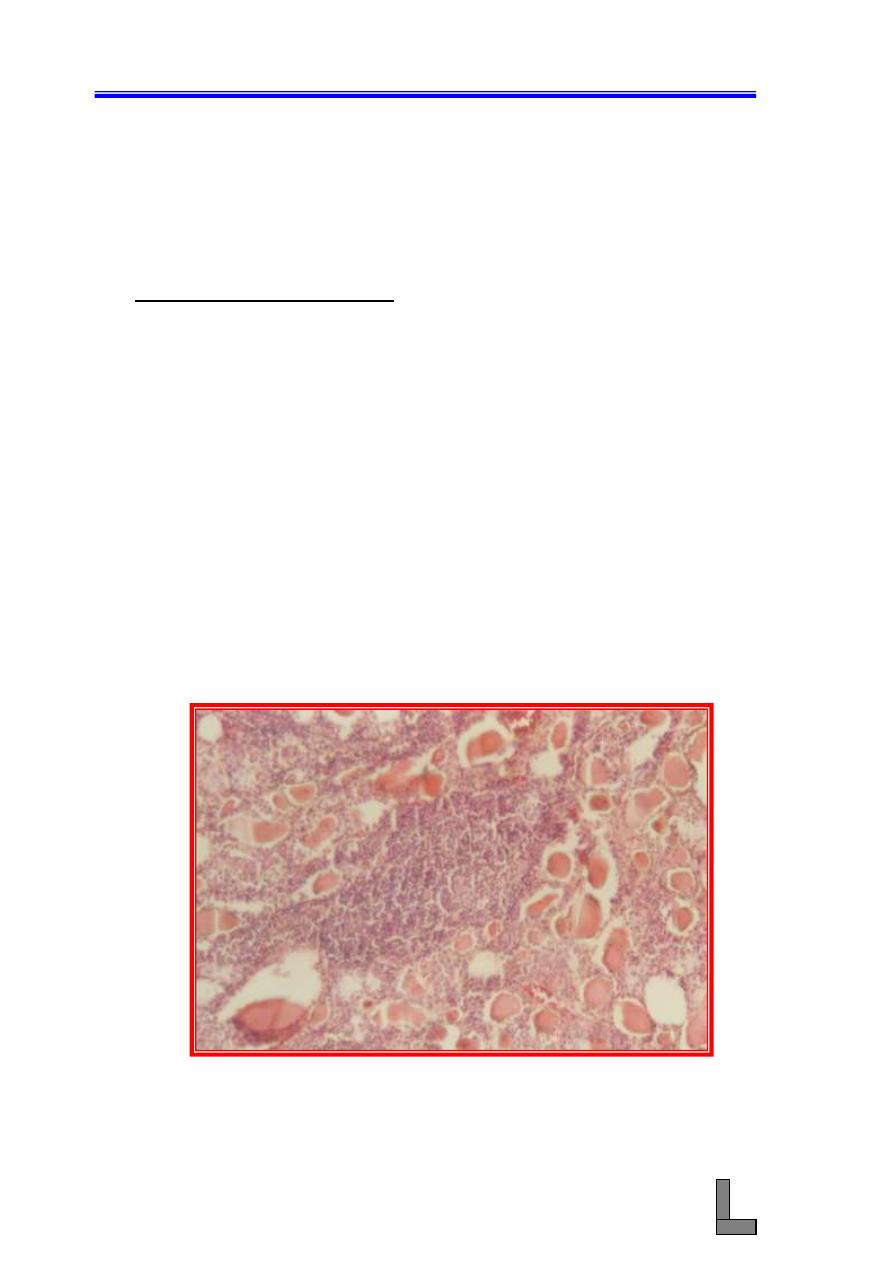
(1) Lymphocytic Thyroiditis: The spontaneous lymphocytic thyroiditis
is seen in dogs, laboratory rats, and monkeys; and it closely resembles
Hashimoto thyroiditis in human beings. The immunologic basis of the
development of chronic lymphocytic thyroiditis in both human beings
and dogs appears to be through production of autoantibodies usually
directed
against
thyroglobulin
or
a
microsomal
antigen
(thyroperoxidase) and infrequently against the TSH receptor protein,
nuclear antigen, and a second colloid antigen from follicular cells.
(A) Multifocal to diffuse infiltrates of lymphocytes, plasma cells, and
macrophages and, sometimes, lymphoid nodules are evident.
(B) Thyroid follicles are small and lined by columnar epithelial cells;
lymphocytes, macrphages, and degenerate follicular cells are
often present in vacuolated colloid.
(C) Thyroid C cells are present as small nests or nodules between
follicles.
Fig. 85 -
Photomicrograph of a thyroid gland showing lymphocytic
thyroiditis. Note the heavy infiltration of lymphocytic
exudate between the thyroid follicles. H&E stain. X 100.
Systemic Pathology (19) : The Endocrine System
99
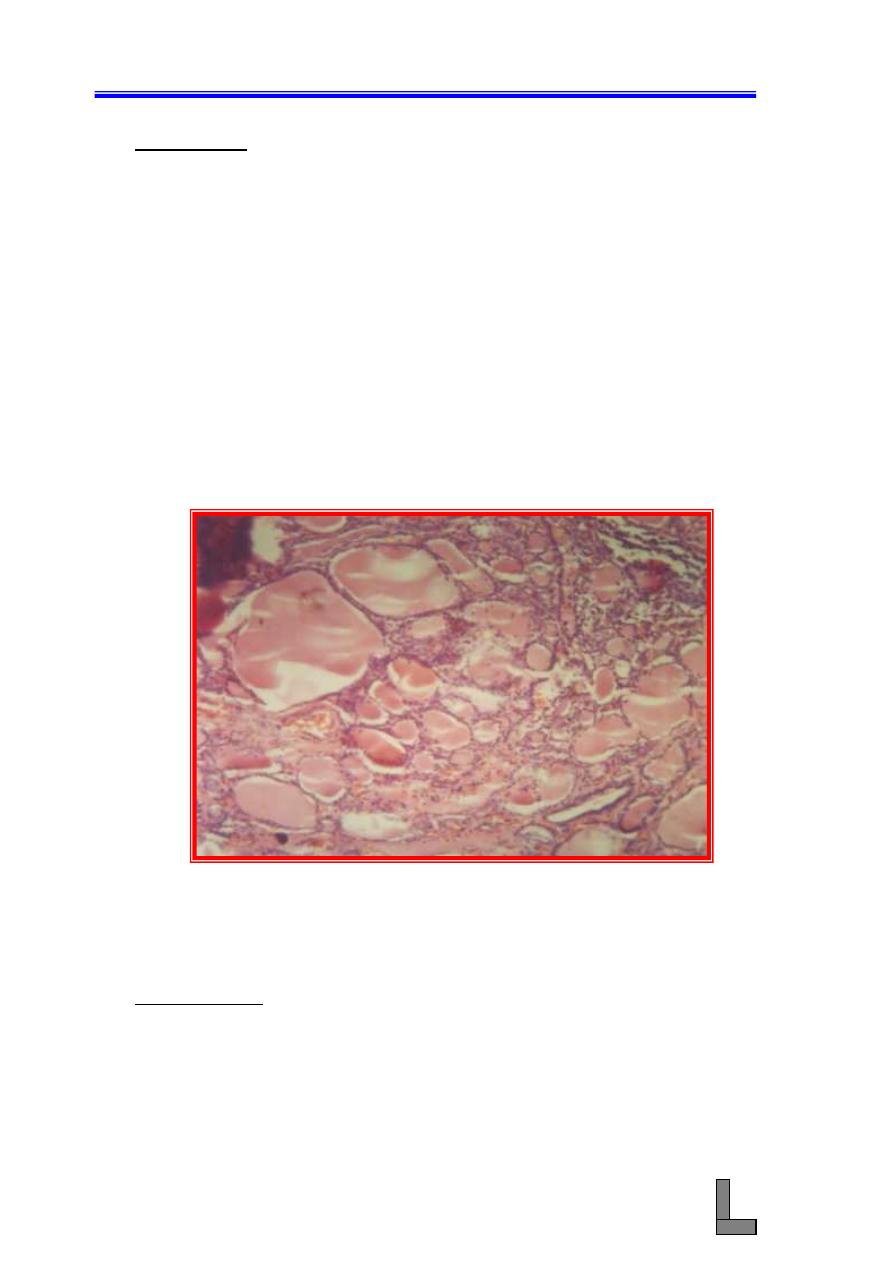
(2) Goiter: Goiter means non-inflammatory and nonneoplatic
enlargement of the thyroid gland. The condition could be
accompanied by either hypo-or hyperthyroidism, or compensated
function. Three general causes exist for goiter: (1) iodine deficiency,
(2) goitrogens, and (3) hereditary biosynthetic defects of thyroid
hormones. Three types of goiter are known to occur: (1) simple goiter
(hyperplastic goiter, colloid goiter, diffuse nontoxic goiter), (2)
Multinodular
goiter
(adenomatous
goiter,
nodular
thyroid
hyperplasia), and (3) exophthalmic goiter (goiter of hyperthyroidism,
toxic goiter, Grave's disease). In simple goiter, the thyroid is
uniformaly enlarged grossly. The microscopical changes include:
(A) The presence of large follicles with colloid, which is lined by
flattedened epithelium.
(B) A great variation in the size of the follicles, and some may retain
papillary projections into the lumens.
Fig. 86 -
Photomicrograph of a thyroid gland from a case of goiter.
Note the variation in the size of the colloidal follicles and the
large quantity of colloidal material in each follicle. H&E
stain. X 100.
References:
Capen CC. Endocrine system. In: McGavin MD, Carlton WW, Zachary JF.
Eds. 3rd ed. Thomson's special veterinary pathology. St. Louis :
Mosby, 2001 : 295 - 305.
Gosselin SJ, Capen CC, Martin SL, Krakowka S. Autoimmune lymphocytic
thyroiditis in dogs. Vet Immunol Immunopathol 1982; 3: 185
–
201.
Jones TC; Hunt RD; King NW. Veterinary Pathology. 6th ed. Philadelphia :
Lippincott Williams & Wilkins ; 1997 : 1236-1237.

Systemic Pathology (20) : Nervous System
100
Systemic Pathology (20)
Nervous System
(1) Spongy Degeneration: This is a group of diseases of young animals
characterized by a spongy lesion (status spongiosus). This lesion can
develop by several different mechanisms that include splitting of the
myelin sheath, accumulation of extracellular fluid, swelling of cellular
(e.g. astrocytic, neuronal) processes, and axonal injury (e.g. Wallerian
degeneration) when swollen axons are no longer detectable within
distended spaces.
(A) Variabley sized empty spaces within the white matter could be
seen.
(B) There is a defect in the formation of myelin.
Fig. 87 -
Photomicrograph of a brain exhibiting spongy degeneration
(status spongiosus). Note the presence of variably sized
empty spaces. H&E stain. X 100.
Systemic Pathology (20) : Nervous System
101
(2) Listeriosis: This is a bacterial infection with particular affinity to
cause CNS disease, mainly in domestic ruminants. Infections also
occur in human beigns. Three forms of the disease are known to occur
and they include meningoencephalitis, abortion and stillbirth, and
septicemia, the later commonly develops in young animals, possibly
from an in utero infection. The encephalitic and genital forms of the
disease rarely occur together in an individual animal or in the same
flock or herd.
(A) The lesion consists of clusters of microglia and variable numbers
of neutrophils. Macrophages, gitter cells, and necrosis could also
be seen.
(B) Gram – positive bacilli can be detected in some lesions.
Fig. 88 -
Photomicrograph of a brain showing microabscesses in the
brain (Listeriosis). Note the presence of focal accumulation
of neutrophils and loss of parenchyma. H&E stain. X 100..
Rferences:
Charlton KM, Garcia MM. Spontaneous listeric encephalitis and neuritis in
sheep. Vet Pathol 1977; 14: 297
– 313.
Storts RW, Montgomery DL. The nervous system. In: McGavin MD, Carlton
WW, Zachary JF. Eds. 3rd ed. Thomson's special veterinary
pathology. St. Louis : Mosby, 2001 : 381
– 459.