Abdulateef
Rand
Dr.
Lecture 3
1
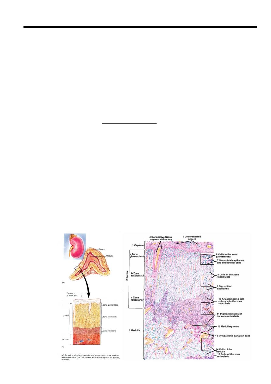
Adrenal glands
• The body has two adrenal (suprarenal) glands, one of which is located superior to
each kidney.
• In the human, they are about 4-6cm long, 1-2cm wide & 0.5cm thick, and both may
weigh about 15gm.
• Each gland is surrounded by a thick connective tissue capsule that sends septa into the
interior of the gland.
• They are divisible by embryonic origin, structure, and function into cortex and
medulla.
Adrenal Cortex
Embryonic origin:
The adrenal cortex derives from mesoderm.
Structure in adults:
Cells of the adrenal cortex have the characteristic steroid-synthesizing cell structure
(polygonal or rounded, with pale- staining central nucleus, acidophilic cytoplasm that
contains many lipid droplets, abundant SER , mitochondria containing enzymes),
The suprarenal cortex consists of three concentric layers or zones:
a) zona glomerulosa.
b) zona fasciculate.
c) zona reticularis.

Abdulateef
Rand
Dr.
Lecture 3
2
a. Zona glomerulosa :
this outermost cortical layer lies directly under the capsule.
constitutes about 15% of adrenal volume.
composed of small cells arranged in form of clusters (glomeruli), separated by thin-
walled capillaries.
ultrastructurally the cells contain well developed SER and comparatively little RER
with scanty lipid droplets.
the cells of this layer secrete mineralocorticoids.
b. Zona fasciculata :
this middle layer of the cortex.
constitutes about 65% of adrenal volume .
•
Its cells form straight cords or columns (usually 2-3cells wide) separated by capillaries
the columns are perpendicular to the organ surface.
•
ultrastructurally, the cells have prominent RER, extensive lipid vacuoles and
characteristic small round or ovoid mitochondria.
•
Its cells produce glucocorticoids and some adrenal androgens.
c. Zona reticularis :
•
this innermost layer of the adrenal cortex.
•
constitutes about 7% of adrenal volume.
•
consists of an anastomosing network of cell cords with capillary network closely
apposed to the cell membranes.
•
Its cells are smaller and more acidophilic than those in the fasciculata and contain fewer
lipid droplets, more mitochondria, many lipofuscin granules and prominent SER.
•
the reticularis and fasciculata seem to constitute a single functional zone, with the
reticularis producing most of the glucocorticoids and adrenal androgens.
Normal function.
The adrenal cortex produces three types of steroid hormones.
1. Mineralocorticoids mainly aldosterone: produced by the zona glomerulosa in
response to angiotensin II, it stimulates sodium absorption by the distal renal tubules.
2. Glucocorticoids mainly cortisol and corticosterone: produced by the zona reticularis
in response to ACTH and by the fasciculata after prolonged stimulation, glucocorticoids
control carbohydrate metabolism and suppress the immune response by decreasing
circulating lymphocytes and eosinophils.
3. Adrenal androgens: secreted by zona reticularis in response to ACTH and by
fasciculata after prolonged stimulation, they produce musculinizing and anabolic effect.
Abdulateef
Rand
Dr.
Lecture 3
3
Adrenal Medulla
Embryonic origin.
The adrenal medulla derives from the neural crest.
Structure.
The adrenal medulla is composed of epithelial cells supported by a delicate connective
tissue richly supplied by blood sinusoids.It contains two major cell types:
a) chromaffin cells.
b) ganglion cells.
a. Chromaffin cells. Also known as pheochromocytes
these are the predominant cell type.
they contain large nuclei and cytoplasmic granules that become brown when exposed
to chromium salts for this reason, they are sometimes referred to as chromaffin cells.
the chromaffin cells synthesize and secrete norepinephrine and epinephrine in
response to stimulation by preganglionic sympathetic nerve fibers, some cells are
thought to produce serotonin in addition to epinephrine, the hormones are released
by process of exocytosis.
with an electron microscope, the cells shows well developed Golgi complex, few
profiles of RER, many oval mitochondria, and membrane bound cytoplasmic
granules.
•
There are two types of chromaffin cells:-
1- cells secrete norepinephrine have dense core granules.
•
2- light cells secrete epinephrine have granules not so dense.
b. Ganglion cells: few parasympathetic ganglion cells present show typical
morphologic characteristics of autonomic ganglion cells (large, multipolar cells,
randomly distributed throughout ganglion, have large and ovoid nuclei mostly
eccentric).
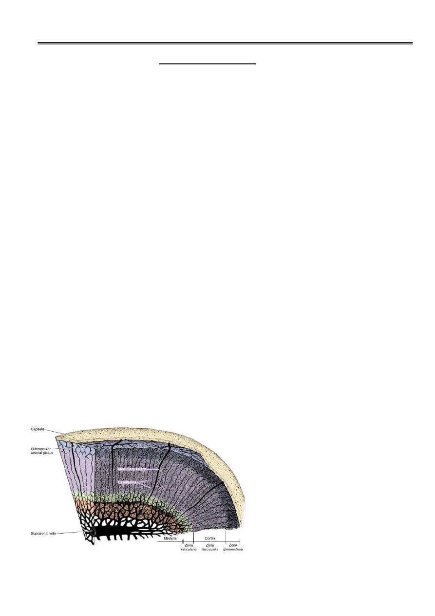
Adrenal Blood Supply :
1. Arteries:Three main arteries supply each adrenal gland :
Abdulateef
Rand
Dr.
Lecture 3
4
a. superior suprarenal from the inferior phrenic artery.
b. middle suprarenal from the aorta artery.
c. inferior suprarenal from the renal artery.
These arteries penetrate the capsule separately, and their branches anastomose to form a
subcapsular arterial plexus, this plexus gives rise to three groups of arteries:
a. arteries of the capsule.
b. arteries of the cortex which branch to form the cortical capillaries that
pass between the secretory cells and drain into the medullary capillaries.
c. arteries of the medulla, which traverse the cortex without branching
until they reach the medulla, where they form the medullary capillaries.
2. Medullary capillaries: receive a double blood supply from both cortex and medulla
and converge to form several medullary veins.
3. Medullary veins converge to form a single large suprarenal vein.
4. Suprarenal vein arises at the core of the medulla and drains into the renal vein or
directly into the inferior vena cava.
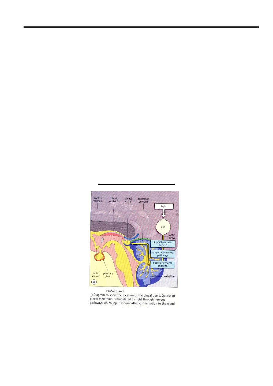
PINEAL GLAND
•
the pineal gland is also known as the epiphysis cerebri, or pineal body.
•
It is small flattened conical structure located just below the posterior end of the corpus
callosum of the brain.
•
covered on its outer surface by pia mater which forms the capsule, from which number
of fibrous trabeculae carrying the blood vessels and unmyelinated nerves extending into
the interior of the gland and incompletely divide the parenchyma into poorly defined
lobules of various size.

Abdulateef
Rand
Dr.
Lecture 3
5
•
The pineal gland is composed of groups of cells called pinealocytes supported by
astroglial cells.
A. Pinealocytes :
Structure.
•
The pinealocytes are large neuron-like cells have extensive processes that are
intertwined with the processes of the astroglial cells.
•
Pinealocytes are often arranged in rosettes, where several cells surround a central
fibrillary area composed of cell processes directed towards a small capillary vessel.
•
With H&E stain, they have pink-staining cytoplasm and dark-staining irregular or lobate
nuclei.
•
With silver stain, they exhibit long cytoplasmic processes that terminate as swellings
close to blood capillaries stored in the terminal swellings are vesicles containing
monoamines and polypeptide hormones.
•
The cells
produce melatonin, which induces rhythmic changes in the secretions of the
hypothalamus, pituitary and gonads, and is said to act as an endocrine transducer.The
concentration of melatonin in the blood conform to a circadian rhythm, being higher
during darkness and lowest during the day.
B. Astroglial Cells:
•
Also known as interstitial cells, these glialike cells have elongated nuclei that stain
more heavily than those of the parenchyma.
•
These cells have long cytoplasmic processes that contain a large number of
intermediate filaments.
•
They are found around blood vessels and between clusters of pinealocytes.
•
They are indistinct unless specially stained.
•
The pineal gland has a rich blood supply and is innervated by sympathetic and
parasympathetic nervous systems. In addition signals from retina arrive indirectly.
•
Concretions of calcified material called "brain sand" (corpora arenacia) accumulate
within the astroglial cells and connective tissue of the pineal gland, progressively with
age, these deposits are useful to radiologists because they serve as a landmark
determine whether the pineal gland has been displaced laterally by a space-occupying
lesion within the skull.