
R
ESPIRATORY
S
YSTEM
1
RESPIRATORY PHYSIOLOGY ……… Proff.Amjad Fawzi
Respiratory System Functions
1. Gas exchanger
2. Regulation of blood pH
3. Voice production
4. Olfaction
5. Protection
Respiration includes 2 processes:
6. External respiration: uptake of O2 and removal of CO2 between lungs and
environment.
7. Internal respiration: uptake of O2 and removal of CO2 between cells and
their fluid medium.
The respiratory system is made up of a gas exchanging organ(the lungs) and a
pump that ventilate the lungs. This pump is made up of:
1. Chest wall muscles which increase and decrease the size of thoracic cavity.
2. least resistance to air flow is in the very small bronchiols and terminal
bronchiols because of their large cross-sectional area.Alveoli are lined
mostly by thin layer of squamous epithelium.
3. Brain centers which control the respiratory muscles.
4. Nerves which connect the brain with respiratory muscles.
Respiratory Airways:
1. Anatomical classification: nose, pharynx (upper respiratory tract),larynx,
trachea, bronchi, bronchioles, terminal bronchioles, respiratory bronchioles,
alveolar ducts, alveolar sacs, and alveoli(lower respiratory tract).
2. Physiological classification: nose, pharynx, larynx, trachea, bronchi,
bronchioles, terminal bronchioles(conducting zone-divided 16 times),
respiratory bronchioles, alveolar ducts, alveolar sacs, and alveoli(respiratory
zone-divided 7 times).These 23 divisions greatly increase the total cross
sectional area thus much reducing air flow through small airways.
To keep the trachea from collapsing multiple cartilage rings extend about five sixth
of the way around the trachea which become less and less extensive and then they
are completely gone in the bronchioles which by now are not prevented from
collapsing by any rigidity of their walls. Instead, they are expanded by the same
transpulmonary pressures that expand the alveoli. In all areas of the trachea and
bronchi not occupied by cartilage plates, the walls are composed mainly of smooth
muscles. The walls of the bronchioles are almost entirely smooth muscles, with the
exception of the respiratory bronchioles that has only a few smooth muscle fibers.
The chief site of airway resistance in the airway passages is at the medium-sized
segmantal bronchi, where the radius of the individual bronchi is decreased. The
typeI) alveolar cells and few thick (type II) surfactant secreting cells in addition to
the alveolar macrophages which engulph small particles reaching the alveoli. Mast
cells also present which contains histamine responsible for allergic
reactions(bronchial asthma).

R
ESPIRATORY
S
YSTEM
2
Respiratory functions of the nose:
[1] Warming the airby the extensive surfaces of the conchae and septum.
[2] The air is almost completely humidified.
[3] The air is filtered.
When a person breathes air through atube directly into the trachea (as through a
tracheostomy), the cooling and especially the drying effect in the lower lung can lead
to serious lung crusting and infection. The nasal filtration of air for removing particles
from air is so effective that almost no particles larger than 4 to 6 microns in diameter
enter the lung through the nose. Of the remaining particles, many of smaller size settle
out in the smaller bronchioles as a result of gravitational precipitation. Some of the
particles smaller than 1 micron in diameter diffuse against the walls of the alveoli and
adhere to the alveolar fluid. But many particles smaller that 0.5 micron in diameter
remain suspended in the alveolar air and are later expelled by expiration. For instance,
the particles of cigarette smoke have a particle size of about 0.3 micron. Almost none
of these are precipitated in the respiratory passageways before they reach the alveoli.
However, up to one third of them do precipitate in the alveoli by the diffusion process,
and the remaining suspended and expelled in the expired air. Particles that become
entrapped in the alveoli are removed mainly byalveolar macrophages. An excess of
particles causes growth of fibrous tissue in the alveolar septa, leading to permanent
debility.
All the respiratory passages are kept moist by a layer of mucus that coats the
entire surface which is secreted by goblet cells in the epithelial lining of the passages
and by small submucous glands. Themucus also traps small particles out of the
inspired air and keeps most of these from ever reaching the alveoli. Then the mucus
itself is removed from the passages by the continual beating of the cilia, which cover
the entire surface of the respiratory passages. The cilia in the lower respiratory
passages beat upward while those in the nose beat downward. This continual beating
causes the coat of mucus to flow slowly toward the pharynx. Then the mucus and its
entrapped particles are either swallowed or coughed to the exterior.
[A]
Nervous control of the bronchioles:
The only important nervous control to
the bronchioles is by way of parasympathetic vagus nerves fibers. These nerves secrete
acetylcholine and when activated cause mild to moderate constriction of the
bronchioles. Irritants entering the airways, such as smoke, dust, sulfur dioxide, and
some of the acidic elements in smog, can all initiate local reactions that cause
obstructive constriction of the bronchioles mediated through a parasympathetic reflex.
[B]
Humoral control of the bronchioles:
several different humoral substances
are often quite active in causing bronchiolar constriction. Two of the most important of
these are histamine and the substance called slow reactive substance of anaphylaxis
(SRA). Both of these are released in the lung tissues by mast cells during allergic
reactions. Therefore, they play key roles in causing the airway obstruction that occurs
R
ESPIRATORY
S
YSTEM
3
in allergic asthma. In addition, the airway smooth muscle is highly responsive to CO
2
,
high blood CO
2
producing bronchodilataion and low
CO
2
bronchoconstriction. In
contrast to the humoral substances that constrict the bronchioles, two other hormones,
epinephrine and norepinephrine, both of which are secreted by the adrenal glands in
response to sympathetic stimulation relax the bronchioles (by activation of
β
2
receptors). Therefore, activation of the sympathetic nervous system is often valuable
in relaxing the airways and preventing obstruction.
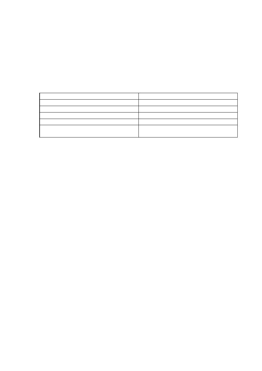
Factor
Effect
Parasympathetic stimulation
Bronchoconstriction
Histamine and SRA
Bronchoconstriction
Low blood PCO2
Bronchoconstriction
High blood PCO2
Bronchodilatation
Sympathetic stimulation to the adrenal glands
(epinephrine and norepinephrine)
Bronchodilatation
The process of respiration can be divided into four major
events:
[1] Pulmonary ventilation which means the inflow and outflow of air between the
atmosphere and the lung alveoli.
[2] Pulmonary diffusion (gas exchange in the lung).
[3] Transport of oxygen and carbon dioxide in the blood and body fluids to and from
the cells.
[4] Regulation of ventilation.
(I)
Pulmonary Ventilation
:
Which includes inspiration and expiration.
[A]Inspiration: Normal inspiration is an active process. The lungs can expand in two
ways:
[1]By downward and upward movement of the muscles of diaphragm (supplied
by phrenic nerves). The downward movement of diaphragm accounts for 75% of
the change in intrathoracic volume during quiet inspiration. In inspiration,
contraction of the diaphragm pulls the lower surfaces of the lungs downward.
[2]By raising the rib cage. In natural resting position, the ribs are extended
forward and downward, thus allowing the sternum to fall backward toward the
spinal column. But when the rib cage is elevated, the ribs project directly forward
so that the sternum now also moves forward away from the spine, making the
anterioposterior thickness of the chest greater during maximum inspiration. The
foreword movement of sternum accounts for 25% of the change in intrathoracic
volume during quiet inspiration. The muscles for raising the rib cage (muscle of
inspiration) are external intercostals, sternocleidomastoid ,and scalene. At rest,

R
ESPIRATORY
S
YSTEM
4
adequate ventilation can be maintained by diaphragm or external intercostals
muscle alone.
[B]Expiration: Normal expiration is a passive process. The lungs can be shrinked or
contracted by two
ways:
[1]Relaxation of diaphragm and the inspiratory muscles which cause compression
on the lungs. In contrast, during heavy breathing, however, the compression
forces are not powerful enough to cause the necessary rapid expiration, so that
abdominal muscles (abdominal recti)and the muscles that pull the rib cage
downward (internal intercostals), i.e. muscles of expiration are contracted and
added to the force needed for rapid expiration.
[2]Elastic recoil tendency of the lung.
Maximal expiratory pressure is achieved by fully contracting the expiratory muscles
with the lungs fully inflated and the glottis- or airway closed. Forced expiration against
a closed airway is termed a valsalva maneuverand is commonly performed when
lifting heavy objects or when defecating. Normally, the maximal expiratory pressure
that can be achieved is 100-150 cm H20 greater than atmospheric pressure. As lung
volume decreases, the maximal achievable expiratory pressure decreases as well.
Elastic recoil tendency of the lungs:
The lungs have a continual elastic tendency
to collapse and therefore to pull away from the chest wall. This is called the elastic
recoil tendency of the lungs and it is caused by two different factors:
[A]The presence of elastic fibers throughout the lungs which are stretched by lung
inflation and therefore attempt to shorten. They account for about one third of the
recoil tendency.
[B]The surface tension of the fluid lining the alveoli which is more important,
accounts for about two third of the recoil tendency,and causes a continual elastic
tendency for the alveoli to collapse. The surface tension is caused by intermolecular
attraction between the surface molecules of the alveolar fluid that is each molecule
pulls on the next one.
Intrapleural(intrathoracic) pressure
:The negative(subatmospheric) pleural
pressure in the pleural space required to prevent collapse of the lungs is called the
pleural pressure (or intrathoracic pressure) which is about – 2.5 mm Hg at the end of
expiration and -12 to -18 mm Hg at the end of inspiration.
Pressure changes during inspiration and expiration:
A thin layer
of fluid normally present between visceral and parietal pleurae. This fluid
helps the lung to slide easily on the chest wall(lubricant) but resist being
pulled awayfrom it(sealant) in the same way that 2 moist pieces of glass
slide on each other but resist separation.
The lungs are stretched when expanded at birth
. At end of quiet expiration,their
tendency to recoil from the chest wall is just balanced by the tendency of the chest
wall to recoil in the opposite direction (neutral point).If the chest wall is opened, the
lungs collapse(pleural pressure becomes atmospheric) and if the lungs lose their

R
ESPIRATORY
S
YSTEM
5
elasticity the chest expands and becomes barrel- shaped(eg: emphysema).Inspiration is
an active process.The contraction of the inspiratory muscles increases the intrathoracic
volume.During quiet breathing, the intrapleuralpressure ,which is about -2.5 mmHg
(relative to atmospheric)at the start of inspiration, decreases to about -6mmHgm and
lungs become more expanded. The pressure in the airway becomes slightly negative,
and air flows into the lungs. At the end of inspiration, the lung recoil pulls the chest
back to the expiratory position, where the recoil pressure of the lungs and chest wall
balance. The pressure in the airway becomes slightly positive, and air flows out of the
lungs. Expiration during quiet breathing is passive since it does not involve expiratory
muscle contraction(depends on lung recoil).Strong inspiratory effort during exercise
reduces pleural pressure to as low as -30 mmHg and correspondingly graeter lung
expansion. Adequate deflation is achieved by contraction of
expiratotymuscles(expiration here is active).
The role of surfactant:
A substance known as surfactant, which is a
lipoprotein secreted from type II alveolar epithelial cells and has many important
functions:
[1]Itreduces the surface tension of the fluid lining the alveoli by decreasing the
forces between the surface molecules of the alveolar fluid, and therefore, allowing the
lungs to expand. In the absence of surfactant, -20 to -30 mm Hg intrapleural pressure
required to overcome the collapse tendency of the alveoli as it occur in some
premature babies who do not secrete adequate quantities of surfactant a condition
known as hyaline membrane disease or respiratory distress syndrome.
[2]Surfactant also plays an important role in stabilizing the sizes of the alveoli. When
the alveolus becomes smaller and the surfactant becomes more concentrated at the
surface of the alveolar lining fluid, the surface tension becomes progressively more
reduced. On the other hand, as an alveolus becomes larger and the surfactant becomes
less concentrated at the surface of the alveolar lining fluid, the surface tension becomes
much greater. Thus, this special characteristic of surfactant helps to stabilize the sizes
of the alveoli, causing the larger alveoli to contract more and the smaller ones to
contract less. This effect helps to ensure that the alveoli in any one area of the lung all
remain approximately the same size.
[3]Surfactant is also playing a role in preventing accumulation of edema fluid in the
alveoli. This can be explained as follow; the surface tension of the fluid in the alveoli
not only tends to cause collapse of the alveoli, but it also tends to pull fluid into the
alveoli from the alveolar wall. In the normal lung, when there is adequate amounts of
surfactant, still the surface tension can pull fluid from the wall with an average
pressure of -3 mm Hg into the alveoli which can reabsorb to interstitium with an
average pressure of-9 mm Hg. This is what keeps the alveoli dry. On the other hand, in
the absence of surfactant, the average surface tension force may becomes as great as -
10 to -20 mm Hg which tends to pull fluid into the alveoli causing massive filtration of
fluid out of the capillaries wall into the alveoli, thus filling the alveoli with fluid
causing sever pulmonary edema.

R
ESPIRATORY
S
YSTEM
6
Expansibility of the lungs and thorax( Compliance):
defined as the volume
increase in the lungs for each unit increase in alveolar pressure.It indicates how easily
a structure can be stretched or inflated. Compliance = [V2-V1] / [P2-P1].
The compliance and elasticity (elastance) are inversely related,i.e., Compliance =
1/elastance.
The Compliance of the normal lungs and thorax combined (total pulmonary
Compliance) is 120-130 ml / cm H
2
O. That is, every time the alveolar pressure is
increased or intrapelural pressure decreased by 1 cm of water, the lungs expand 130
ml.Any condition that restrict expansion of the lungs (restrictive lung diseases') causes
abnormal low compliance such as:
[1]fibrotic or edematouslung.
[2] Any abnormality that reduces the expansibility of the thoracic cage like
neuromuscular or musculoskeletal diseases such as deformities of the chest cage (as
kyphosis, sever scoliosis).
Increased compliance is produced by the pathological processes that occur in
emphysema and also result of the aging process. In both condition, there is a decrease
in the retractive force in the lungs with consequent increase in compliance.
The Work Of Breathing:
During normal quiet respiration, respiratory muscle
contraction occurs only during inspiration, whereas expiration is entirely a passive
process caused by elastic recoil of the lung and chest cage structures. Thus, the
respiratory muscles normally perform work only to cause inspiration and not at all to
cause expiration. During normal quiet breathing most of the work performed by the
respiratory muscles is used to expand the lungs against its elastic forces (compliance
work). A small amount of only few per cent of the total work is used to overcome
tissue resistance (tissue resistance work) which is due to the viscosity of the lungs
and chest wall structures and somewhat more is used to overcome airway resistance
(airway resistance work).
The work required to expand the lungs is greater in adults than in children
because greater volumes of gas have to be shifted in adults than in children.
Compliance work and tissue resistance works are especially increased by diseases
that cause fibrosis of the lungs. On the other hand, airway resistance work is increased
in heavy breathing and in obstructive airway diseases in which air must flow through
the respiratory passageways at a very high velocity and greater proportion of the work
is then used to overcome airway resistance. During normal quiet respiration (at a basal
level of total energy production by the body) or even during heavy exercise (at a high
level of total energy production by the body), only 2-3% of the total energy (total
O2consumption) expended by the body is required to energize the pulmonary
ventilatory process. On the other hand, pulmonary diseases that decrease the
pulmonary compliance, or that increases airway resistance, or that increase the
viscosity of the lung or chest wall can at times increase the work of breathing up to
R
ESPIRATORY
S
YSTEM
7
30% or more of the total energy expended by the body is for respiration alone which
may in certain circumstances lead to death.
The dead space
:
It is the space in which the gas exchange is not taking place.
Some of the air that a person breathes never reaches the gas exchange areas but instead
goes to fill the respiratory passages. The respiratory passages where no gas exchange
takes place is called the anatomical dead space (which consist of nose, pharynx,
larynx, trachea, bronchi, bronchioles). The normal anatomical dead space air in the
young adult is about 150 ml. This increases slightly with age. It also increases by more
than half during a maximal inspiration because the trachea and bronchi expand
markedly as the lungs expand. There is another type of dead space and is called
physiological dead space. This is due to some alveoli are not functional or are only
partially functional because of absent or poor blood flow through adjacent pulmonary
capillaries. Therefore, from a functional point of view, these alveoli must also be
considered to be dead space. In the normal person, all the alveoli are functional in the
normal lung. Therefore, the volume of physiological dead space is equal to zero.
Total dead space = anatomical D.S. + physiological D.S.
= 150 + 0 = 150 ml. i.e. equal to anatomical dead space.
In person with partially functional or nonfunctional alveoli in some part of lungs,
the physiologic dead space is sometimes as much as ten times the anatomical dead
space. If the tidal volume is 500 ml, a normal dead space of 150 ml, and a respiratory
rate of 12 times per minute, alveolar ventilation equals 12 x (500 - 150) = 4200ml /
min.
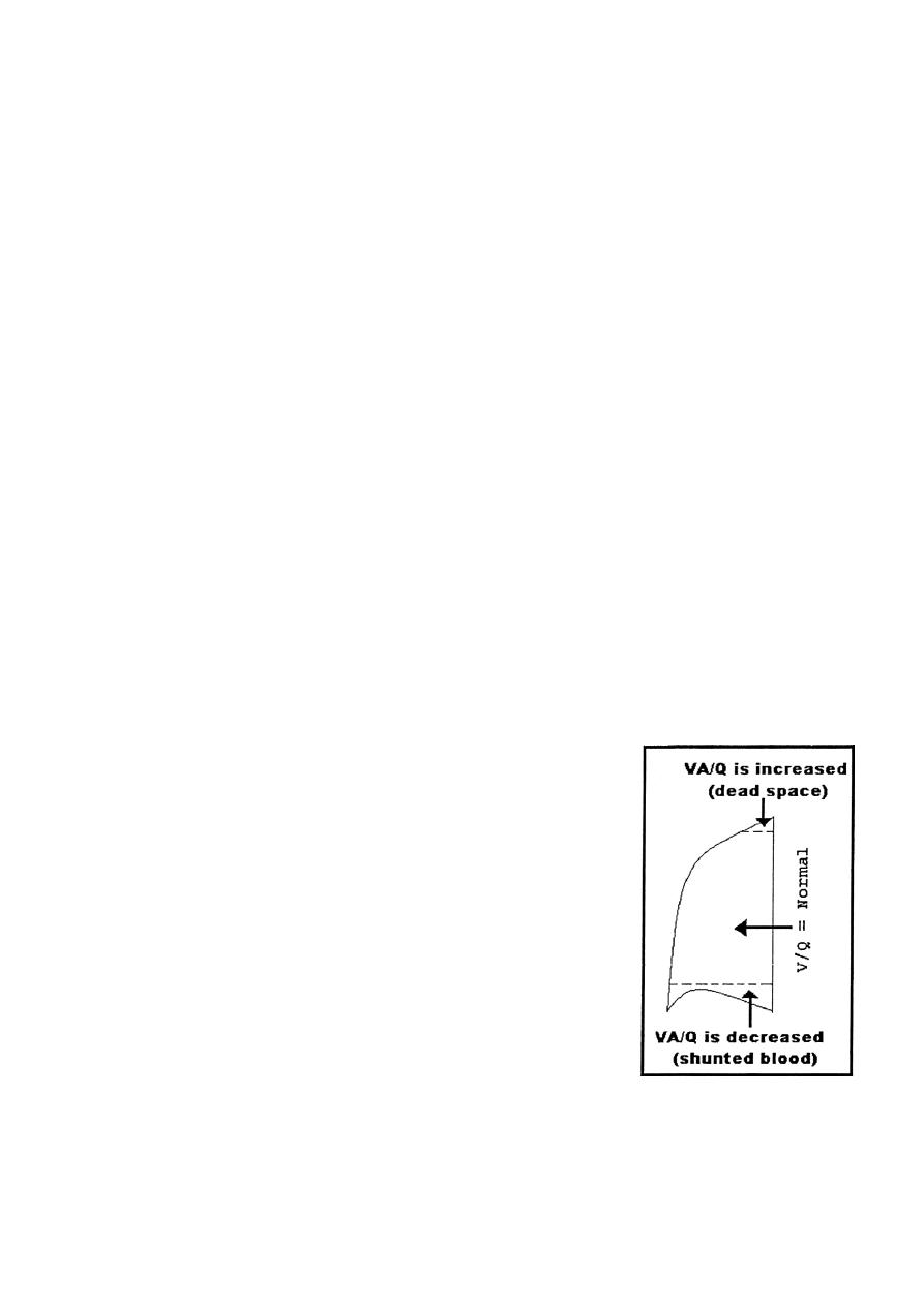
Ventilation — Perfusion Ratio (VA/Q):
[1]If some areas of the lungs are well ventilated but have no
or almost no blood flow, VA/Q = infinity. Therefore, the
alveolar air has the same composition and concentration of
the humidified inspired air (pO
2
= 149 mm Hg, PCO
2
=
0.3mm
Hg). In normal person in the upright position, both
blood flow and alveolarventilation are considerably less in
the upper part of the lung than in the lower part. However,
blood flow is decreased considerably more than ventilation
because the low-pressure pulmonary capillaries at the lung
apices are compressed by the higher-pressure lung alveoli.
Therefore, at the top of the lung, VA/Q is as much as three
times as great as the ideal value, which causes a moderate
degree of physiologic dead space in this area of the lung.
VA/Q equal to infinity does not occur in the normal lung
but instead occurs only in abnormal conditions such as in some lung diseases, a fall in
arterial pressure (following haemorrhage) or breathing against a high pressure as
occurs when a person is blowing on a musical instrument. Breathing against a high
pressure causes a compression of the pulmonary capillaries by the high alveolar

R
ESPIRATORY
S
YSTEM
8
pressure. In some chronic obstructive lung diseases such as emphysema, some areas of
the lungs exhibit very serious physiologic shunt and other areas very serious
physiologic dead space. Both of these tremendously decrease the effectiveness of the
lungs as gas exchange organ.
[2]If some areas of the lung have excellent blood flow but little or no ventilation,
VA/Q = zero. Therefore, the alveolar air comes to equilibrium with the venous blood
gases (PO
2
= 40 mm Hg, PCO
2
= 45 mm Hg) without further gases exchange because
there is no new gas coming from the exterior air to the alveoli. Whenever VA/Q is
below normal (i.e. low ventilation and normal perfusion), the ventilation is not enough
to provide the O
2
needed to oxygenate the blood flowing through the alveolar
capillaries and consequently leads to hypoxemia. Therefore, a certain fraction of the
venous blood passing through the pulmonary capillaries does not become oxygenated.
This fraction is called shunted bloodas it occurs normally in the bottom of the lung
with VA/Q as low as 0.6 times the ideal value. Also, some additional blood flows
through the bronchial vessels rather than through the alveolar capillaries, normally
about 2% of the cardiac output, this too is unoxygenated, i.e. shunted blood. The total
quantitative amount of shunted blood per minute is called the physiologic shunt
At a ratio of either zero(shunt) or infinity(dead space), there will be no proper
exchange of gases through the respiratory membranes of the affected alveoli.When
alveolar ventilation is normal for a given alveolus and blood flow is also normal for
the same alveolus, the VA/Q is also said to be normal(0.8).Therefore, PCO
2
(40 mm
Hg) and PO
2
(104 mm Hg) in the alveoli lie somewhere between that of the inspired air
and that of venous blood.
Compensatory Mechanisms For Matching The Ventilation And
Perfusion:
For proper blood oxygenation, the right proportion of air and capillary blood should be
available to each alveolus, local changes in the tone of smooth muscles of bronchioles
and pulmonary vessels help to maintain this equilibrium through two mechanisms:
[1] Local blood PCO
2
.if an alveolus is receiving too much air for its blood supply,
the blood CO
2
will be washed out and the concentration of CO
2
in the blood and in the
surrounding tissue will be low. Consequently the airways supplying the alveolus are
exposed to this low tissue CO
2
concentration and become constricted and vice versa.
By this local mechanism, ventilation can be matched to blood supply.
[2] Local blood PO
2
:
if an alveolus is receiving too little air for its blood supply,
the blood and tissue O2 will be decreased. A decreased O
2
concentration in the
pulmonary vessel causes a constriction to these vessels and vice versa (the opposite
effect that exerted on systemic arteries). By this local mechanism, perfusion can match
ventilation.
(II)
Pulmonary Diffusion (Gas Exchange in the Lungs):

R
ESPIRATORY
S
YSTEM
9
After the alveoli are ventilated with fresh air, the next step is diffusion of oxygen from
the alveoli into the pulmonary blood and diffusion of CO
2
in the opposite direction,
from the pulmonary blood into the alveoli.At rest, during each minute, body cells
consume about 200ml of oxygen and produce about the same amount of CO
2
.The
relative amounts of these two gases depend primarily upon what nutrients are being
used for energy; e.g., when glucose is utilized, one molecule of CO
2
is produced for
every molecule of oxygen consumed. The ration of CO
2
produced / O
2
consumed is
known as the respiratory quotient (RQ);accordingly for glucose RQ = 1.When fat is
utilized, only 7 molecules of CO
2
are produced for every 10molecules of O
2
consumed,
and RQ = 0.7.On mixed diet, the RQ is between these values.
Composition of alveolar air:
Alveolar air does not have the same concentrations
of gases as atmospheric air and there are several reasons for this difference:
[1]The alveolar air is only partially replaced by atmospheric air with each breath. This
is because that the amount of alveolar air replaced by new atmospheric air with each
breath (tidal volume - dead space) is only one several of functional residual capacity.
So that many breaths are required to exchange most of the alveolar air. This slow
replacement of alveolar air is of particular importance in preventing sudden changes in
gaseous concentrations in the blood. This makes the respiratory control mechanism
much more stable and helps to prevent excessive increases and decreases in tissue
oxygenation, tissue CO
2
concentration, and tissue pH when respiration is temporarily
interrupted.
[2]O
2
is constantly being absorbed from the alveolar air into the blood of the lungs,
and new O
2
is continually entering the alveoli from the atmosphere. Therefore, O
2
concentration in the alveoli, and therefore, its partial pressure as well, is controlled by:
[a]- the rate of absorption of O
2
into the blood, [b]-the rate of entry of new O
2
into the
lungs by ventilatory process.
[3]CO
2
is constantly diffusing from the pulmonary blood into the alveoli. The two
factors that determine alveolar concentration of CO
2
and also its partial pressure are (a)
the rate of excretion of CO
2
from the blood into the alveoli and (b)the rate at which
CO
2
are removed from the alveoli out by alveolar ventilation.
[4]Dry atmospheric air that enters the respiratory passage is humidified even before it
reaches the alveoli. This water vapor
simply dilutes all the
other gases in the inspired air as
shown in the table.
Composition
Atmospheric air
(mm Hg)
Humidified air
(mm Hg)
Alveolar air
(mm Hg)
Expired air
(mm Hg)
N
2
567
563.7
569
566
O
2
159
149
104
120
CO
2
0.3
0.3
40
27
H
2
O
37
47
47
47
Total
760
760
760
760

R
ESPIRATORY
S
YSTEM
10
The Respiratory Unit:
It is composed of a respiratory bronchiole, alveolar
ducts, alveolar sacs, and alveoli (about 300 million in the two lungs). Each alveolus
having an average diameter of about 0.2 mm. the alveolar gases are in close proximity
to the blood of the capillaries. The gaseous exchange between the alveolar air and the
pulmonary blood occurs through the membrane of all the terminal portions of the
lungs. These membranes are collectively called the respiratory membrane (pulmonary
membrane)which consist of the following layers:
[1] Alayer of fluid lining the alveolus and containingsurfactant.
[2] The alveolar epithelium.
[3] The epithelial basement membrane.
[4] Avery thin interstitial space.
[5] Acapillary basement membrane that in many placesfuses with the epithelial
basement membrane and
obliterating the interstitial space.
[6] The capillary endothelial membrane.
The average thickness of these layers is about 0.63 micron. The total surface area
of the respiratory membrane is estimated to be about 160 square meters, over which a
quantity of blood of about 60-140 ml only (the quantity of blood in the capillaries if
the lung at any given instant) is spread, which explain the rapidity of respiratory
exchange of gases. In addition, the diameter of the pulmonary capillaries is about 8
microns which is about the same diameter of RBC, therefore, RBC as it pass through
these capillaries are in fact in close contact with the endothelial membrane. This also
help in making the gas exchange rapid because the gases can pass directly from RBC
to the alveoli without passing through significant plasma.
Factors That Affect Rate Of Gas Diffusion Through The
Respiratory Membrane:
[1]
The thickness of the membrane:
any factor that increases the thickness to
more than two or three times the normal can decrease significantly the rate of gases
diffusion. This can occur in edema of the interstitial space of the membrane, and in
some fibrotic diseases of the lung.
[2]
The surface area of respiratory membrane:
when the total surface area is
decreased to about one third to one fourth normal, exchange of gases through the
membrane is impeded to a significant degree even under resting conditions. This can
occur in emphysema of the lung in which many alveoli coalesce with dissolution of
many alveolar walls.
[3] The diffusion coefficient:
depends proportionally on the solubility of the gas
in the membrane and inversely on the square root of its molecular weight. Therefore,
for a given pressuredifference, CO
2
diffuse through the membrane about
20
times as
rapidly as O
2
. Oxygenin turn diffuses about two times as rapidly as nitrogen.

R
ESPIRATORY
S
YSTEM
11
[4] The pressure difference
between the two sides of the membrane, which tends
to move the gas from area of higher partial pressure to an area of low partial pressure.
(III)
Transport of oxygen and carbon dioxide in the blood
and body fluids
:
Blood transports O2 and CO2 between the lungs and other body
tissues.Gases are transported in different forms:
1. Dissolved in plasma
2. Chemical combination with Hb
3. Converted into different molecule.
It is important to understand the difference between the partial pressure of a gas
and the gas content of a liquid. The partial pressure of the gas represents the
pressure it would exert in the gas phase. The gas content represents the volume of
the gas per unit volume of liquid that is present. Liquids must be exposed to a gas
tension for a finite time for the gases to dissolve in the liquid phase. If the
exposure time is long enough, the gas tension in the liquid will become equal to
that of the gas phase and an equilibrium will exist between the gases and the
liquid phases. Gases can move from one point to another by diffusion, which is
driven by the pressure difference between the two points. Thus, O
2
diffuses from
the alveoli (PO
2
= 104 mm Hg) into the pulmonary capillary blood (PO
2
= 40 mm
Hg) where it combines with Hb. Then from the systemic capillaries (PO
2
= 95
mm Hg) O
2
diffuses to and equilibrate with interstitial fluid of 40 mm Hg and
then diffuses to the cells (PO
2
= 23 mm Hg). Therefore, PO
2
of the blood leaving
the tissues capillaries and entering the veins is about 40 mm Hg. Conversely,
when O
2
is metabolized in the cells, the PCO
2
rises to a high value (PCO
2
= 46
mm Hg), which causes CO
2
to diffuse into the interstitial fluid with PCO
2
of 45
mm Hg and then it diffuses to and equilibrate with CO
2
of blood in tissue
capillaries (PCO
2
= 40 mm Hg) and combines with chemical substances in the
blood that increase CO
2
transport. Therefore, PCO
2
of the blood leaving the tissue
capillaries and entering the veins is about 45 mm Hg. Similarly, it diffuses out of
the blood into the alveoli because the PCO
2
in the alveoli (40 mm Hg) is lower
than that in the pulmonary capillary blood (45 mm Hg).
About 98% of the blood that enters the left atrium from the lungs passes through
the alveolar capillaries and becomes fully oxygenated (PO
2
= 104 mm Hg) and
2% passes through the bronchial circulation (dead space), which represents the
shunted blood by passing the gas exchange areas and has a PO
2
about the same of
the normal venous blood (PO
2
= 40 mm Hg). This blood combines in the
pulmonary veins with the oxygenated blood from the alveolar capillaries. This
mixing of the blood is called venous admixture of blood, and it causes the PO
2
of
the blood pumped by the left heart into the aorta to fall to about 95 mm Hg.
The PO
2
in the interstitial fluids is affected by:
R
ESPIRATORY
S
YSTEM
12
1- The blood flow: As the blood flow increases, the O
2
delivery to the tissues
increases.
2- Tissue metabolism; if the cells utilize more O
2
for metabolism than normally,
this tends to reduce the interstitial fluid PO
2
.
3- Hb concentration; because about 97% of the O
2
transported in the blood is
carried by Hb, a decrease in Hb concentration reduces the O
2
delivery to the
interstitial fluid causing a reduction in PO
2
in the interstitial fluid.
Since only 3 mm Hg of O
2
pressure is normally required for full support of the
metabolic processes of the cell, one can see that even this low cellular PO
2
, 23 mm Hg,
is more than adequate and actually provides a considerable safety factor.
The PCO
2
in the interstitial fluid can be affected by:
1- the decrease in blood flow which causes an increase in the PCO
2
.
2- increase in metabolic rate greatly elevates the PCO
2
at all levels of blood flow.
Oxygen Transport
Each RBC molecule contains about 250 million Hb molecules.Each Hb
molecule consists of:
1. globin portion(4 polypeptide chains)
2. Four iron containing pigmets(heme groups)
Each iron atom binds one oxygen molecule thus Hb molecule can bind up
to 4 oxygen molecules(100% saturated) or fewer o2 molecules(partially
saturated).
R
ESPIRATORY
S
YSTEM
13
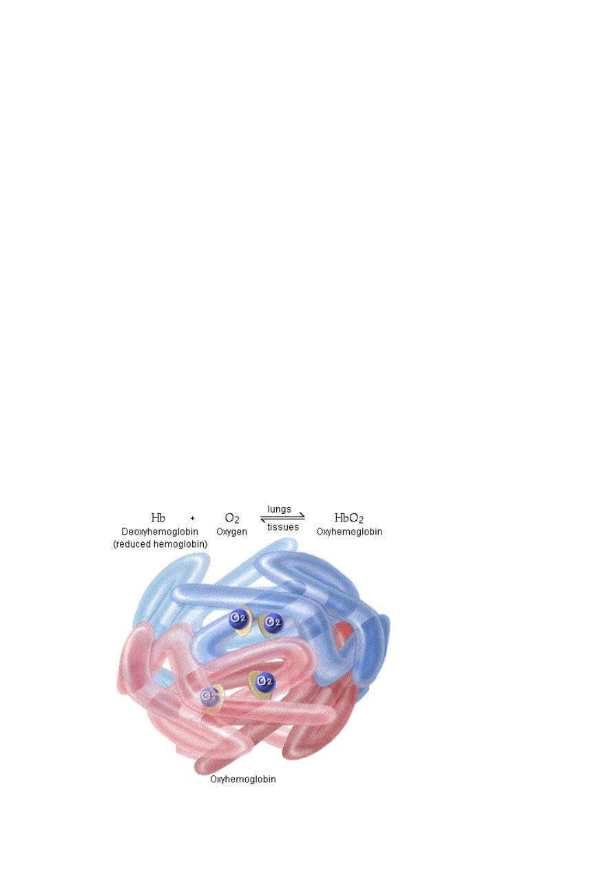
Oxygen binding occurs in response to the high PO2 in the lungs
forming oxyhemoglobin.
Once O2 binds to Hb,other molecules binds more readily because Hb
affinity for O2 increases as its saturation increases(co-operative binding).
The formation of oxyHb is the reversable reaction(Hb +O2 = Hb02)
depending on concentration of reactants and products of reaction.
In the lungs, when PO2 is high the reaction procedes to the right forming
HbO2(O2 loading).
In the tissuess, when PO2 is high the reaction procedes to the left ;0xyHb
is dissociated releasing O2 (O2 unloading).
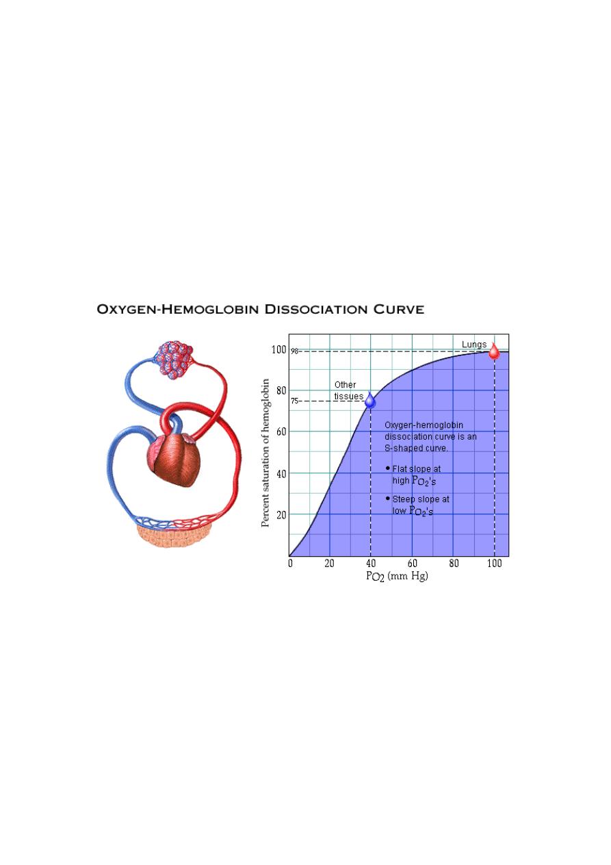
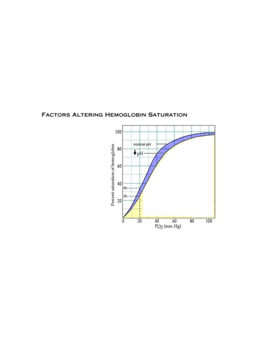
This is a curve which correlates between partial pressure of oxygen and
hemoglobin saturation with oxygen.
The degree of Hb saturation deppends on PO2. In the lungs,the PO2 is
about 100 mmHg,Hb has high affinity for O2 and it is 98% saturated with
O2.In the tissues,PO2 is 40 mmHg, Hb has lower affinity for O2 and it is
75% saturated with O2 when leaving the tissues.
The curve is "S" shaped with flat slope at higher PO2s (80-
100mmHg)and steep slope at lower PO2(10-60 mmHg).
At see level,PO2 in the lungs=100mmHg and Hb is 98% saturated.
R
ESPIRATORY
S
YSTEM
14
At higher altitude or cardiopulmonary diseases PO2 decreases by 20%
(PO2=80%) but Hb is still highly saturated(95%).
In the tissues where PO2= 40 mmHg, Hb has a lower affinity for O2 and
it is only 75% saturated.During vigorus exercise,PO2 decreases(20
mmHg) because muscles use more O2 and Hb is 35% saturated.As PO2
decreases, Hb releases much more O2 to the tissues. This allows the body
to math closely between O2 delivery by Hb and the O2 utilization by
tissues.
In addition to PO2,Hb saturation is altered by four other factors:
1. pH
2. temperature
3. PCO2
4. BPG(Biphosphoglycerate)
Any of these factors or all of them togetherplay a role during exercise.
During vigorous exercise,contracting muscle produce more metabolic
acids like lactic acid which lowers pH,more temperature and more

R
ESPIRATORY
S
YSTEM
15
CO2..In addition, high temp and lower PO2 increase BPG production by
RBC which decreases Hb affinity for O2 thus releasing more O2 to active
cells.When pH decreases, the curve shifts to right(increased O2
unloading).A similar shift occurs in response to high temp,high
POC2,high BPG.
At decreased temperatures,the Hb affinity for O2 is higher(less O2 is
released);the curve shifts to left.Similar shift occurs in response to high
pH,low PCO2,low BPG.
The role of 2,3 — PPG:
It is highly charged anion that binds to the Bchains of
deoxygenated Hb but not to those of oxyHb as follow: HbO
2
+ 2,3-DPG→ Hb-2,3-
DPG + O
2
. In this equilibrium, an increase in the concentration of 2,3-DPG shifts the
reaction to the right, causing more O
2
to be liberated.
The normal 2,3-DPG in the RBC keeps the O
2
-Hb dissociation curve shifted slightly to
the right all the time. In hypoxic conditions that last longer than a few hours, the
quantity of 2,3-DPG in the RBC increases considerably, thus shifting the curve even
farther to the right.This mechanism might be important for adaptation to hypoxia.
However, the presence of the excess 2,3-DPG also makes it difficult for the Hb to
combine with O
2
in the lungs when the alveolar PO
2
is reduced, thereby often creating
as much harm as good. Thyroid hormones, growth hormone, and androgens increase
the concentration of 2,3-DPG in the RBC and hence P
50
. 2,3-DPG is very plentiful in
RBC.
The role of Hb-F:
The greater affinity of Hb-F than Hb-A for O
2
facilitates the
movement of O
2
from the mother to the fetus. The cause of this greater affinity is the
poor binding of 2,3-DPG by the y polypeptide chains that replace Bchains in Hb-F.
Transport of O
2
in the dissolved state
: 0.17 ml of O
2
is normally transported in
the dissolved state to the tissues by each 100 ml of blood (3% of the total transported
O
2
). If a person breathsO
2
in high concentration ( very high alveolar PO
2
), the amount
then transported in the dissolved state can become very high.
Myoglobin:
It is an iron-containing pigment found in skeletal muscle. It resembles
Hb but binds one rather 4 mol of O
2
per mol. Its dissociation curve is a rectangular
rather than a sigmoid curve. Because its curve is to the left of the Hb curve, it takes up
O
2
from Hb in the blood. It releases O
2
only at low PO2 values, but the PO
2
in
exercising muscle is close to zero. The muscle blood supply is compressed during
contractions, and myoglobin may provide O
2
when blood flow is cut off.
Combination of Hb with CO:
CO has affinity of 230 times to combine with Hb
than O
2
do and form carboxyHb. A patient poisoned with CO can be treated by

R
ESPIRATORY
S
YSTEM
16
administration pure O
2
, for O
2
at high alveolar pressure displaces CO from its
combination with Hb far more rapidly than can O
2
at the low pressure of atmospheric
O
2
. The patient can also be benefited by simultaneous administration of a few per cent
CO
2
because this strongly stimulates the respiratory center. This increases alveolar
ventilation and reduces the alveolar CO concentration, which allows increased CO
release from the blood.
CO2 Transport:
7% dissolved in plasma and 93% carried by RBC(23% combined with
Hb forming carbamino-Hb, 70% converted into bicarbonate ions).
Carbamino-Hb
: CO2 combines reversibly with the globin
portion of Hb forming carbamino-Hb.
a. In the tissues where PCO2 is high, Hb-CO2 is formed.
b. In the lungs where PCO2 is low, Hb-CO2 is dissociated and
CO2 is exhaled.
Bicarbonate ions
: 70% of CO2 is converted into bicarbonate
ions (faster than CO2) within the RBC in a sequence of reversible
reactions then tranported in plasma:
a. In the tissues( regions with high PCO2) CO2 enters RBC
combines with water to form carbonic acid.This reaction is
catalyzed by carbonic anhydrase enzyme. Similar reaction occurs
in plasma but without enzyme it is very slow.Carbonic acid
dissociates into H+ and bicarbonate.Hydrogen ions is buffered by
Hb forming(H-HB).To maintain electrical neutrality,bicarbonate
ions diffuse out of RBC in exchange with chloride ions(chloride
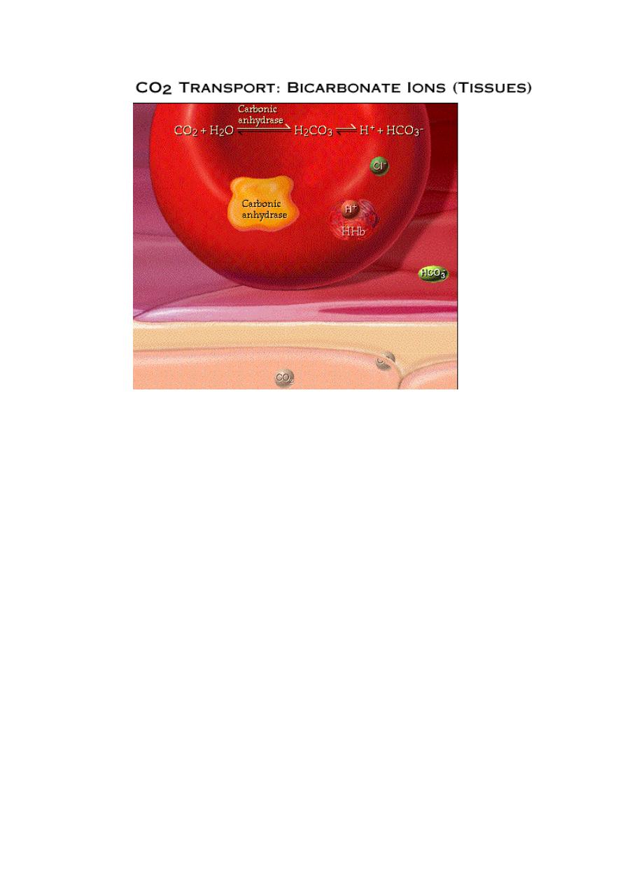
R
ESPIRATORY
S
YSTEM
17
shift).Within the plasma,bicarbonate ions act as pHbuffer. .
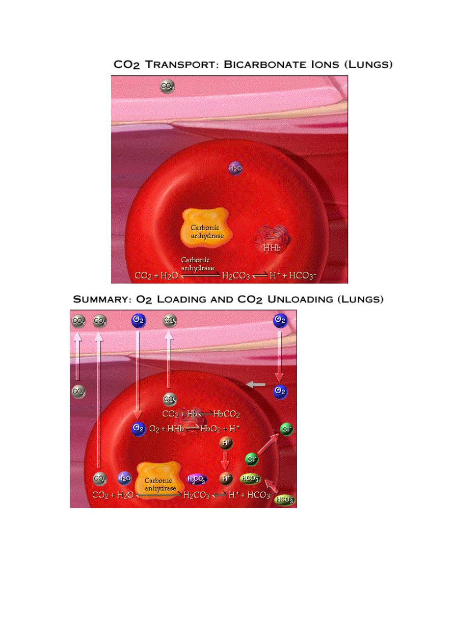
b. In the lungs, CO2 diffuses out of the plasma into the
alveoli.This lowers PCO2in the blood causing the chemical
reactions to reverse.Bicarbonate ions diffuse back into RBC and
chloride ions diffuse out(chloride shift).Hydrogen ions combine
with bicarbonate ions to form carbonic acid. Carbonic acid
breaksdown into CO2 and H2O.This reverse reaction is also
R
ESPIRATORY
S
YSTEM
18
catalyzed
by
carbonic
anhydrase
enzyme.
During external respiration,small amounts of O2 remains
dissolved in plasma with the majority in combination with Hb
forming oxy-hemoglobin(Hb-O2).when Hb is saturated with
O2,its affinity for CO2 decreases.Any CO2 combined with Hb
R
ESPIRATORY
S
YSTEM
19
,dissociates and diffuses out of RBC through plasma and then to
alveoli.The H+ ions released from Hb is combined with
bicarbonate ions which diffuses into RBC from plasma in
exchange for chloride(chloride shift).Then reaction between
H+and bicarbonate to form carbonic acid which breaks down
into CO2 and H2O.This CO2 plus small amounts transported in
the dissolved form diffuse into alveoli.
In other wards,O2 loading facilitates CO2 unloading from
Hb.This is called
Haldane effect
.
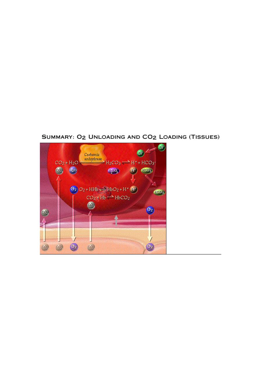
During internal respiration,small amounts of CO2 remains dissolved in
plasma with the majourity within RBC reacting with water to form carbonic
acid which dissociates into H+ and Bicarbonate ions.within RBC, H+ ions
are buffered by Hb forming H-Hb.
When Hb is bound to H+ it has lower affinity
for O2 thus O2 dissociates from Hb and diffuses out of RBC to the tissue.The
interaction between H+ binding and Hb affinity for O2 is called
Bohr effect.By
forming H+ ions,CO2 loading facilitates O2 unloading.Small amounts of
O2 transported in dissolved state also diffuses out into tissue cells.DeoxyHb
has high affinity to CO2
.

R
ESPIRATORY
S
YSTEM
20
The Response of Respiratory System to Exercise and
Stress:
During exercise or other stressful conditions, the body may require as much
as
20
times of the normal amount of O
2.
The increased cardiac output causes blood to
stay half normal time in the pulmonary capillary. Yet, the blood is still almost
completely saturated with O
2
when it leaves the pulmonary capillaries due to an
increase in the diffusing capacity of the respiratory membrane for O
2
about threefolds
during exercise, the reasons for this are:
[A] The average RBC spends approximately 0.75 sec in the pulmonary capillary.
If O
2
equilibration occurs in 0.25 sec, then there is normally no increase in the
O
2
content for the last 0.50 sec of transit through the pulmonary capillary. This
time provides a safety margin that ensures an adequate O
2
uptake during periods
of stress.
[B] The opening up a number of previously dormant pulmonary capillaries, and
dilatation of already functioning pulmonary capillaries thereby increases the
surface area of blood into which the oxygen can diffuse.
[C] Increased alveolar ventilation.
[D] More ideal ventilation-perfusion ratio in the upper part of the lungs.
[E] During exercise, there is a considerable shift of the Hb-O
2
dissociation curve
to the right (i.e. decrease in the affinity of Hb to combine with O
2
) in the muscle
capillary blood due to the release of large amounts of CO
2
, acids, and phosphate
compounds, in addition to high temperature of the muscles. Then in the lungs, the
events are reversed, thus, the shift occurs in the opposite direction (i.e. to the left,
which means an increase in the affinity of Hb to combine with O
2
), thus allowing
pickup of extra amounts of O
2
from the alveoli.
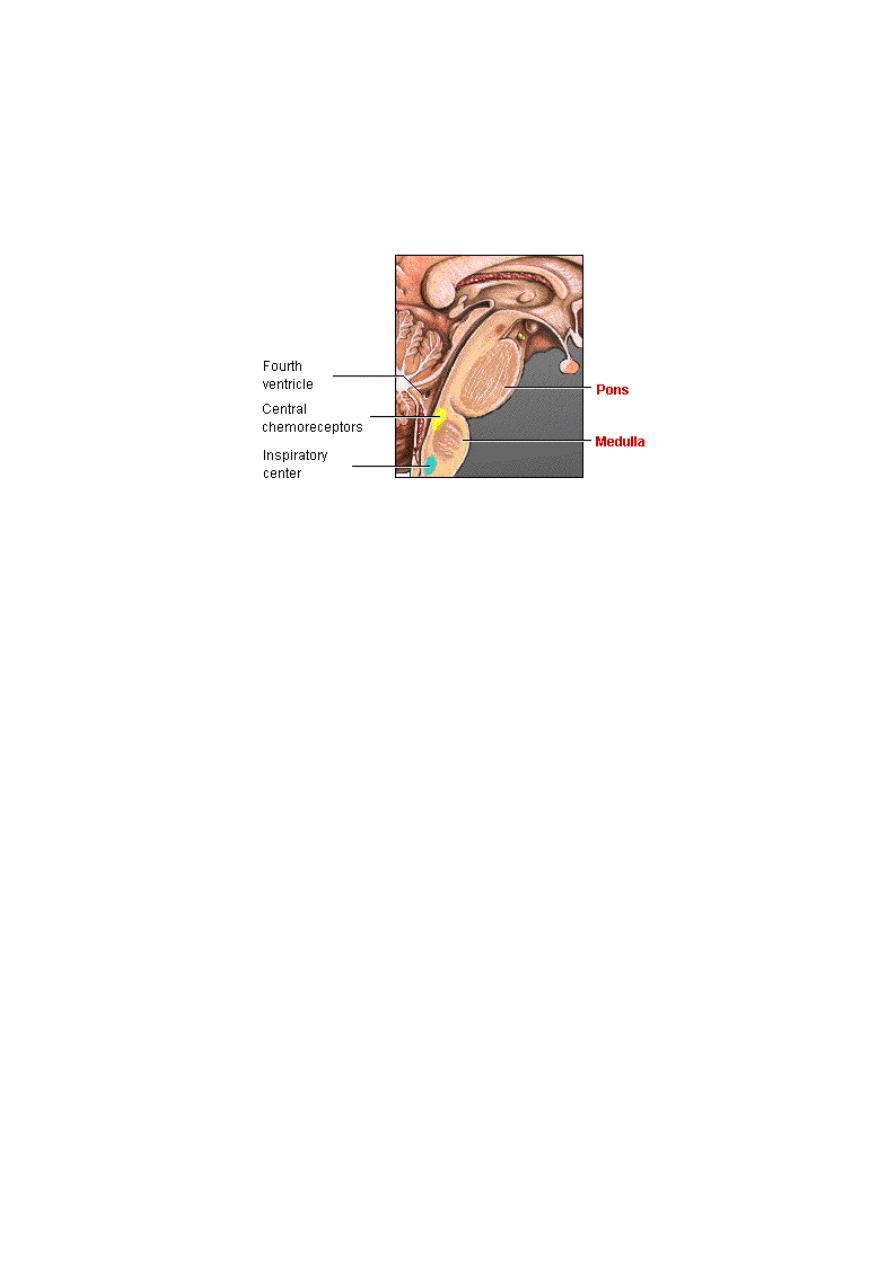
(IV)Regulation of Respiration
The respiratory centre
:
The respiratory cycle (inspiration and expiration) is
regulated by the respiratory center in the brain which is composed of three major
groups of neurons located bilaterally in the medulla oblongata and pons and these are:
[1] The dorsal group of neurons:
This group of neurons is located in the medulla
within the nucleus of the tractussolitorius which is also the sensory termination of both
the vagal and glossopharyngeal nerves transmitting sensory signals into the
respiratory center from the peripheral chemoreceptors, baroreceptors and several
different types of receptors in the lung. They are responsible for the basic rhythm of
respiration by autonomous repetitive bursts of inspiratory action potentials. The nerve
signal that is transmitted to the inspiratory muscles (through contralateral phrenic and
intercostal motoneurons and to the ventral respiratory group) begins very weakly at
first and increase steadily in a ramp fashion for about 2-sec. Then it abruptly ceases for
approximately the next 3 sec, and then begins again for still another cycle, and again

R
ESPIRATORY
S
YSTEM
21
and again. The advantage of this is that it causes a steady increase in the volume of the
lungs during inspiration, rather than inspiratory gasps.
[2] The pneumotaxic group:
This group of neurons is located within the pons in
the nucleus parabrachialis and they transmit impulses continuously to the dorsal
respiratory group of neurons. The primary effect of these is to control the switch off
point of the inspiratory ramp, thus controlling the duration of the filling phase of the
lung cycle.
[3] The ventral group:
These neurons are located in the nucleus ambiguus and
nucleus retroambiguus and is comprised of the upper motor neurons of the vagus and
the nerves to accessory muscles of respiration.These neurons contribute to both
inspiration and expiration, however, they are especially important inproviding the
powerful expiratory forces during expiration. Thus, this area operates more or less as
an overdrive mechanism when high levels of pulmonary ventilation are required.
Control Of Respiration:
The respiratory centers and consequently the
ventilation can be controlled by the following chemical and neural factors:
1. Chemical control: mediated through changes in PCO
2
, [H
+
], and PO
2
.
2. Neural control:
a. Higher brain centers and peripheral proprioceptors control of respiration.
b. Motor cortex control ofrespiration.
c. Vasomotor center control of respiration.
d. Body temperature control of respiration.
1. Chemical control of respiration:
A: Central chemoreceptors:
Surplus of CO
2
or H
+
affected respiration mainly by
excitatory effects on the respiratory center itself, causing greatly increased strength of
both the inspiratory and expiratory signals to the respiratory muscles. The resulting
increase in ventilation (4-fold increase caused by decreasing the blood pH or 11-fold
increase caused by increasing PCO
2
) increases the elimination of CO
2
from the blood,
this also removes H
+
from the blood because decreased CO
2
also decreases the blood
carbonic acid. It is believed that the blood CO
2
and H
+
do-not affect the respiratory
centers directly. Instead, achemosensitive area located bilaterally only a few microns
beneath the surface of themedulla ventral to the entry of the glossopharyngeal and
vagal nerves into the medulla, this area is highly sensitive to changes in H ion
concentration, and it in turn excites the other portions of the respiratory center. It has
especially potent effects on increasing the degree of activity of the inspiratory center,
increasing both the rate of rise of the inspiratory ramp signals and also the intensity of
the signal. However, H
+
do not easily cross either the blood-brain barrier or the blood-
cerebrospinal fluid barrier. For this reason, changes in H
+
concentration in the blood
actually have considerably less effect in stimulating the chemosensitive neurons than
do changes in CO
2
. This is because CO
2
passes through blood-brain barrier and blood-
cerebrospinal fluid barrier very easily. Consequently, whenever the blood CO
2
concentration increases, the PCO
2
in both the interstitial fluid of the medulla and in the
R
ESPIRATORY
S
YSTEM
22
cerebrospinal fluid also increase. In both of these fluids the CO
2
immediately reacts
with the water to form carbonic acid which dissociates into H
+
and bicarbonates. Thus,
paradoxically, more H
+
are released into the respiratory chemosensitive sensory area
when the blood CO
2
concentration increases than when the blood H
+
concentration
increases. For this reason, respiratory center activity is affected considerably more by
changes in blood CO
2
than by changes in blood H
+
.
The stimulatory effect of increased CO
2
on respiration reaches its peak within a
few minutes after an increase in blood PCO
2
. Thereafter, the stimulation gradually
declines for the next one to two days to as little as one-fifth the initial effect due to
adaptation of the receptors. Therefore, A change in blood CO
2
concentration has a
very potent acute effect on controlling respiration but only a weak chronic effect after
a few days' adaptation.
PO
2
: Arterial PO
2
does not have a significant direct effect on the respiratory center of
the brain in controlling respiration. Instead, it acts almost entirely on peripheral
chemoreceptors located in the carotid and aortic bodies, and these in turn transmit
appropriate nervous signals to the respiratory center for control of respiration.
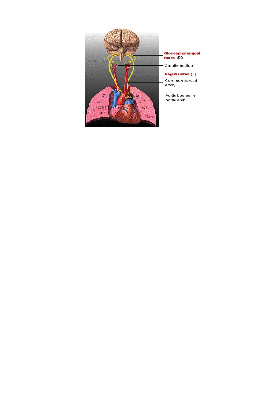
B: Peripheral chemoreceptors(carotid and aortic bodies):
Carotid
bodiesare located bilaterally in the bifurcations of the common carotid arteries, and
their afferent never fibers pass to the glossopharyngeal nerves and thence to the dorsal
respiratory area of the medulla. Aortic bodies are located along the arch of the aorta,
and their afferent fibers pass through the vagito the dorsal respiratory area.
The blood flow through the carotid and aortic bodies is extremely high. Because
of this, arteriovenousoxygen difference is very small, which means that the
venous blood leaving these bodies still has a PO
2
nearly equal to that of the
arterial blood. It also means that PO
2
of the tissues in these bodies remains atall

R
ESPIRATORY
S
YSTEM
23
times almost equal to that of the arterial blood. Thesebodies are more influenced
by :
(a)arterial PO
2:
which is determined by the amount of dissolved O
2
ratherthan by
arterial oxygen content.Hence, they are not influenced by a low Hb level.When
the arterial PO
2
falls below normal (especially PO
2
in the range between 60 and 30
mm Hg), or when the blood pressure sufficiently low causing a low blood flow
(even though constituents of blood do not change), the chemoreceptors become
strongly stimulated.
(b)increase in CO
2
or H
+
concentration: also excites the chemoreceptors and in
this way indirectly increases respiratory activity. However, the direct effects of
both these factors in the respiratoiy center itself are so much powerful.
The cause of the poor effect of low PO
2
on respiratory control in comparison to
those of CO
2
and H
+
concentration can be explained as follow: The increase in
ventilation that does occur when the PO
2
falls blows off the respiratory stimulants
(CO
2
and consequently H+). However, overseveral days, the respiratory center
gradually becomes adapted to the diminished CO
2
and alveolar ventilation then
rises to as high as five to seven times normal. This is part of the acclimatization
that occurs as a person slowly ascends a mountain. Yet, under some abnormal
conditions, as occurs in pneumonia, emphysema in which gas exchange is
impaired, the PCO
2
and H
+
concentrations increase at the same time that the
arterial PO
2
decrease. Under these conditions, all three of the feedback
mechanisms work together, and the PO
2
mechanism then exerts its full share of
respiratory stimulation, sometimes becoming even more potent as a controller of
respiration than the PCO
2
and H
+
mechanisms.
R
ESPIRATORY
S
YSTEM
24
2.Neural control:
A: Peripheral receptors
:
Stretch receptors:
in the wall of the bronchi and bronchiole transmit signals
through the vagi into the dorsal respiratory group of neurons when the lungs
become overstretched(the tidal volume increases to greater than approximately
1.5 liters). Therefore, when the lungs become overly inflated, the stretch
receptors activate an appropriate feedback response that switches off the
inspiratory ramp and thus limits further inspiration. This is called the Hering-
Breuer inflation reflex,this reflex appears to be mainly a protective mechanism
for preventing excess lung inflation rather than important ingredient in the
normal control of ventilation.
J receptors:
These receptors are located in the pulmonary interstitium at the
level of the pulmonary capillaries and are stimulated by distension of the
pulmonary vessels (e.g., as caused by left ventricular failure, pulmonary
embolization, and certain chemicals or drugs). These receptors initiate reflexes
causing rapid, shallow breathing (tachypnea).
Chest wall receptors:
which can detect the force generated by the respiratory
muscle during breathing. If the force required to distend the lungs becomes
excessive (either as a result of high airway resistance or low compliance), the
information from these receptors gives rise to the sensation of dyspnea
(difficulty in breathing).
Irritant receptors:located in the large airways and are stimulated by smoke,
noxious gases, and particulates in the inspired air. These receptors initiate
reflexes that cause coughing, bronchconstriction, mucus secretion, and breath
holding (i.e., apnea).
Joint proprioceptors During exercise, the body movements, especially of the
limbs, are believed to increase pulmonary ventilation by exciting that then
transmit excitatory impulses to the respiratory center.

R
ESPIRATORY
S
YSTEM
25
B.
Motor cortex:
Respiration can be controlled voluntarily, and that one can
hyperventilated or hyporventilated to such an extent that serious derangements in
PCO
2
, pH and PO
2
can occur in the blood. This is mediated by the nervous pathway for
voluntary control passes directly from the motor cortex and other higher centers
downward through the corticospinal tract to the spinal neurons that drive the
respiratory muscles.
C.
Vasomotor center:
The
vasomotor
center
that
control
peripheral
vasoconstriction and heart activity is closely related to respiratory center in the
medulla. A moderate degree of spillover of nerve signals occurs between the two
centers. Therefore, almost any factor that increase the activity of the vasomotor center
also has at least a moderate effect on increasing respiration.
D.
Body temperature:
An increase in body temperature increases the rate of
respiration directly by increasing respiratory center activity and indirectly by
increasing the cellular metabolism and eventually enhances the chemical stimuli for
increased respiration.
In strenuous exercise, O
2
utilization and CO
2
formation can increase as much as
twentyfold associated with increase in alveolar ventilation. This increase in alveolar
ventilation is not mainly due to change in blood PO
2
, PCO
2
,and H
+
concentration
which all remain almost exactly normal, There are at least two different effects seem to
be predominantly concerned :
[A] The brain, on transmitting impulses to the contracting muscles, is believed to
transmit collateral impulses into the brain stem to excite the respiratory center .But,
occasionally, the nervous signals are either too strong or two weak in their stimulation
of the respiratory center, then the chemical factors play a very significant role in
bringing about the final adjustment in respiration required to keep the CO
2
and H
+
concentrations of the body fluids as nearly normal as possible.
[B]Joint proporioceptors transmit excitatory impulses to the respiratory center.
Pulmonary Blood Flow:
The pulmonary circulation is basically low-pressure,
low-resistance, highly compliant system. Pressure in the pulmonary artery is about 25
mmHg systolic and 8 mmHg diastolic (a mean of about 14 mmHg). Pressure in the left
atrium is about 5 mmHg, resulting in pressure drop across the pulmonary circulation of
about 9 mmHg. Pulmonary vascular resistance is 1.8 mmHg/L/min which is about
10% of the systemic vascular rersistance(18 mmHg/L/min).
Abnormalities Of Respiratory Control:
1.Respiratory center depression:the activity of respiratory center may be depressed
or even totally inactivated by cerebrovascular disease, acute brain edema, anesthesia
and overdose of narcotics.

R
ESPIRATORY
S
YSTEM
26
2.Periodic breathing:The most common type of periodic breathing is called cheyne-
stokes breathing which is characterized by slowly waxing and waning respiration
separated by periods of apnea and can seen in heart failure or brain stem lesions.
3.Kussmaul's respirationwhich is a rapid, deep breathing often seen in patients
suffering from diabetic ketoacidosis. It occurs as the body tries to compensate for
metabolic acidosis by increasing the rate of CO
2
excretion.
Hypoxia:
Hypoxia is cellular deficiency of
O
2
. Traditionally hypoxia has been
divided into 4 types:
[1] Hypoxic hypoxia:
In which the PO
2
of the arterial blood is reduced due to low
atmospheric O2, inadequate ventilation of the alveoli or insufficient diffusion of O
2
through the respiratory membrane.
[2] Anaemic hypoxia:
In which the arterial PO
2
is normal but the amount of Hb
available to carry O
2
is reduced.
[3] Ischaemic hypoxia:
In which the blood flow to a tissue is so low that adequate
O
2
is not delivered to it despite a normal PO
2
and Hb concentration.
[4] Histotoxic hypoxia:
because of the action of certain toxic agents, the tissue
cells cannot make use of the O
2
supplied to them such as in cyanide poisoning, in
which the action of cytochrome oxidase(respiratory chain enzyme in the mitochondria)
is completely blocked.Also, deficiency of oxidative enzymes such as vitamin B
deficiency (Beriberi).
Cyanosis:
It is a dusky bluish discoloration of the tissues and appears when the
reduced Hb concentration of the blood in the capillaries is more than 5 gm/dl.
Cyanosis is easily seen in mucus membrane and thin skin areas like lips, fingers, ear
lobes, also nail bed. In polycythaemia, cyanosis is very common because of the large
amount of Hb in the blood whereas in anaemia, cyanosis is rare because it is difficult
for there to be enough deoxygenated Hb to produce the cyanotic color. Cyanosis
divided into 2 types:
[1]
Central cyanosis: In which there is Hb-undersaturation or an abnormal Hb
derivative, and the mucous membranes and skin are both affected.
[2]
Peripheral cyanosis: Which is due to a slowing of blood flow to an area and
abnormally great extraction of O
2
from normally saturated arterial blood. It result
from vasoconstriction and diminished peripheral blood flow, such as occurs in
moderate cold exposure, shock, heart failure, and peripheral vascular disease.
Often, in these conditions, the mucous membranes of the oral cavity may be

R
ESPIRATORY
S
YSTEM
27
spared. In very cold weather cyanosis does not developed, because the drop in
skin temperature inhibits the dissociation of oxy Hb and the O
2
consumption of
the cold tissues is decreased. Cyanosis does not occur in anaemic or histotoxic
hypoxia. In CO poisoning, the color of reduced Hb is obscured by the cherry red
color of carboxyHb. A discoloration of the skin and mucous membranes similar
to cyanosis is produced by high circulating levels of met Hb.
Hypercapnia:
It means excess CO
2
in the body fluids. Hypercapnia is association
with hypoxia when hypoxia is caused by hypoventilation or by circulatory deficiency.
Hypoxia resulting from poor diffusion through pulmonary membrane, serious
hypercapnia usually does not occur because CO
2
diffuses 20 times as rapidly as O
2
.
O
2
Therapy
: In hypoxia, O
2
therapy is of great value, especially in certain types of
hypoxia (such as law atmospheric O2, hypoventilation hypoxia, diffusional hypoxia")
and of slight value in hypoxia due to other causes. inchronic hypoxia,O
2
lack becomes
a far more powerful stimulus to respiration than usual, sometimes increasing the
ventilation as much 5-7 times. Therefore, during O
2
therapy, relief of the hypoxia
occasionally causes the level of pulmonary ventilation to decrease so low that lethal
levels of hypercapnia develop. For this reason, O
2
therapy in hypoxia is sometimes
contraindicated.
O
2
Toxicity:
Administration of 100% O
2
has been demonstrated to exert toxic
effects. The toxicity seems to be due to the production of the superoxide anion (O
2-
)
and H
2
O
2
. When 80-100% O
2
administered for periods of 8 hours or more the
respiratory passages become irritated, causing substernal distress, nasal congestion,
sore throat and coughing. Exposure for 24-48 hours causes lung damage as well. The
reason O
2
produce the irritation is probably due to inhibition the ability of lung
macrophages to kill bacteria, and surfactant production is reduced.

R
ESPIRATORY
S
YSTEM
28
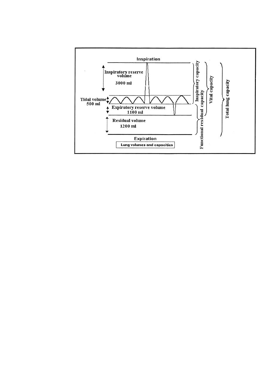
Lung Volumes And Capacities
Pulmonary ventilation can be recorded by using the spirometer and the process called
spirometry by which volume of air that is moved in and out of the lung can be
recorded. The volumes and capacities of lungs are:
[1] The tidal volume (TV):Is the volume of air inspired or expired with each normal
breath and it is about 500
ml
in average young adult man.
[2]
The inspiratory reserve volume (IRV):
Is the extra volume of air that can be inspired
over and beyond tidal volume and it is about 3000 ml.
[3]
The expiratory reserve volume (ERV):
Is the amount of air that can be expired after the
normal tidal expiration, which is about 1100 ml.
[4]
The residual volume (RV):
Is the volume of air still remaining in the lungs after the most
forceful expiration, which is about 1200 ml.This is important because it provides air in the alveoli to
aerate the blood even between breaths which otherwise the concentration of oxygen and carbon
dioxide in the blood would rise and fall markedly with each respiration, which would certainly be
disadvantageous to the respiratory process.
This volume cannot be measured directly by spirometer. Therefore, an indirect method must be
used usually the helium dilution method. Once the functional residual capacity (FRC) has been
determined, the residual volume can then be determined by subtracting the expiratory reserve volume
from the functional residual capacity, i.e. RV = FRC - ERV.
The other way for determination of lung RV is by using body plethysmogrph.
[5]The inspiratory capacity (IC)
= TV +IRV = 500 +3000 = 3500
ml.
This is the amount of
air that a person cans breath beginning at the normal expiratory level and distending the lungs to the
maximum amount.
[6] The functional residual capacity (FRC)
=
ERV + RV = 1100 + 1200 = 2300
ml.
This is the amount of air remaining in the lungs at the end of normal expiration.
[7] The vital capacity (VC)= IRV + TV + ERV = 3000 + 500 + 1100 = 4600
ml.
This is the maximum amount of air that a person can expel from the lungs after filling
the lungs first to their maximum extent, and then expiring to the maximum extent.
Vital capacity can be decreased markedly in restrictive lung diseases (paralysis of
the respiratory muscles, tuberculosis, lung cancer, fibrotic pleurisy, pulmonary
vascular congestion and edema as in left sided heart failure) and may be normal in
obstructive lung diseases (asthma, chronic bronchitis, emphysema). When the vital
capacity is reduced to about 40% of normal, the patient can no longer perform even the
simplest movements without becoming breathless.
[8] The total lung capacity (TLC)= VC + RV = 4600 + 1200 = 5800 ml
.
This is the
maximum volume to which the lungs can be expanded with the greatest possible
inspiratory effort.
All pulmonary volumes and capacities are about 20-25% less in women than
men, and they are greater in large athletic persons than in small and asthenic persons.
Pulmonary volumes and capacities change with the position of the body, most of them
decreasing when the person lies down and increasing on standing, this change with
position is caused by two factors:
R
ESPIRATORY
S
YSTEM
29
[A] - a tendency for the abdominal contents to press upward against the diaphragm in
the lying position.
[B]-an increases in the pulmonary blood volume in the lying position, which
correspondingly
decreases
the
space available
for
pulmonary
air.
Lung Function Tests
1.Forced vital capacity (FVC): It is the maximum volume of air
expired forcefully following maximum inspiration, a normal
subject this is accomplished in 3-4 sec.
2.Timed vital capacity (forced expiratory volume-1 sec, FEV1):
It is the volume of air expired during the first second of forceful
expiration.
3.Percent vital capacity (FEV1%): [FEV1/VC] x 100. In normal
subject, the FEV1% is at least 80%. However, in obstructive lung
diseases like asthma, FEV1% is markedly reduced while normal in
restrictive lung diseases.

R
ESPIRATORY
S
YSTEM
30
4.Peak expiratory flow (PEF):It is the maximum airflow obtained
during maximum expiratory effort after maximum inspiration.
When a person expires with great force through Wright peak flow
meter, the expiratory airflow reaches a maximum flow beyond
which the flow cannot be increased even with greatly increased
additional force. This is because the pressure that force the air
outside also tends to collapse the bronchioles at the same time, thus
greatly increasing the airway resistance and opposing the
movement of the air to the exterior. The maximum expiratory flow
is much greater when the lungs are filled with a large volume of air
than when they are almost empty. The curve recorded for
maximum expiratory flow was achieved by asking the subject first
to inhale as much air as possible and then expires with maximum
expiratory effort until he can expire no more. A normal subject
cans quickly reaches a maximum expiratory airflow over 400
liters/min.Maximum expiratory flow is reduced in cases of
restrictive (constrictive) lung diseases like fibrotic diseases of
lungs, kyphosis, scoliosis, fibrotic pleurisy, and in obstructive lung
diseases like asthma, and emphysema. In restrictive lung diseases
there is a reduction in the compliance of the lungs and
consequently there is a reduction in total lung capacity. Therefore,
the maximal expiratory flow cannot rise to equal that of the normal
curve.
In obstructive lung diseases, there is much more difficulty in
expiration than on inspiration, because the closing tendency of the
airways is greatly increased by positive pressure in the chest during
expiration, while negative pleural pressure of inspiration actually
pulls the airway open at the same time that it expands the alveoli.
Therefore, because of the obstruction of the airways and its
tendency to collapse easily during expiration, the maximum
expiratory flow is greatly reduced.
5.TheForced Expiratory Flow
25%–75%
(FEF
25%–75%
): The FEF
25%–
75%
is the average flow rate that occurs during the middle 50 percent

R
ESPIRATORY
S
YSTEM
31
of an FVC measurement. This average measurement reflects the
condition of medium- to small-sized airways. The average FEF
25%–
75%
for normal healthy men aged 20 to 30 years is about 4.5 L/sec
(270 L/min), and for women of the same age, about 3.5 L/sec (210
L/min). The FEF
25%–75%
decreases with age and in obstructive lung
disease. In obstructive lung disease, flow rates as low as 0.3 L/sec
18 L/min) have been reported. The FEF
25%–75%
is also decreased in
patients with restrictive lung disorders, primarily because of the
low vital capacity associated with restrictive lung disorders.
Although the FEF
25%–75%
has no value in distinguishing between
obstructive and restrictive disease, it is helpful in further
confirming—or ruling out—an obstructive pulmonary disease in
patients with borderline low FEV1%.
6.The minute respiratory volume (the minute ventilation):The
minute respiratory volume is the total amount of new air moved
into the respiratory passages each minute and this is equal to TV
(500 ml) x respiratory rate (about 12 breaths / min) = 6000 ml.
Respiratory rate is between 14-34 breaths / min between 2-4 years
of age, 20-25 breaths/min between 5-14 years of life, and 10-18
breaths/min in adult subject.
7.Test For Lung Diffusing Capacity: The ability of the
respiratory membrane to exchange a gas between the alveoli and
the pulmonary blood can be expressed in quantitative terms by its
diffusing capacity ,which defined as the volume of a gas that
diffuses through the membrane each minute for a pressure
difference of 1 mm Hg.
Gas dilution method ( Helium and CO mixture in low
concentrations) is used to test lung diffusing capacity(DLCO-
single breath test). In average young male adult, the diffusing
capacity for oxygen under resting conditions average 21-25
ml/min/mm Hg. Since the diffusion coefficient of CO
2
is 20 times
that of O
2
, one would expect a diffusing capacity for CO
2
under
resting conditions of about 400-450 ml/min/mm Hg and during

R
ESPIRATORY
S
YSTEM
32
exercise of about 1200-1300 ml/min/mm Hg. The importance of
this high diffusing capacity for CO
2
is that: when the respiratory
membrane becomes progressively damaged, its capacity for
transmitting O
2
into the blood is often impaired enough to cause
death of the person while CO
2
diffusion can still occur in adequate
amounts. However, the patient's life can be maintained by intensive
O
2
therapy that overcomes the reduction in O
2
diffusing capacity.