1
L2
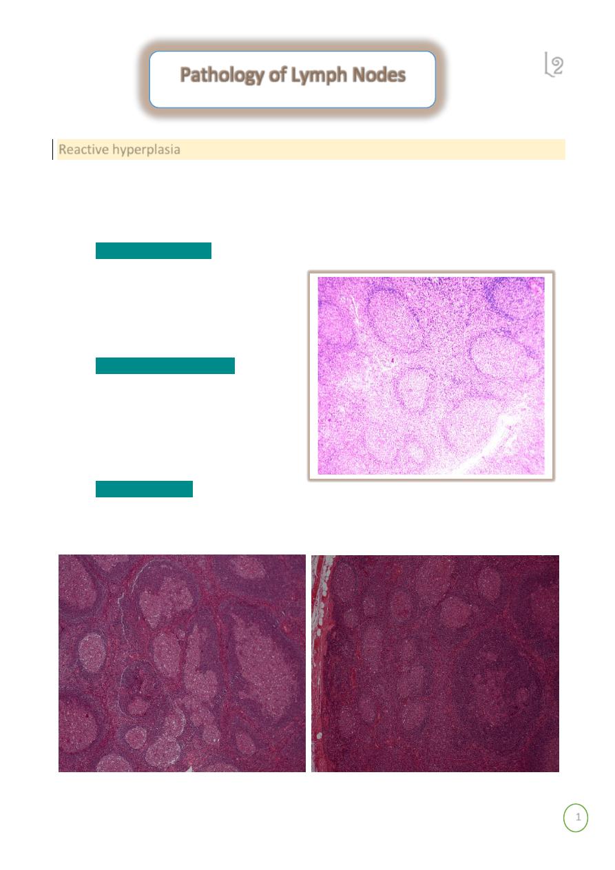
Reactive hyperplasia
Exaggerations of normal histology.
o Expansion of all regions or selective expansion
o Some types characteristic of certain diseases, but most not
Follicular hyperplasia
- increase in number and size of germinal centers, spread into
paracortex, medullary areas
o Collagen vascular diseases
o Systemic toxoplasmosis
o Syphillis
Interfollicular hyperplasia
- paracortex
o Skin diseases
o Viral infections
o Drug reactions
Sinus histiocytosis
- expansion of the medullary sinus histiocytes-
o Adjacent cancer
o Infections
Pathology of Lymph Nodes
2
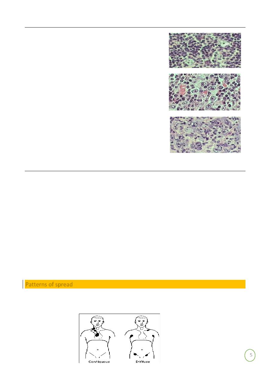
There is marked differences in size of germinal centers, their well-circumscribed character, and the
fact that they are surrounded by a well-defined mantle of lymphocytes
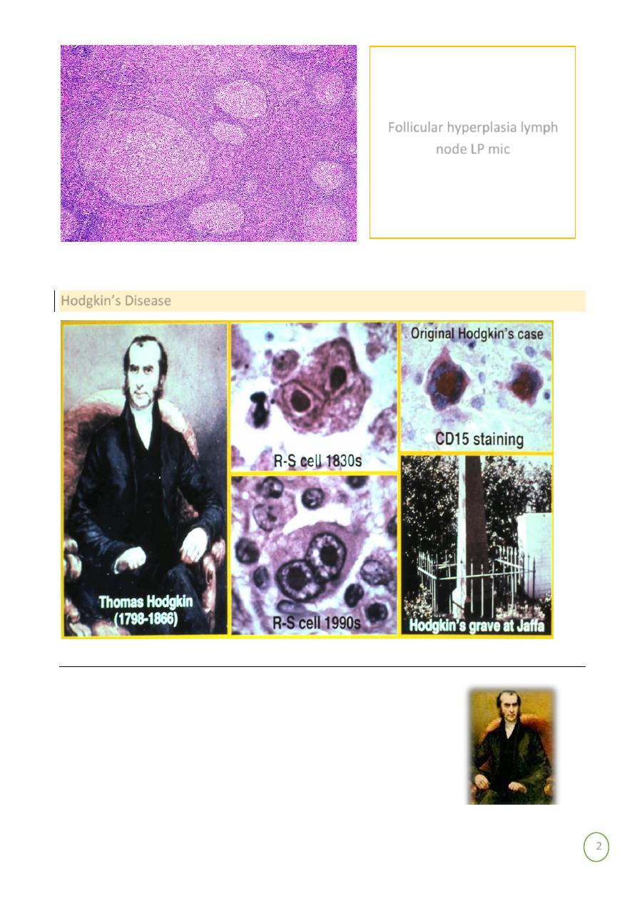
Hodgkin’s Disease
Hodgkin Lymphoma
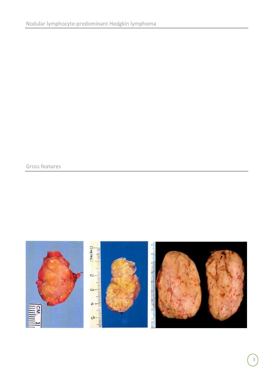
Nodular lymphocyte-predominant Hodgkin lymphoma
Classical Hodgkin lymphoma
Nodular sclerosis classical Hodgkin lymphoma
Lymphocyte-rich classical Hodgkin lymphoma
Mixed cellularity classical Hodgkin lymphoma
Lymphocyte-depleted classical Hodgkin lymphoma
Follicular hyperplasia lymph
node LP mic
3
Nodular lymphocyte-predominant Hodgkin lymphoma
Clinical features
ⱴ Peak age incidence in fourth decade of life
ⱴ Male predominance
ⱴ Most common presentation with enlarged cervical ,axillary lymph node
ⱴ Lymphadenopathy may be of long duration
ⱴ Most patient have stage 1 or stage 11disease
ⱴ Recurrence common
ⱴ 3-5% develop diffuse large B-cell lymphoma
ⱴ Neoplastic cell(L&H cell) often have multilobated nuclei (popcorn)
ⱴ CD45 positive
ⱴ CD20 ,CD79a positive
ⱴ CD15 negative, CD 30 negative
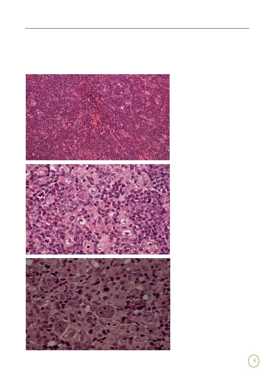
Gross features
ⱴ lymph nodes involved by Hodgkin's lymphoma are enlarged, sometimes massively
ⱴ So. The gross appearance depend on the microscopic subtypes The consistency
ⱴ varies from soft to hard depending on the amount of fibrosis
ⱴ Some degree of nodularity is often appreciated, particularly in the nodular sclerosis
ⱴ Foci of necrosis may be present the cut surface of the node has a more heterogeneous
ⱴ Appearance than most non-Hodgkin's lymphoma.
In advanced cases, several nodes from the same group
ⱴ become matted together
4
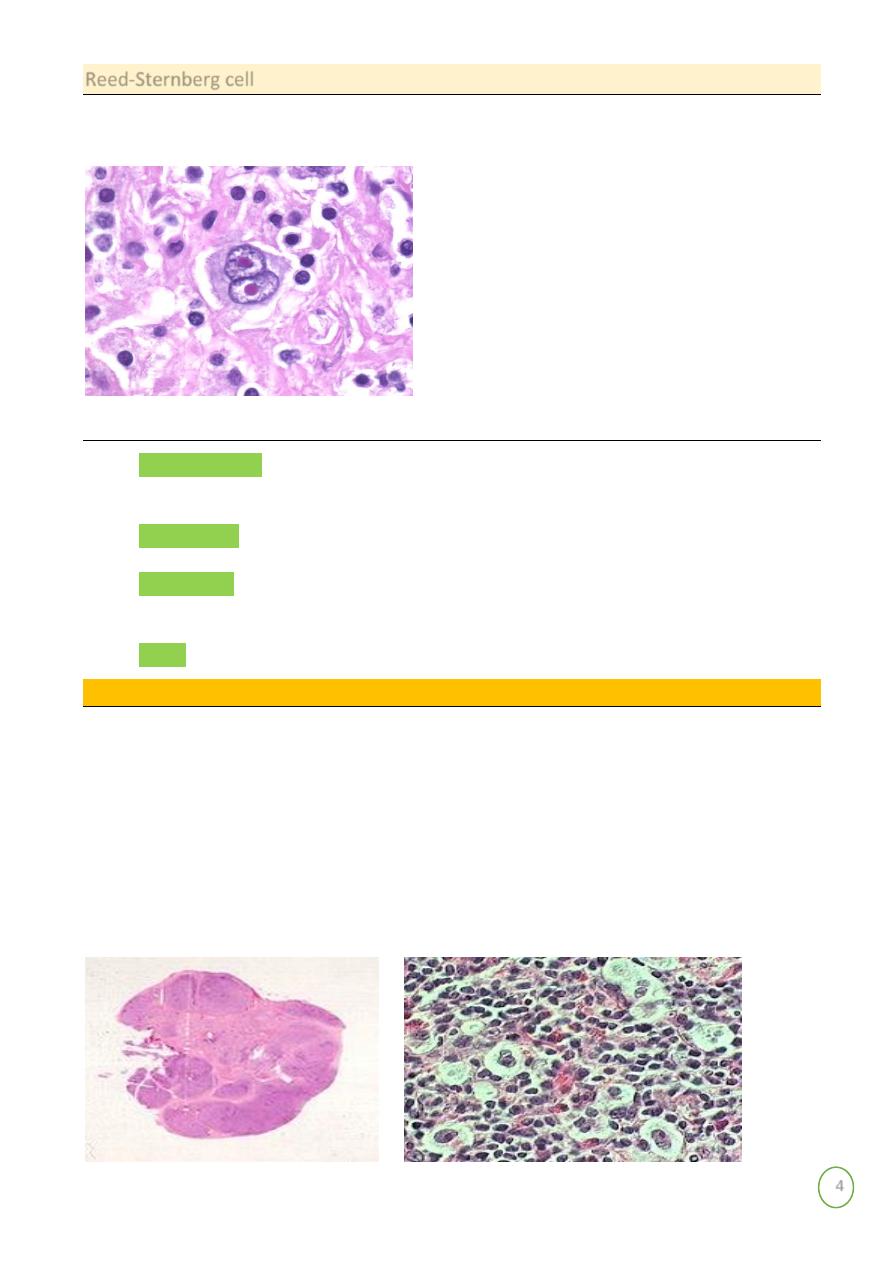
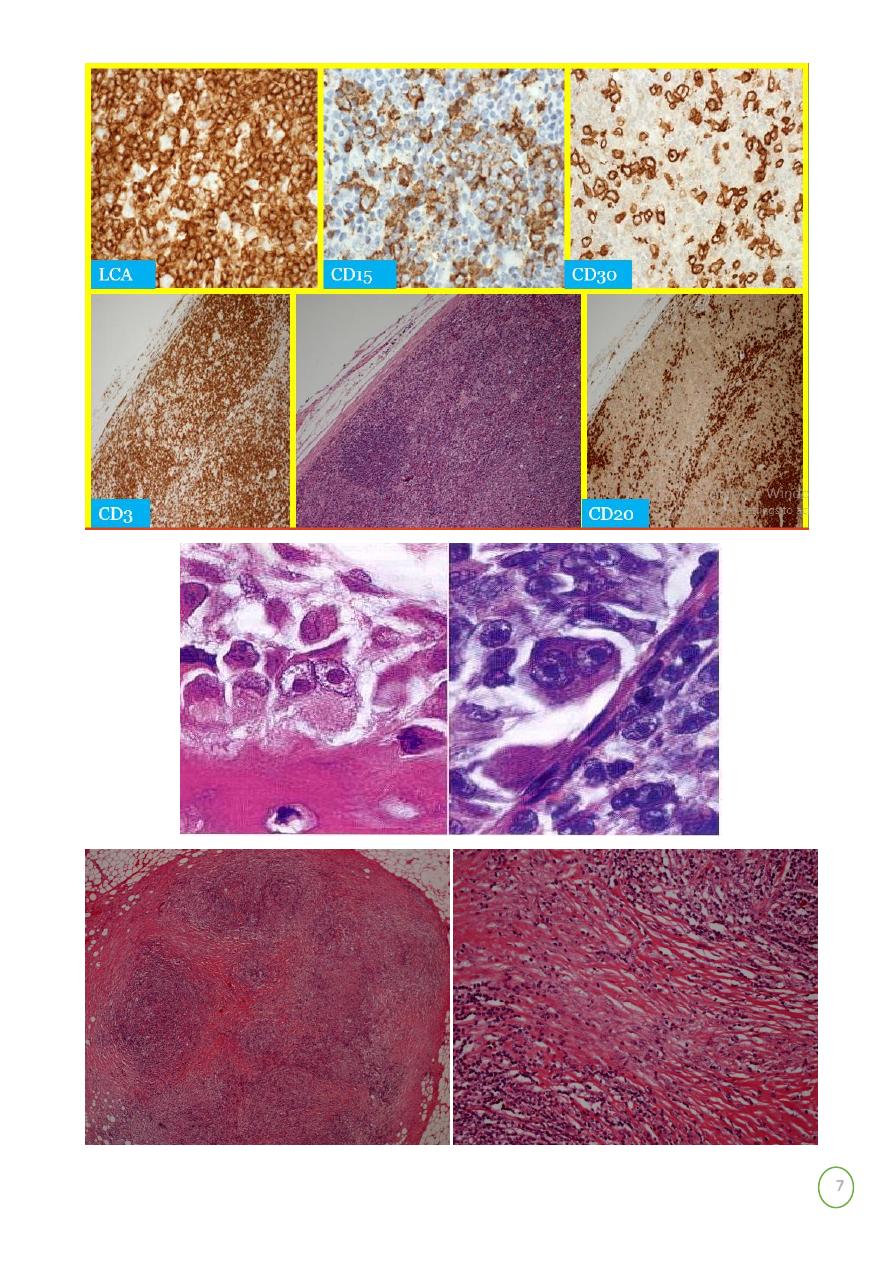
Reed-Sternberg cell
The classic Reed-Sternberg cell, as seen in all subtypes of classical Hodgkin's lymphoma, but
not in NLPHL.
CHD
Most important immunocytochemical profile of the Reed-Sternberg cell is:
ⱴ CD15 (Leu-Ml): This is expressed in over 80% of the cases; the pattern may be
paranuclear (corresponding to the Golgi region), diffuse cytoplasmic, and/or
corresponding to the cell membrane.
ⱴ CD30 (Ki-1): As recognized by the monoclonal antibody Be-Hz, this is found in
approximately 90% of the cases,
ⱴ CD45 (1CA): This is expressed in less than 10% of the cases.
ⱴ CD40 (a protein present in B cells and nerve growth factor receptor): This is expressed
in approximately 70%.
ⱴ CD74: This is expressed in over 75%.
Hodgkin's Histologic subtypes
ⱴ Are characteristic patterns of involvement, and characteristic variants of Reed
Sternberg cell associated with different subtypes
ⱴ Nodular sclerosing HL
o Most common type Hodgkin's lymphoma in US/Europe
o Usually presents in the anterior mediastinum and neck of young adult females
o Characterized by fibrotic capsule and bands subdividing tissue and
o
Lacunar
variant Reed Sternberg cell
5
Histologic Subtypes 2
ⱴ Lymphocyte predominant
o Usually presents with limited disease in the
neck of young adults
o Associated with L and H (lymphocytic and
histiocytic) or "popcorn cell" variant RS cell
ⱴ Mixed cellularity
o More extensive disease
o Older patients than NS and LP
o More R-S cells, eosinophils, plasma cells
o Mononuclear variant R-S cells
o more aggressive disease
ⱴ Lymphocyte depleted
o Often presents in retroperitoneum, older
patients
o Accompanied by loss lymphocytes, sclerosis
and pleomorphic RS cell variants
o Also more aggressive disease
General and clinical features
Hodgkin's lymphoma comprises approximately 20% to 30% of all malignant lymphomas in the
United States and Western Europe but a much lower percentage in Japan and other Oriental
countries. With a peak at 15 to 35years and a second, smaller peak in more than 50 years.
In poorly developed countries, there is a high incidence in children, a relatively low incidence
in the 15- to 35-year age group.
ⱴ The is a male predominant (approximately 1.5 to 1) in all microscopic types except
nodular sclerosis.
ⱴ The disease may present in a variety of ways
ⱴ The most common (approximately 90% of the cases) being
painless enlargement
of
superficial (usually cervical) lymph node
ⱴ
Fever, night sweats, and loss of weight
(so-called "B symptoms") occur in
approximately 25% of the cases; their presence influences the clinical staging
ⱴ Pruritus is also frequent.
Patterns of spread
ⱴ Hodgkin's lymphoma spreads contiguously via lymphatics
ⱴ Staging as in NHL- may or may not include laparotomy/splenectomy
6
Prognosis
ⱴ Hodgkin's lymphoma is a curable malignancy
ⱴ Overall cure rate approximately 80%
ⱴ With modern therapy, prognosis based more on staging, bulk of disease, than
morphologic subtype
M. 63Y. PELVIC AND
GROIN
LYMPHADENOPATHY
M.57Y. ISOLATED
CERVICAL
LYMPHADENOPATHY
F. 41Y.CERVICAL
LYMPHADEOPATHY AND
SPLEENOMEGALY
7
8
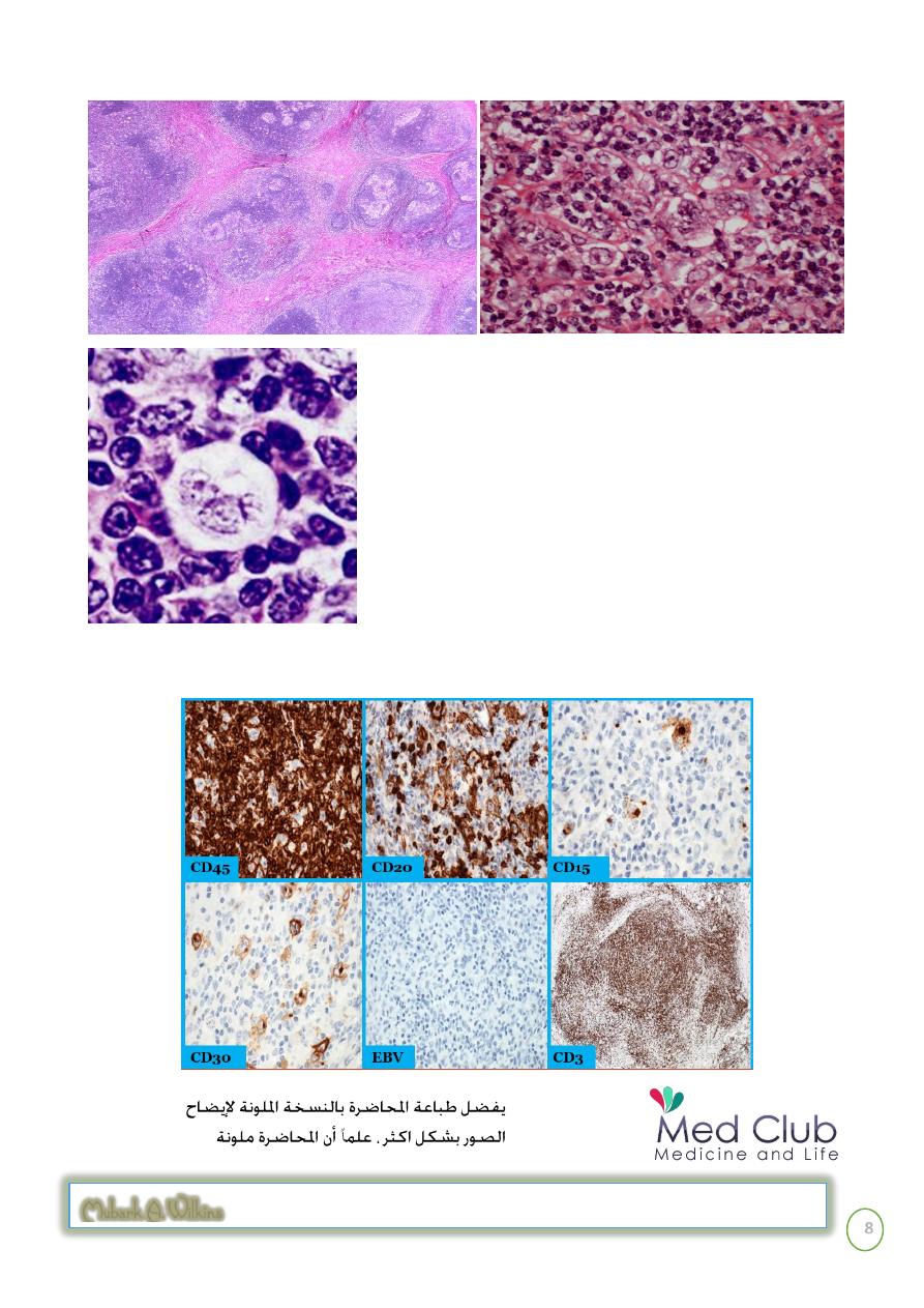
large cells with a surrounding prominent clear space, an artefact of formalin fixation. These are the
lacunar cells characteristic for the nodular sclerosis type of Hodgkin's disease.
Mubark A. Wilkins
Hodgkin's disease, nodular
sclerosis type, lacunar cell
ⱴ