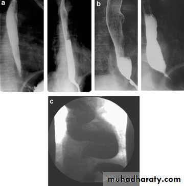
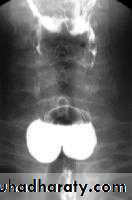
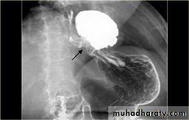
A chalasia Cardia
A : AbsenceChalasia : Relaxation
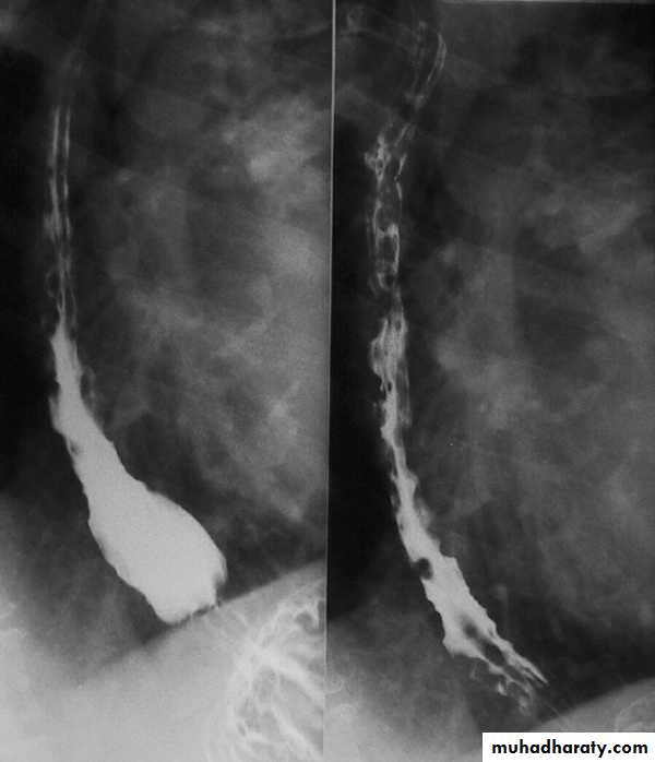
Narrowing :
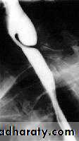
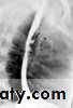
1-the narrowing is Constant Short length (confined to cardia).
2-Regular and smooth.
3- No shouldering sign.
4-Tapering (Tip of pencil , cigar shape) Under left dome of diaphragm.
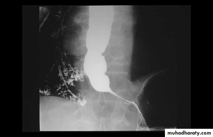
Achalasia continue
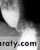
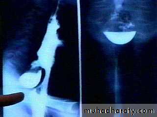
5. DILATATION (Sac like in proximal part )6-Undulating or spiky out line due to sluggish peristalsis.
7 Non- homogeneity of Barium due to food particles.
8-Air Barium level.
9- CXR shows widening of mediastinum.
10-Absence of fundal gas shadow.
7-Basal fibrosis in lungs due to repeated aspiration pneumonia .
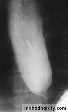
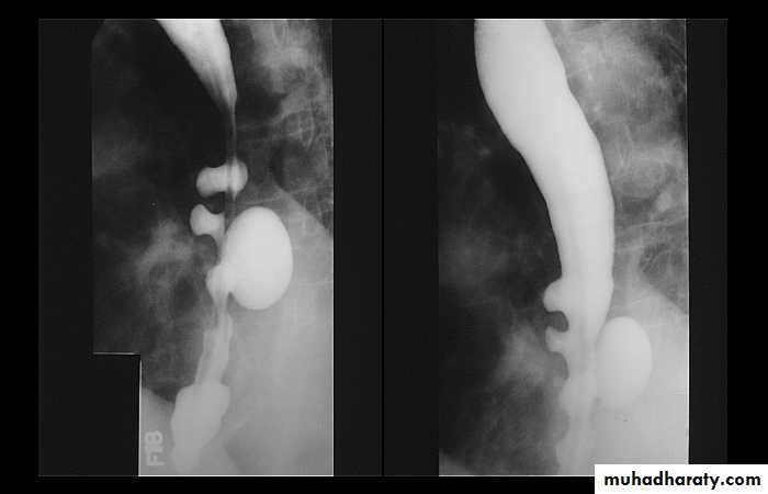
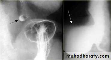
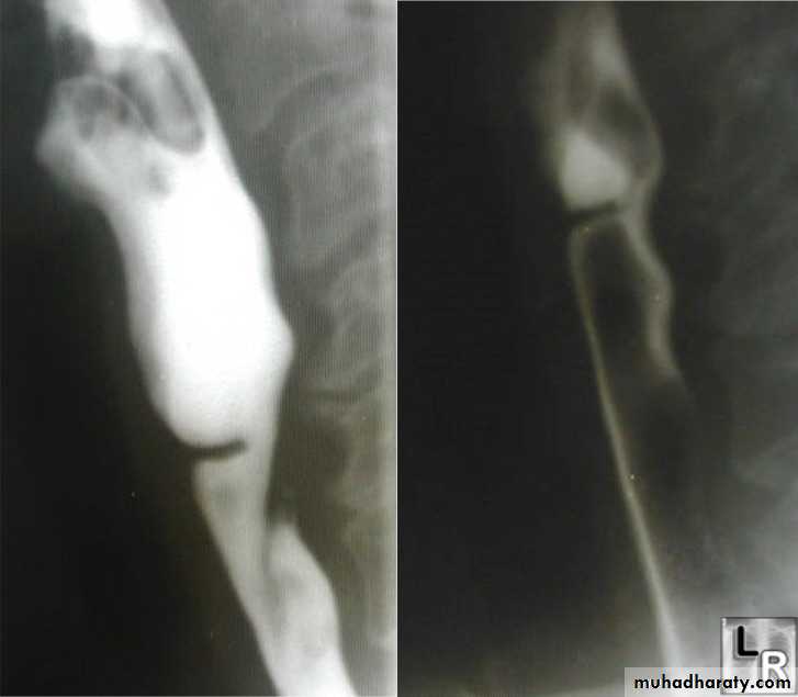
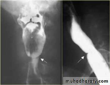
LEFT: Dilated esophagus (arrows) appears as long, well-defined structure paralleling heart RIGHT: Dilated esophagus usually deviates to right. Narrowing (arrow) at hiatus.
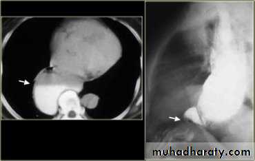
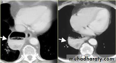
LEFT: CT shows dilated esophagus (arrow) that led to esophagram.RIGHT: Esophagram shows narrowing (arrow) at level of hiatus.
• z
PULSION DIVERTICULUM• Due to raised intra-luminal tension
• 2- Chocking after meal .
• 3- In cervical portion at level of C5
• 4- Posteriorly (Killience dehiscent)
• 5- Lateral view show increased pre-vertebral space with air fluid level.
• 6- Confirmed by Ba. Swallow.
TRACTION DIVERTICULUM
Out pouching of lumen laterally due to fibrosis & adhesions( post-Tb.)
2-In the middle third at level of hilum
3- Up ward direction of diverticulum
4- Irregular base
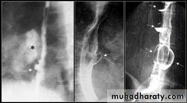
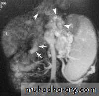
On the far left a traction diverticulum (arrow) due to hilar granulomatous disease. Calcified adenopathy (asterisk). In the middle a pulsion diverticulum (arrow) due to high intra luminal pressure.On the right multiple pulsion diverticula (arrows)
CONGENITAL DIVERTICULUM
1-Asymtomatic unless complicated.2-At lower part of esophagus above the diaphragm (Epi-phrenic)
3- Lateral or posterior in position.
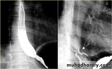
Sliding herniaOn the left initially, GE junction is below the esophageal hiatus. Later, stomach protrudes through hiatus
Para esophageal hernia
On the far left gas filled gastric funds (asterisk) protrudes through hiatus but GE junction (arrow) is below diaphragm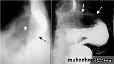
• Thin mucosal fold (membrane)
• 2- Arise from anterior wall and extend Posteriorly .• 3- Lateral view Ba. Swallow show self like filling defect with proximal dilatation.
• 4-Single or multiple.
ESOPHAGEAL WEB
10% incidence at autopsy
Can be congenital or acquiredMost in hypopharynx and proximal esophagus
Majority protrude from anterior esophageal wall
Symptoms if lumen > 50% compromised
Sideropenic dysphagia (Plummer-Vinson syndrome)
Iron deficiency anemia
Esophageal web with dysphagia
Increased incidence of carcinoma
Validity of syndrome debatable
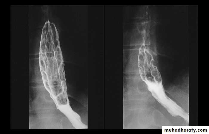
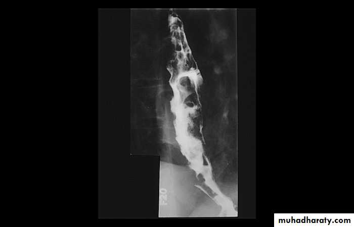
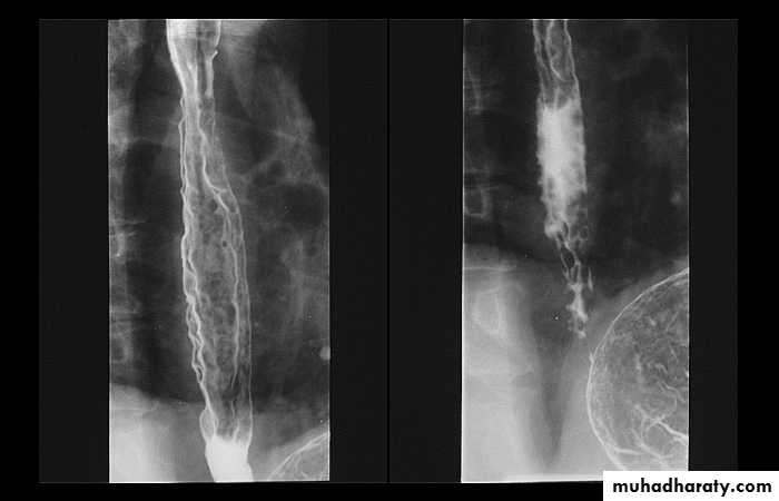
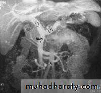
1-Dilatation of venous plexus in the wall of the esophagus due to increased pressure ( portal H.T.).
2-Important cause of Hematemesis .
3-Early changes seen in the mucosa (D.C.) loss of parallelism with thick and tortuous folds.
4-Later multiple small filling defects (fine cobble stone).
5-In advanced stage large filling defects ( coarse cobble stone ) .
6- More advanced stage elongated and worm like filling defect .
7-The changes are seen at lower third and gastric fundus.
Esophageal Varieces