Vision
- Visible light is a frequency range of the electromagnetic radiation which evokes light sensation in the eye.
- For humans, the wavelength of visible light is between 380 nm and 780 nm.
- Vision is the detection and perception of these electromagnetic radiations.
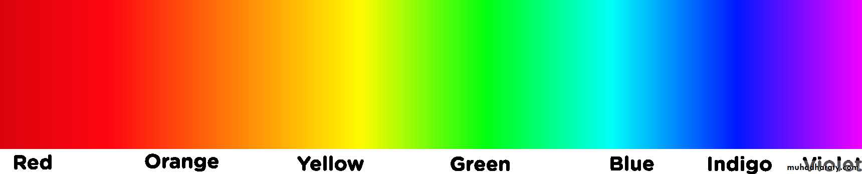
- Sunlight contains waves with all the different wavelengths which make up visible light.
- These light waves with different wavelengths elicit different colour sensations in the eye.
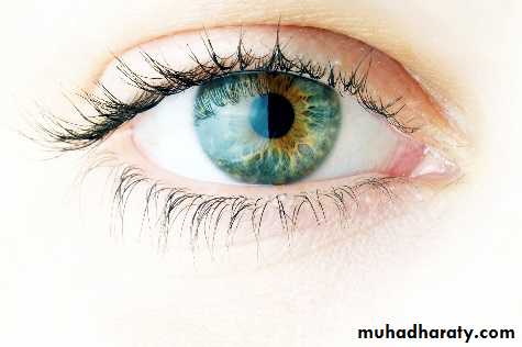
How the Human Eye Works?
In a number of ways, the human eye works much like a digital camera:
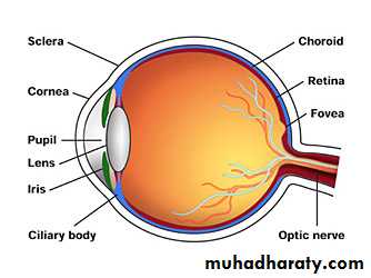
Light is focused primarily by the cornea — the clear front surface of the eye, which acts like a camera lens.
The iris of the eye functions like the diaphragm of a camera, controlling the amount of light reaching the back of the eye by automatically adjusting the size of the pupil (aperture).
The eye's crystalline lens is located directly behind the pupil and further focuses light. Through a process called accommodation, this lens helps the eye automatically focus on near and approaching objects, like an autofocus camera lens.
Light focused by the cornea and crystalline lens (and limited by the iris and pupil) then reaches the retina — the light-sensitive inner lining of the back of the eye.
- The retina acts, like an electronic image sensor of a digital camera, converting optical images into electronic signals.
- The optic nerve then transmits these signals to the visual cortex — the part of the brain that controls our sense of sight.
Retina
- The photoreceptor cells of the retina are the rods and the cones.
a. The cones
- responsible for photopic vision that is for color vision in daylight.
- Cones may belong to three groups according to their photopigment type.
- “green” cones: (M-cones from medium wavelength)
have an absorption maximum of about 530 nm
- “Red” cones: (L-cones from long wavelength)
most sensitive to light with a longer wavelength (560 nm),
i.e. with yellow-red colour,
- “blue” cones: (S-cones from short wavelength)
most sensitive to light with a short wavelength (420 nm),
i.e. with violet-bluish colour.
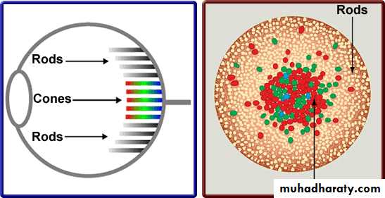
b. The rods
- responsible for scotopic (twilight/night) vision.- distributed mostly on the periphery of the retina.
- As all rods contain the same photopigment (rhodopsin), they cannot
distinguish colours.
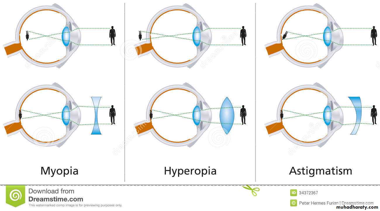
Myopia and Hypermetropia
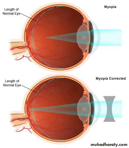
a. Myopia (Short Sighted):
- The term used to define short sightedness.
- Light from a distant object forms an image before
it reaches the retina, which could be due to:
- the eye is too long, or
- the cornea or crystalline lens is too strong.
- A myopic person has clear vision when looking at
objects close to them, but distant objects will appear
Corrected by
Corrected by
blurred
A concave lens (minus powered) is placed in front of a myopic eye, moving the image back to the retina and clarifying the image.
A concave lens (minus powered) is placed in front of a myopic eye, moving the image back to the retina and clarifying the image.
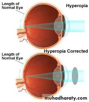
b. Hypermetropia (long sight)
- The sight where the image of a nearby object is
formed behind the retina, this could be because
- the eye is too short, or
- the cornea or crystalline lens does not refract the
light enough.
- A hypermetropic person may have blurred vision when looking at objects close to them, and clearer vision when looking at objects in the distance.
Corrected by
Corrected by
a convex (plus powered) lens is placed in front of
a hypermetropic eye,. the image is moved forward and focuses correctly on the retina
c. Astigmatism
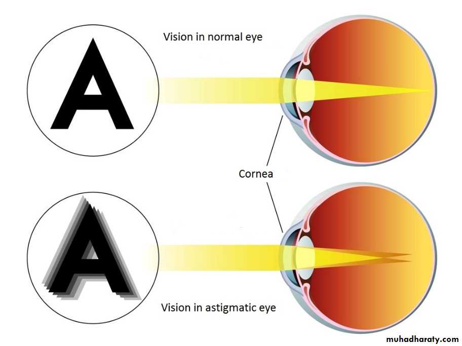
c. Astigmatism
- Astigmatism is the optical term for more than one point of focus.
- It occurs when the surface of the cornea or crystalline lens is not spherical.
Light from an object does not focus exactly
on the retina but at two separate points.
The eye is unable to focus a point or object
into a sharp focused image on the retina- There are two types of astigmatism, regular and irregular.
- Regular astigmatism arising from either the cornea or crystalline lenscan be corrected by a toric lens.
- Irregular astigmatism is often caused by a corneal scar or scattering in the crystalline lens and cannot be corrected by standard prescription lenses, but may be corrected by contact lenses or mild astigmatisms can be treated by laser eye surgery.
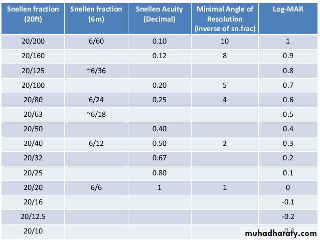
E or C Snellen chart for measuring visual acuity
- It is used for distance vision testing- aid examination and diagnosis of eye disease or refractive error
Instructions before the test:
- Ensure good natural light or illumination on the chart
- Test each eye separately – the ‘bad’ eye first
- Position the patient, sitting or standing, at a distance of 6 meters from the chart
. The accuracy will be read as a ratio: (VA)
Procedure- Ask the patient to wear any current distance spectacles, to cover one eye with his/her hand (or with a plain occluder), and to start reading from the top of the chart
- The smallest line he/she can read (the visual acuity (VA)) will be expressed as a fraction, e.g. 6/18 or 6/24 (usually written on the chart).
Distance from the chart
. This ratio equal to =
Distance from where a normal eye can read
- if the patient cannot read the largest (top) letter at 6 metres, move him/her closer, one metre at a time, until the top letter can be seen – the VA will then be recorded as 5/60 or 4/60, etc.
- If the top letter cannot be read at 1 metre (1/60), hold up your fingers at varying distances of less than 1 metre and check whether the patient can count them
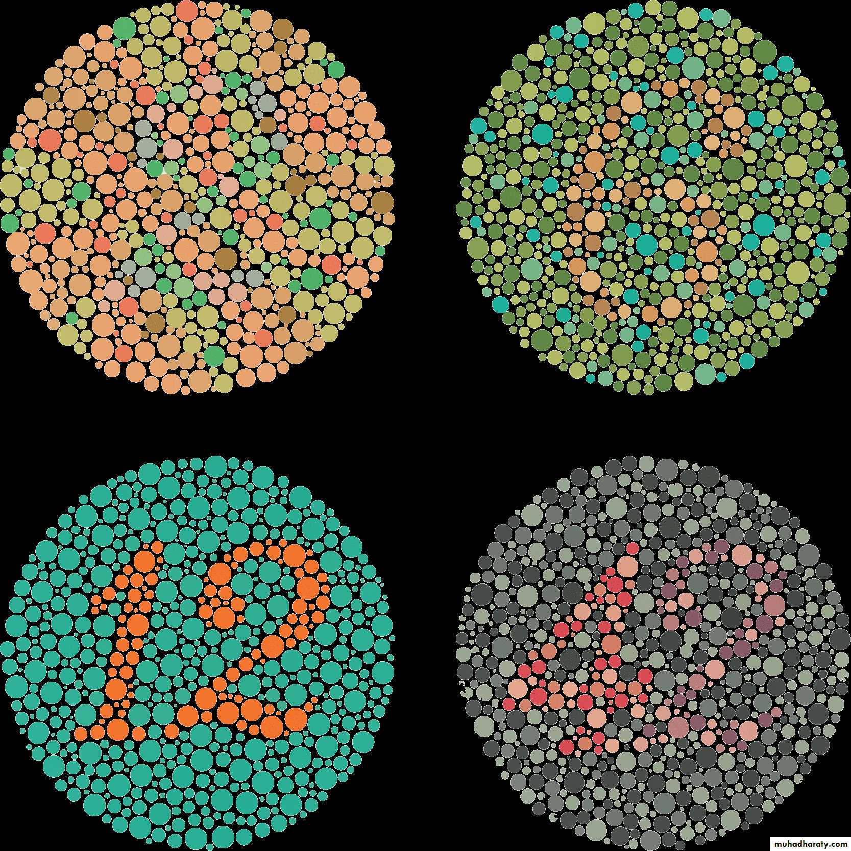
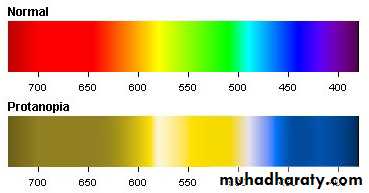
Colour Blindness
- There are several types of inherited colour blindness.
- Normal colour vision uses all three types of light cones correctly and is known as trichromacy. People with normal colour vision are known as trichromats.
A- Protanopia:
- have difficulties to distinguish between blue and green colors and also between red and green colors.- have either defective long-wavelength cones (L-cones) or
the L-cones are missing at all. If they are missing it is called protanopia or
sometimes red-dichromacy.
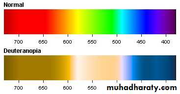
B- Deuteranopes: of two types
1. Dichromats: Deuteranopia (also called green-blind).- In this case the medium wavelength sensitive cones (green) are missing
at all.
2. Anomalous Trichromats: Deuteranomaly (green-weak). This can be
everything between almost normal color vision and deuteranopia.
. The green sensitive cones are not missing in this case, but the peak of
sensitivity is moved towards the red sensitive cones.
3-Tritanopes
- People affected by tritan color blindness confuse blue with green and yellow with violet.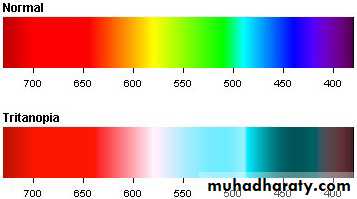
Colour Vision Testing Chart