Antibody-Antigen Reactions
Introduction. Microorganisms can be identified by:
Morphology
Isolation and culturing of causative M.OsDetection of Ags or meaningful Abs titre by different serological tests
Nuclear acid detection whether bacterial or viral DNA e.g. by PCRAntibody-Antigen Reactions
Antibody-antigen complexes can be detected by differentserological methods including:
1. Precipitation tests : Antibodies react with soluble antigens. A. Single radial immunodiffusion (Mancini).
B. Double immunodiffusion (Ouchterlony).
C. Immunoelectrophoresis (IEP).
2. Agglutination tests: Antibodies react with insoluble antigens.
A. Latex agglutination test.
B. Agglutination of bacterial antigen.
C. Haemagglutination.
3. Labeled Antibody serology like ELISA and RIA
Principle
1. Gell is incorporated with an antibody against the antigen to be detected (e.g anti-x if we want to detect x antigen)2.Precipitation ring is resulted from the reaction between
the Ab incorporated into gell and Ag of the sample.3. Square diameter of the ring is proportional to the
concentration of Ag.Precipitation tests
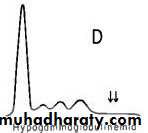
Single radial immuno-diffusion (Mancini test)Single radial immuno-diffusion (Mancini test)

Applications
1. Measure the concentrations of immunoglobulins e.g IgM,IgD etc ….2. Measure the concentrations of complements.
3. Measure the concentrations of any antigen when its antibody is avaliable as albumen, alpha-fetoprotein and alpha -1- antitrypsin.
Ring diameter (mm)
Ag mg/ml concentrationS
S/2
S/4
S /8
S/16
? 2
?1
?
?= Unknown conc.
1
2
Abs
Double immunodiffusion ( Ouchterlony)
Principle
Samples containing antigen and antibody are placed in opposing
wells to diffuse toward one another in a moist chamber for 18-24
hours. The resultant precipitation lines that represent antigen-
antibody complexes are analyzed visually. Three types reactions
can occur.
1. Reaction of identity.
R= antigen.
αR= antibody.
R
R
α R
2. Reaction of nonidentity.
R, S=antigen.α R=antibody to R antigen.
α S=antibody to S antigen.
3. Reaction of partial identity.
R=antigen.
R1=antigen.
α R=antibody to both R
R1 antigens.
α R
α SS
R
α R
RR1
Immunoelectrophoresis (IEP)
The IEP combines both electrophoretic separation andimmune precipitation of proteins.
Proteins are separated in an electrical field according to their charge. e.g. Albumen is of negative charge so it migrate to the positive pole (anode) while Igs are of positive charge so migrate to the negative pole(cathod)
antiserum
serum
serum
antiserum
serumserum
+ ve
(anode)- ve
(cathod)
Applying the electrical field)
Trough
Alb
α
β
γ
Alb
α
β
γ
Cont/...Precipitin lines
Applications of IEP
1. Determination of the exact H &L chains of paraproteins
2. Distinguishing polyclonal from monoclonal increase in γ-
globulins.
3. Diagnosis of panhypogammaglobuliemia (decrease in IgG, IgA,
and IgM).
4. Identifying L chains in the urine of patients with plasma cell
dyscrasias.


MM (multiple myeloma)
Hypogammaglobulinemia

Polyclonal and monoclonal gammopathies
Normal
Agglutination tests
1. Agglutination of RBC2. Agglutination of latex particles
3. Agglutination of bacterial AgsAgglutination tests: Agglutination of RBCs (Haemoagglutination)
Direct (Simple)Indirect (Passive)

Blood grouping
AB
AB
O
Viral haemoagglutination
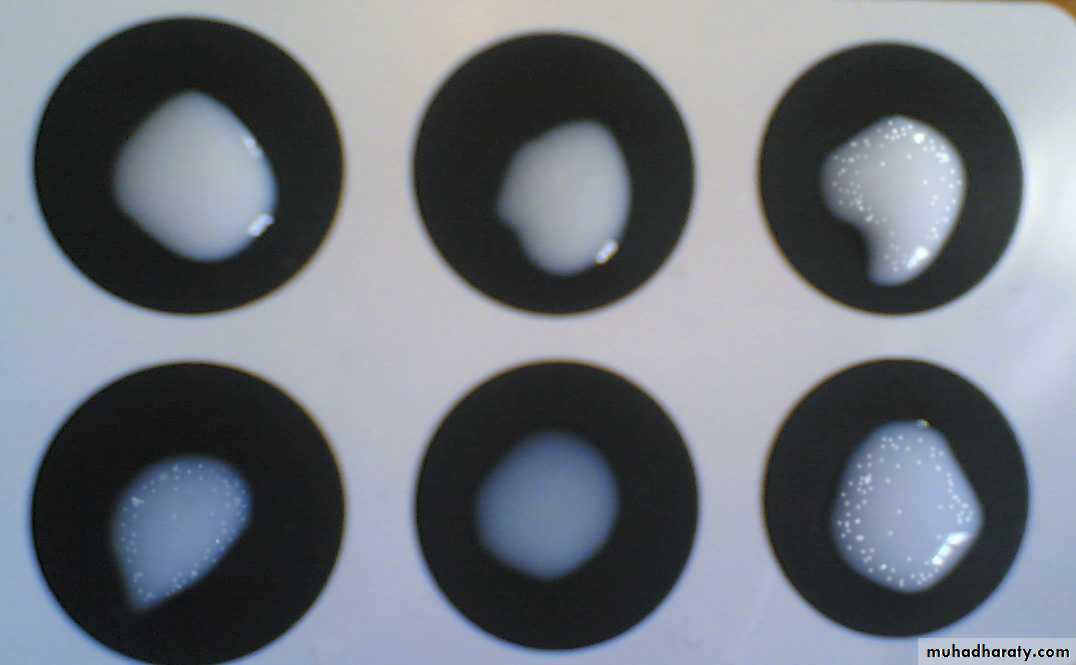
Agglutination tests: latex agglutination test
Principle:
latex particles are coated with antigens in order to detect the presence of antibodies and vice versa.
Slide method:
•
• • •
• • •
• •
• • • •
•
•
•
•
•
- ve
+ve
Applications:
1. Detection of Rheumatoid factor (IgM).
2. Detection of ANF (anti-nuclear factor).
3. Pregnancy test (detection of HCG)
1
2
3
6
5
4
+
+
+
Agglutination of bacterial Ags: Brucella agglutination testWidal test
Two methodsSlide agglutination method
Tube dilution method
Slide agglutination test

Agglutination of bacterial Ags: Widal test: Tube agglutination method
Serial dilutions of the serum from 1/20 to 1/640.Add one drop of salmonella Ag.
Incubate and examine for agglutination at the bottom of the test tube.
The last tube showing sign of agglutination should be taken as the titer for the test.
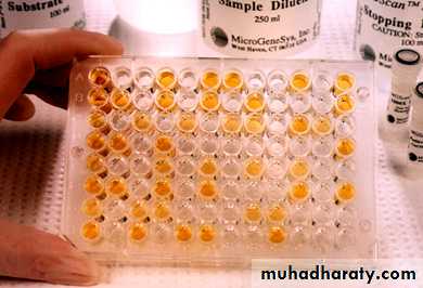
Labelled Antibody serology: Enzyme-Linked Immunosorbent Assay (ELISA)
The ELISA is a common serological test for the presence of particular antigens or antibodies, there are two forms of this assay:1. Direct ELISA employs monoclonal antibodies to detect
the presence of a particular antigen in a sample.
2. Indirect ELISA is used to determine the presence of a
specific antibody ( e.g., HIV antibodies) in a specimen such
as serum.


Y Y Y Y Y Y
In the Direct ELISA method, a specific monoclonal antibody is first attached onto the walls of a microtiter plateA suspension of serum or other fluid is added to the well to test for the presence of a complementary antigen. In this example, the sample on the left contains the complementary antigen whereas the sample on the right does not.
If the antigen is present in the specimen, it will bind to the antibodies that are attached to the wall of the well.
Binding of the antigen to the antibody is strong enough to washstand rinsing that removes the test fluid and unbound antigen.
After rinsing to remove unbound antigen, another aliquot of the monoclonal antibody is added to the well. The antibodies in this aliquot are modified so that they carry what is known as a reporter enzyme. A reporter enzyme is designed to produce a color change when the enzyme reacts with its substrate.
The sample is again rinsed to remove any unbound antibodies. If the antigen is present, a complex will have formed that includes the antibody bound to the well, the antigen, and the enzyme-conjugated antibody. If the antigen is not present, the enzyme-bound antibody will have been washed away.
The enzyme’s substrate is now added. A color change reveals the presence of enzyme–labeled antibody as well as its bound antigen. No color change indicates that antigen was not present in the tested fluid.
Direct ELISA
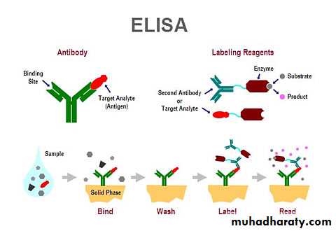
The indirect ELISA test is one that determines whether a specific
antibody is present in a sample such as serum. In this case, theappropriate antigen is first absorbed to the walls of a microtiter
plate.
Serum that might contain antibodies against the antigen is added
to the well. If the antibodies are present in the sample, they will
bind to the antigens that are absorbed to the wall of the well. If
the sample contains only non –specific antibodies, they will not
bind to the antigen.
Indirect ELISA

Thank you