Histology:2nd stage
2016-2017Department of Anatomy &Histology Dr. Raja Ali
--------------------------------------------------------------------------------------------
Nerve Tissue & the Nervous system II:
Myelinated and Unmyelinated Axons:
In the PNS, all axons are enveloped by Schwann cells which provide both structural and metabolic support.
Many axons with small diameter invaginate into one Schwann cell longitudinally and are simply surrounded by the cytoplasm of Schwann cells. They are called unmyelinated nerve fibres.
Other axons, especially the ones with larger diameter, invaginate into the Schwann cell and are wrapped by concentric layers of the Schwann cell plasma membrane forming myelin sheath. These axons are called myelinated nerve fibres (Fig.1a ).
There are gaps (areas of axon not covered by myelin) along the length of myelin sheath at regular intervals called nodes of Ranvier.
In large myelinated axons, the nerve impulse jumps from node to node resulting in faster conduction (salutatory conduction).
The segment of myelin between two nodes of Ranvier is called internode.
The myelin of one internode is formed by a single Schwann cell.
The myelin sheath shows cone-shaped clefts called Schmidt-Lantermann clefts. They are areas of remnants of cytoplasm of Schwann cells present within the myelin sheath.
In the PNS, the myelin sheath of an individual axon is provided by many Schwann cells lying along the length of the axon.
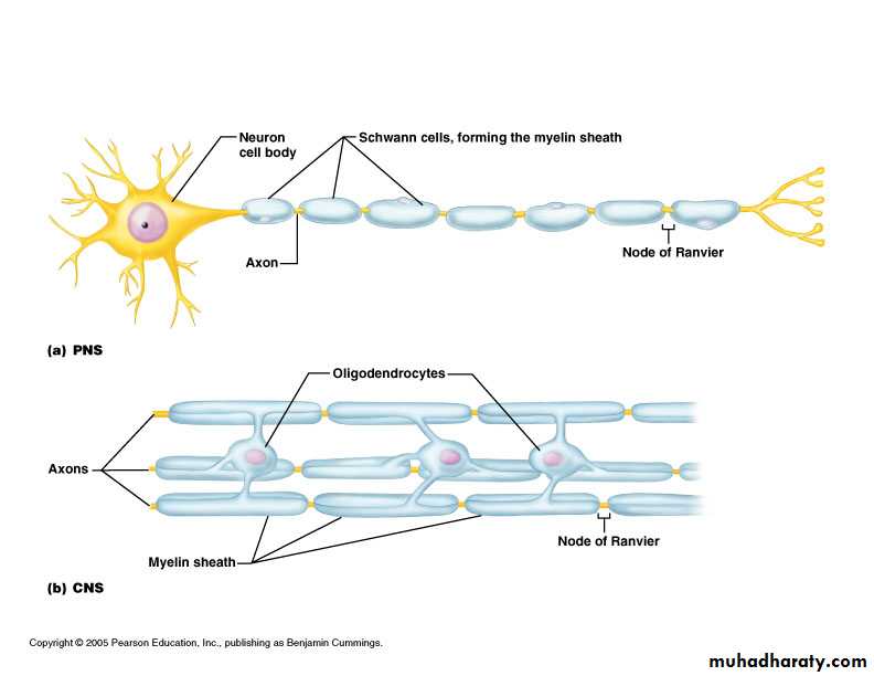
In the CNS, the myelin sheath is formed by processes of oligodendrocytes Fig.(1b) .
Fig.(1 ):Myelin in the Peripheral and Central Nervous Systems
Myelination:
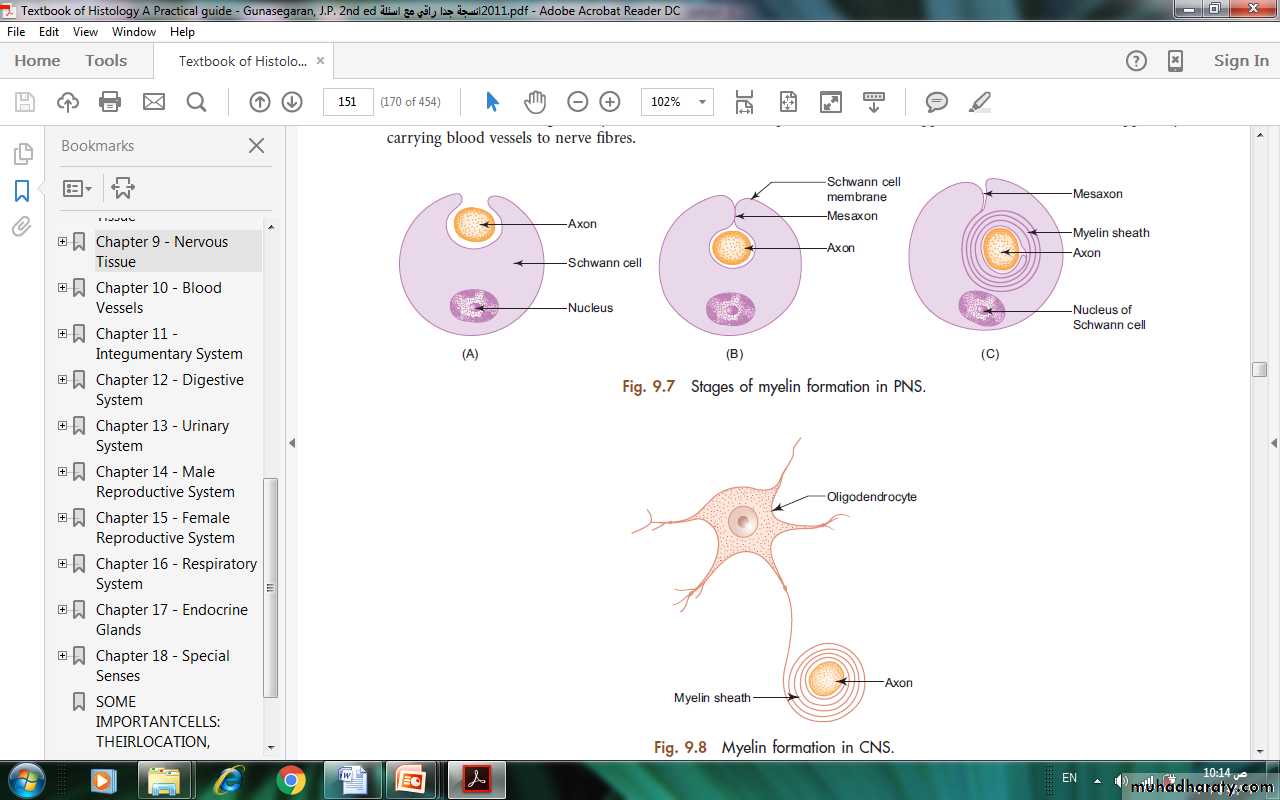
Myelination begins with the invagination of the axon into the Schwann cell. The invaginated axon is suspended from the periphery of the cell by a fold of fused plasma membrane called mesaxon (Fig.2a).
As myelination proceeds the Schwann cell and mesaxon rotates itself around the axon several times resulting in enveloping the axon in concentric layers of Schwann cell cytoplasm and plasma membrane alternately (Fig.2b&2c).
With further rotation cytoplasm between the concentric layers of plasma membrane is squeezed out and the opposing inner surfaces of the plasma membrane fuse with each other forming myelin sheath.
Thus myelin sheath is actually composed of many layers of modified cell membrane of Schwann cell.
Fig.(2): Stages of myelin formation in PNS.
Peripheral Nerve:Each peripheral nerve (spinal or cranial) is made of bundles (fascicles) of nerve fibres (axons) which may be myelinated and/or unmyelinated.
The bundles are held together by connective tissue which provides structural support as well as nutritional support by carrying blood vessels to nerve fibres.
The connective tissue framework is well appreciated in cross section of a nerve (Fig. 3&4 ), where following structures can be observed:
Epineurium: Dense connective tissue sheath surrounding the entire nerve.
Perineurium: A sleeve of fl attened specialised epithelial cells surrounding the bundles of nerve fi bres.
Endoneurium: Loose connective tissue composed of reticular fibers supporting individual nerve fibers.

In the case of optic nerve, it is surrounded by meninges of brain.
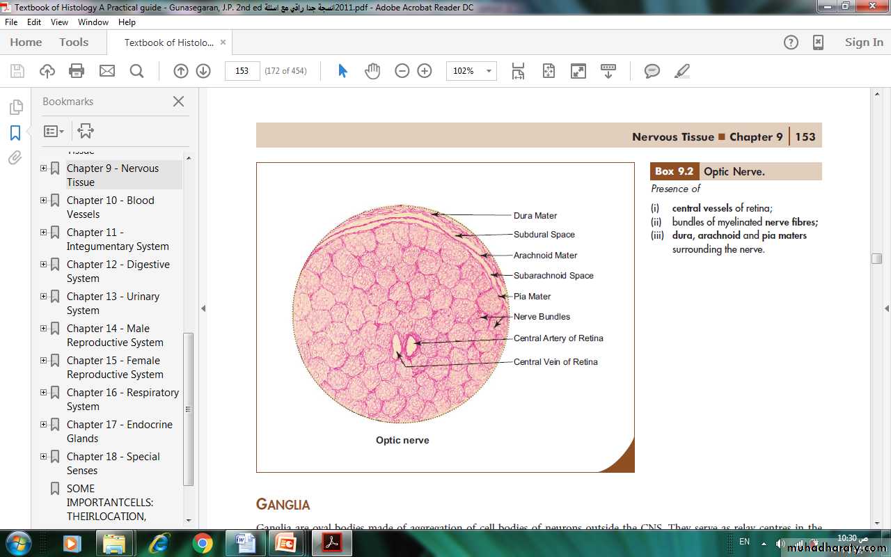
Fig.(3): Peripheral nerve.
Fig.(4): Optic nerve.
Ganglia:
Ganglia are oval bodies made of aggregation of cell bodies of neurons outside the CNS.
They serve as relay centres in the neuronal pathway.
They are usually covered by a dense connective tissue capsule known as epineurium.
The cell bodies of the neurons are enveloped by a layer of cuboidal cells called satellite cells.
Two types of ganglia can be distinguished on the basis of morphology and function; sensory and motor ganglia (Fig.5&6 ). Their distinguishing features are enumerated in table (1):
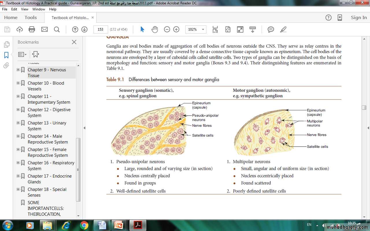
Table (1)::Differences between sensory and motor ganglia.
Fig.(6): Sympathetic ganglion.
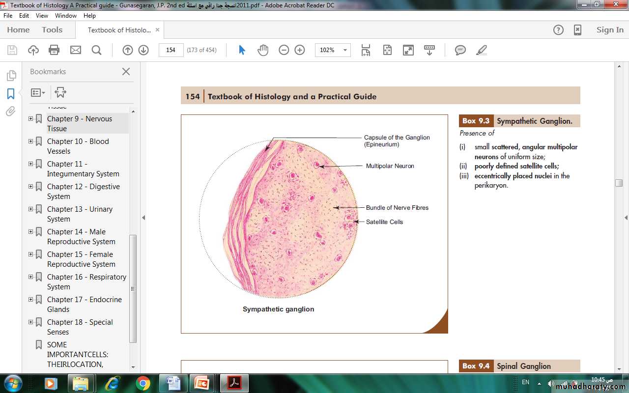
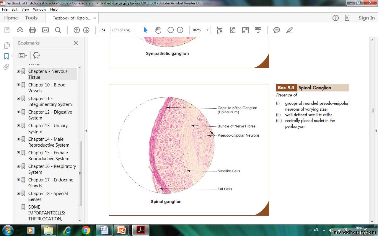
Fig.(5): Spinal ganglion.
Neuroglia (in CNS):
There are six kinds of glial cells
Neuroglia are highly branched cells that support the neurons by occupying the spaces between them.
Providing both structural and metabolic support.
There are four principal types of neuroglia in the CNS; namely astrocytes, oligodendrocytes, microglia and ependymal cells.
Of the four types, ependymal cells form a specialized simple low columnar epithelium which lines the ventricles of brain and central canal of spinal cord. The epithelium lacks a basement membrane.
The characteristic features and functions of the other three neuroglial cells are presented in Table (2):
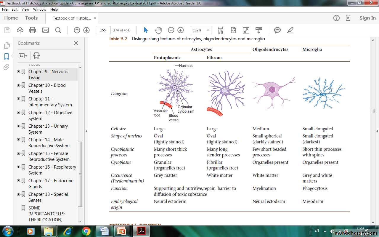
Table (2): Distinguishing features of astrocytes, oligodendrocytes and microglia
There are two type of neuroglia in the PNS:
Shwann cells &stellate cells
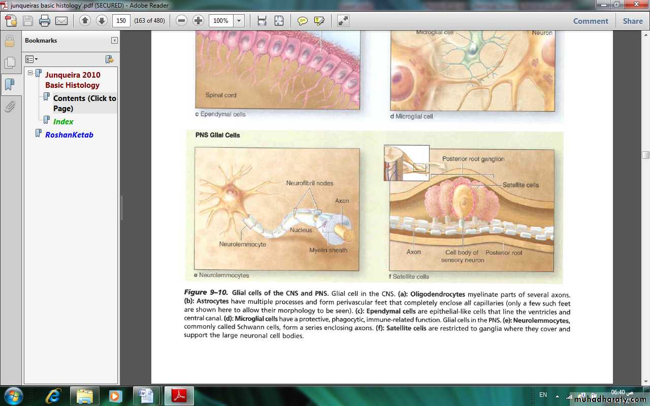
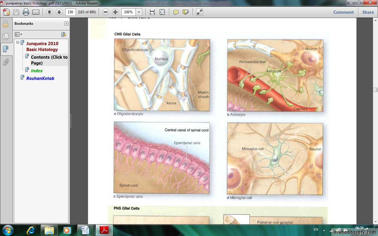
Figurc (7). Glial cells of the CNS and PNS
The principal functions of neuroglia are summarized in table (3)Table (3): Principal functions of neuroglial cells.
Glial Cell TypeLocation
Main Functions
Oligodendrocyte
Central nervous system
Myelin production, electric insulation.
Neurolemmocyte
Peripheral nerves
Myelin production, electric insulation.
Astrocyte
Central nervous system
Blood-brain barrier, metabolic exchanges.
Ependymal cells
Central nervous system
Lining cavities of central nervous system.
Microgl ia
Central nervous system
lmmune-related activity
Satellite Cells
Peripheral nerves
Supportive role.