Dr.
Shayma’a Jamal Ahmed
Prof. Genetic Engineering
& Biotechnology

At the end of this lecture the student will be able to:
Define the
Nucleic Acids.
Recognize to the DNA.
Recognize to the RNA.
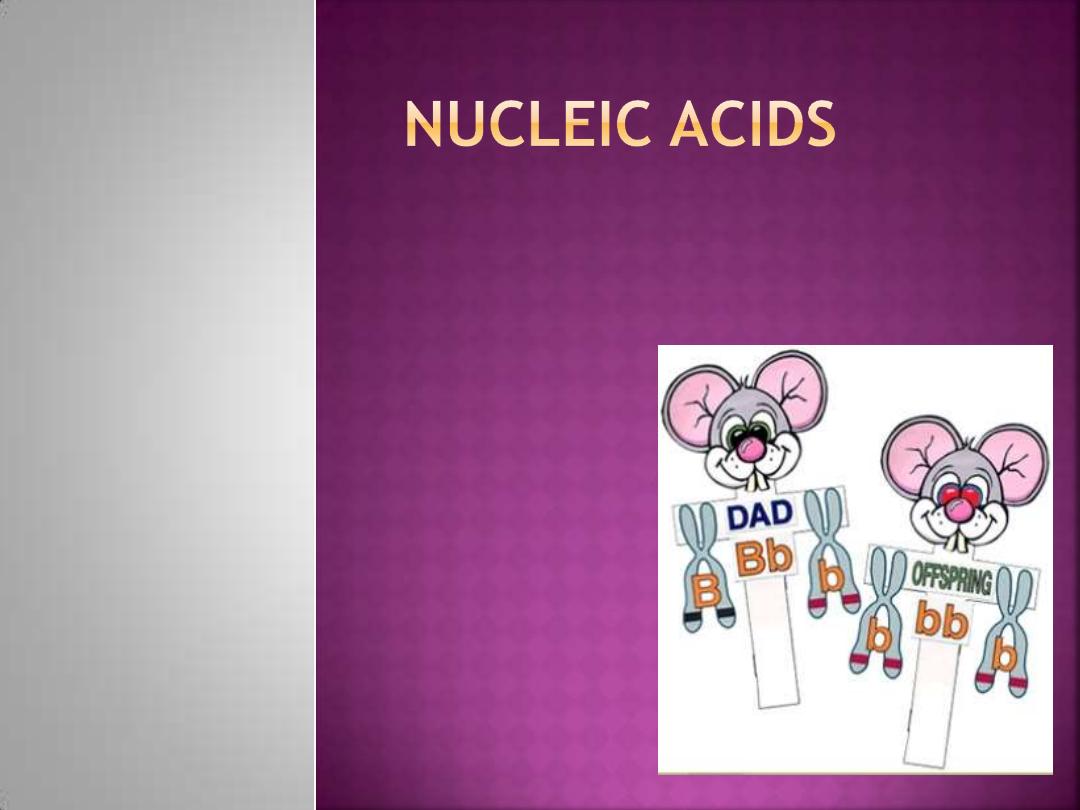
Compare between DNA & RNA.
Describe the Characteristics of DNA Coiling.
Describe the Characteristics of Gene Expression.
Are macromolecules
Strongly acidic
Carry a high density of negative charge at
physiological pH.
That is why they are associated with basic proteins
like histones and with alkaline cations like Mg
⁺²
The essential function of nucleic acids:
The storage & transmission of it’s genetic
information.
The expression of this information in the form of
synthesis of cellular proteins for building &
maintaining the life.
Ribonucleic acid or
RNA
:
which contains
ribose sugar
Deoxyribonucleic acid or
DNA
:
which contains
deoxyribose sugar
RNA
DNA
Position
Nucleus, cytoplasm
Nucleus
Types
Three
One
Helix
No
Yes
Strands
Single
Double
Sugar
Ribose
Deoxyribose
Bases
Adenine (A),
Guanine(G),
Cytosine(C), Uracil(U)
Adenine (A), Guanine(G),
Cytosine(C), Thymine(T)
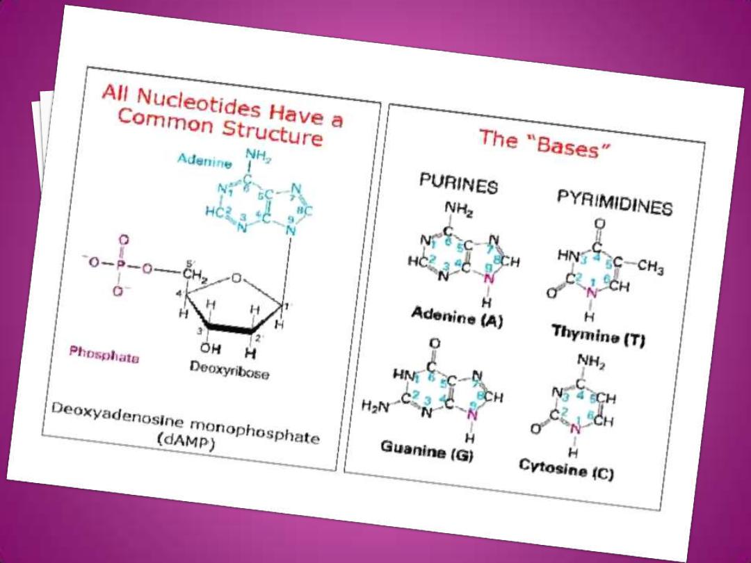
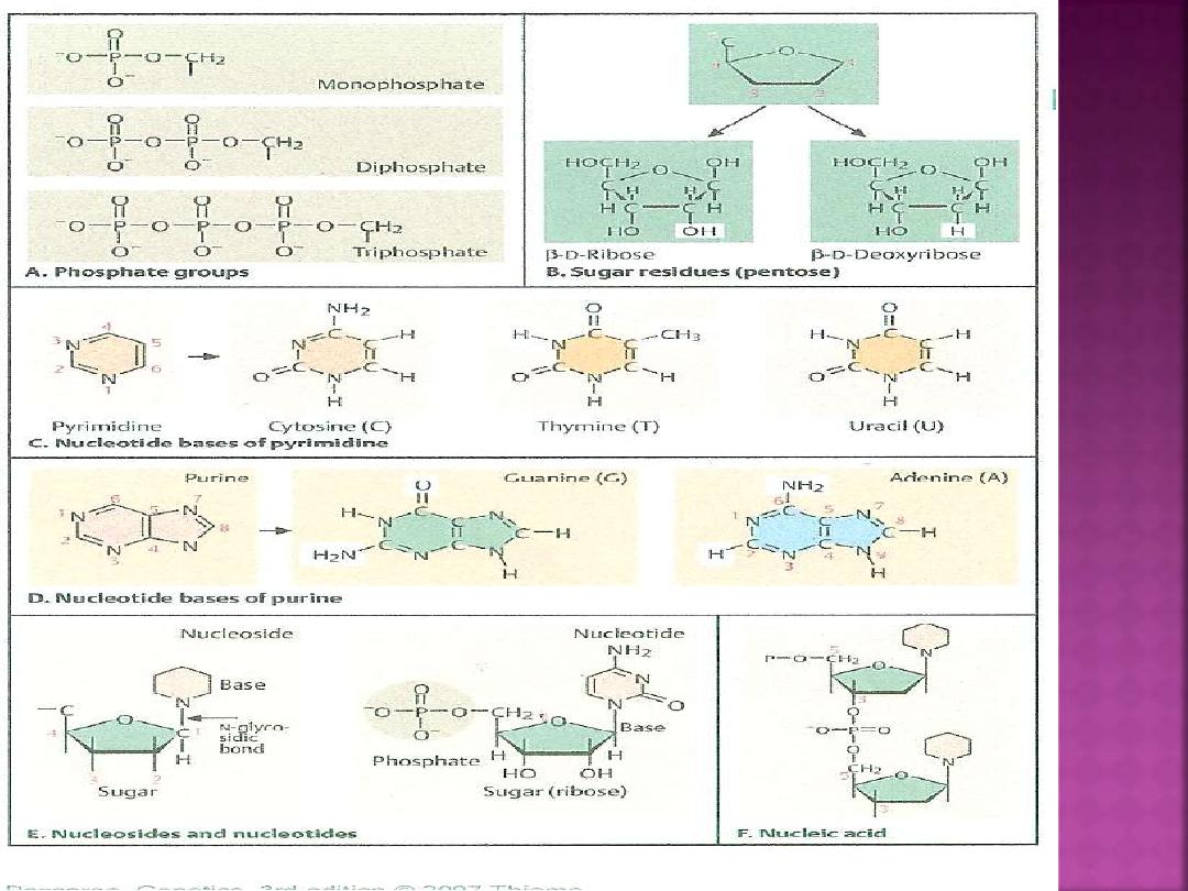
Is a polymer of units called
Nucleotides
which are a complex of a:
Sugar
is 5-carbon (pentoses) which called
dexoyribose.
Nitrogen bases
are two types
(Purines &
Pyrimidines ).
Phosphate group
(po4
⁻)
Nucleoside :
when a base linked to sugar
Purines:
which are 2-carbon-nitrogen ring
structures.
The common purines present in nucleic acid are:
1.
Adenine (A)
2.
Guanine (G)
Pyrimidines:
which are 1-carbon-nitrogen ring
structures.
The common pyrimidines present in DNA
1.
Thymine (T)
2.
Cytosine (C)
The common pyrimidines present in RNA
1.
Uracil (U)
2.
Cytosine (C)
..
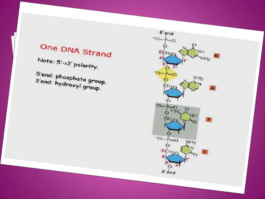
The C-1 (carbon atom) of deoxyribose is bounded to
N-1 (nitrogen atom) of pyimidine or N-9 of purine
these linkage called
glycosidic bond.
The phosphate group is attached to the number C-5 of
the sugar, often designated as the 5
ʹposition.
The nucleotides are
linked together in to a
polynucleotide chain by a backbone consisting of an
alternating series of sugar & phosphate residues.
Specifically, the 3
ʹ-OH (3ʹ-hydroxyl) of sugar of one
deoxyribonucleotide is joined to the 5
ʹ-OH of adjacent
sugar by a
phosphatediester bridge
.
Thus the phosphate-sugar backbone is said to consist
of 5
ʹ―3ʹ linkages. The nitrogenous base “stick out”
from the phosphate-sugar backbone.
The terminal nucleotide at one end of the
chain has a free 5
ʹ group, the terminal
nucleotide at the other end has 3
ʹgroup
(hydroxyl group). It is conventional to write
nucleic acid sequences in the 5
ʹ―3ʹ direction
, that is, from the 5
ʹ terminus at the left to the
3
ʹ terminus at the right thus a DNA chain has
polarity.
.
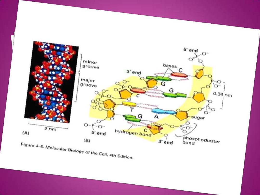
In 1953, James Watson & Francis Crick deduced
the three dimensional structure of DNA, the important features of
their model of DNA are:
I. X-ray diffraction data showed the DNA molecule consist of:
1.
Two right-handed helical polynucleotide chains.
2.
These two chains are of opposite polarity (antiparallel) .
3.
The two chains are coiled around the same axis to form a
double helix.
4.
The diameter of double helix is approximately 20-22
A˚.
5.
Each turn of the double helix covers a distance of 34
A˚ (3.4
nm).
6.
The distance between adjacent nucleotides is 3.4
A˚,
there must
be (10) nucleotide per turn.
7.
Each nucleotide is turned 36
A˚
8.
The twisting of the two strands around one another forms a
double helix with
narrow groove (12
A˚across)
called
Minor
groove
&
a wide groove (22
A˚across)
called
Major groove.
.

II. The density of DNA suggests that:
1.
The constant
diameter of the helix can be explained if
the bases in each chain face inward & are restricted
so that a
purine is always opposite a pyrimidine ,
avoiding purine-purine (too thick) or pyrimidine-
pyrimidine (too thin)
partnership.
2.
The bases are on the inside of the helix in pairs
arranged in such a fashion that apyrimidine of one
chain always pairs with a purine of the opposite
strand, & vice versa.
III. Inrrespective of the actual amount of each bases:
1.
Only certain bases pairs can be accommodated.
These are
A with T (2 hydrogen bonds)
&
G with C
( 3 hydrogen bonds)
.
2.
The proportion of G&C is always the same in DNA
and the proportion of A&T is always the same, thus
the composition of any DNA can be described by the
proportion of it’s bases that is
G+C which ranges
from 26% -74% for different species
.
1.
The pairing rules (base pairing) require that
the bases in the two chain are
complementary
so that the sequence of one
chain is completely determined by the
sequence of it’s partner.
2.
The highly negatively charged
phosphodiester backbone face outward &
it’s strongly polar groups can interact with
the aqueous environment.
3.
When DNA is in solution
in vitro ,
the charge
are neutralization by binding of metal ions ,
usually Na
⁺ is provided in the natural state
in
vivo ,
positively charged proteins provide
some of neutralizing force.
1.
The base composition is characteristic of an
organism, species or strain.
2.
All the cell of organism or a tissue have identical
or closely similar base composition, age, phase
of growth environmental or physiological factors
do not have any effect on this (thinking that
there are no mutations in any of the cells).
3.
Different organisms exhibit wide variation in the
base composition which is expressed by the
ratio (A+T)/(G+C).
4.
Similar base compositions are exhibited by
closely related organisms.
5.
According to Chargaff ‘s rule A&T,G&C or
A+G=T+C or A+C=G+T.
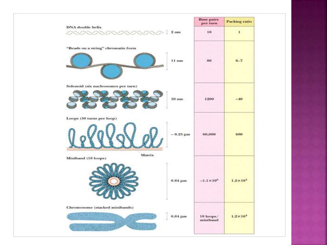
The length of DNA about (2) meters in straight line, so to
package all of this DNA into a tiny cell nucleus, it’s coiled at
several levels:
First,
the DNA is wound around
a histone protein
core to
form a
nucleosome
. A bout (140-150) DNA bases are
wound a round each histone core, and then (20-60) bases
form
a spacer
element before the nuclosome complex.
The nucleosomes are turn form
a helical solenoid
, each
turn of solenoid includes about (6) nucleosomes. The
solenoids, themselves are organized into
chromatin loop
,
which are attached to a protein scaffold.
Each of these loops contains a pproximataly
100,000 base
pairs (100 kilo bases or Kb )
of DNA.
The end,
result of this coiling & looping is that the DNA ,
when at it’s maximum stage of condensation is only about
1/10,000 .

Like DNA, RNA is a polymer of nucleotides . The
nucleotide in RNA, however, contain the sugar
ribose & the bases Adenine (A),Guanine (G),
Cytosine (C)& Uracil (U) . In other words, the
bases Uracil replace the Thymine found in DNA.
Finally, RNA is single stranded and does not form
a double helix in the same manner as DNA.
There are three major classes of RNA :
Messenger RNA(mRNA):
takes a message from
DNA in the nucleus to the ribosomes in the
cytoplasm.
Ribosomal RNA(rRNA):
along with proteins,
makes up the ribosomes, where proteins are
synthesized.
Transfer RNA(tRNA):
transfers amino acids to the
ribosomes.
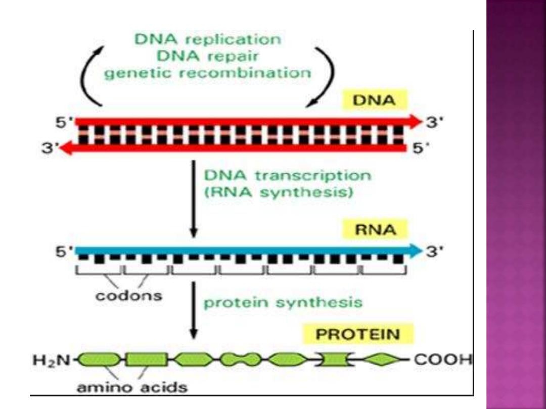
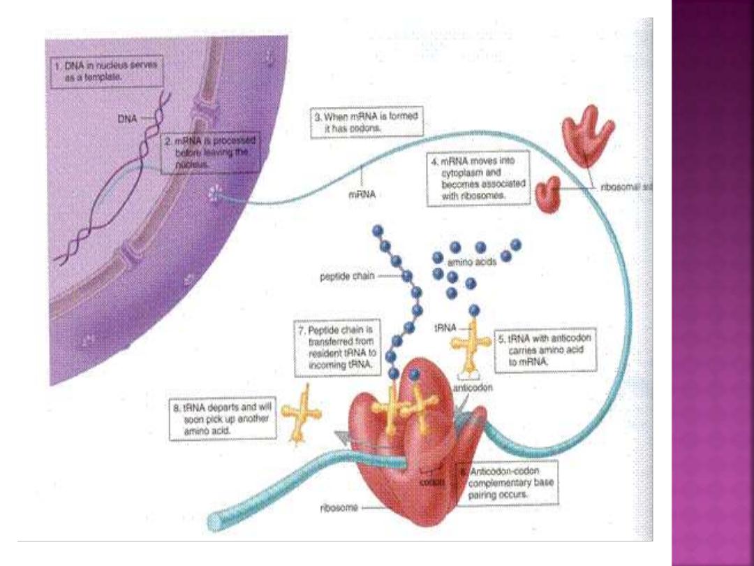
1- DNA contains genetic information. The
sequence of it base determines the sequence of
amino acids in a polypeptide.
2- during transcription, one strand of DNA serves
as a template for the formation of mRNA. The
bases in mRNA are complementary to those in
DNA; every three bases is a codon that codes for
amino acid.
3- the mRNA is processed before it leaves the
nucleus, during which time the introns are
removed.

4- the mRNA carries a sequence of codons to
the ribosomes, which are composed of rRNA
and proteins.
5- the tRNA molecules, each of which is
bonded to a particular amino acid, have anti
codons that pair complementarily to the
codons in mRNA.
6- during translation, tRNA molecules and
their attached amino acids arrive at the
ribosomes, and the linear sequence of codons
of the mRNA determines the order in which
the amino acids become incorporated into a
protein.

Thank
you