Dr. Shayma`a Jamal Ahmed
Prof. Genetic Engineering
& Biotechnology

At the end of this lecture the student will
be able to:
Define the cell division.
List the cell division vocabulary.
Describe the Life Span &Cell cycle .
Determine the features of Cell cycle.
Describe the Stages of cell cycle.
List the Phases of Mitosis.
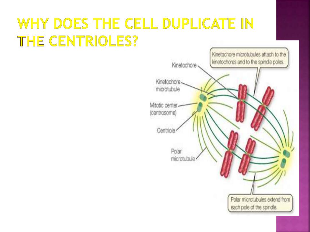
Why does the cell duplicate the centrioles?
Recognize who the Mitosis is inhibited by
suppressor genes.
All of the living organisms on Earth are made
up of one or more cells, which are the
simplest units of life capable of independent
existence and reproduction.
Cells have the extraordinary ability to make
nearly identical copies of themselves by the
process of cell division. Since new cells are
only produced by existing cells, cell division
is essential for the continuation of life.
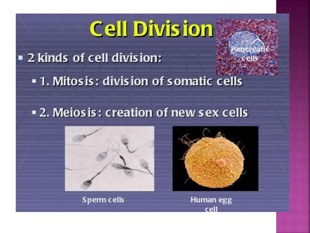
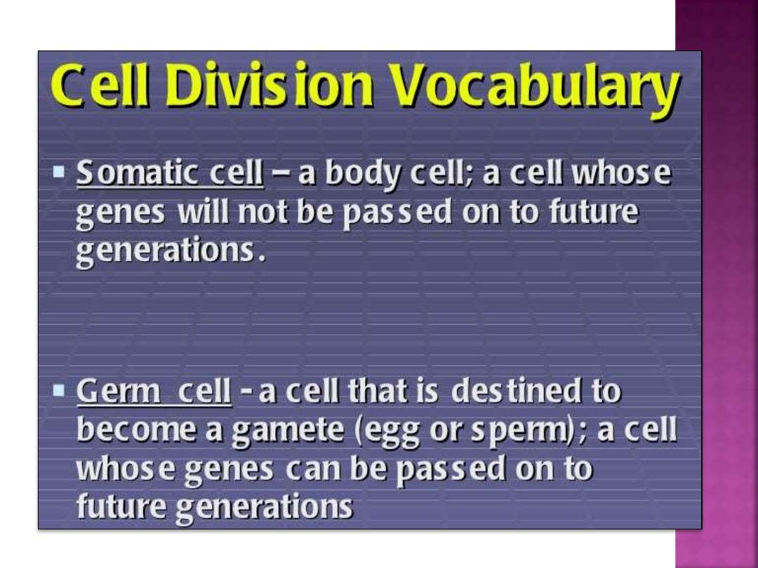
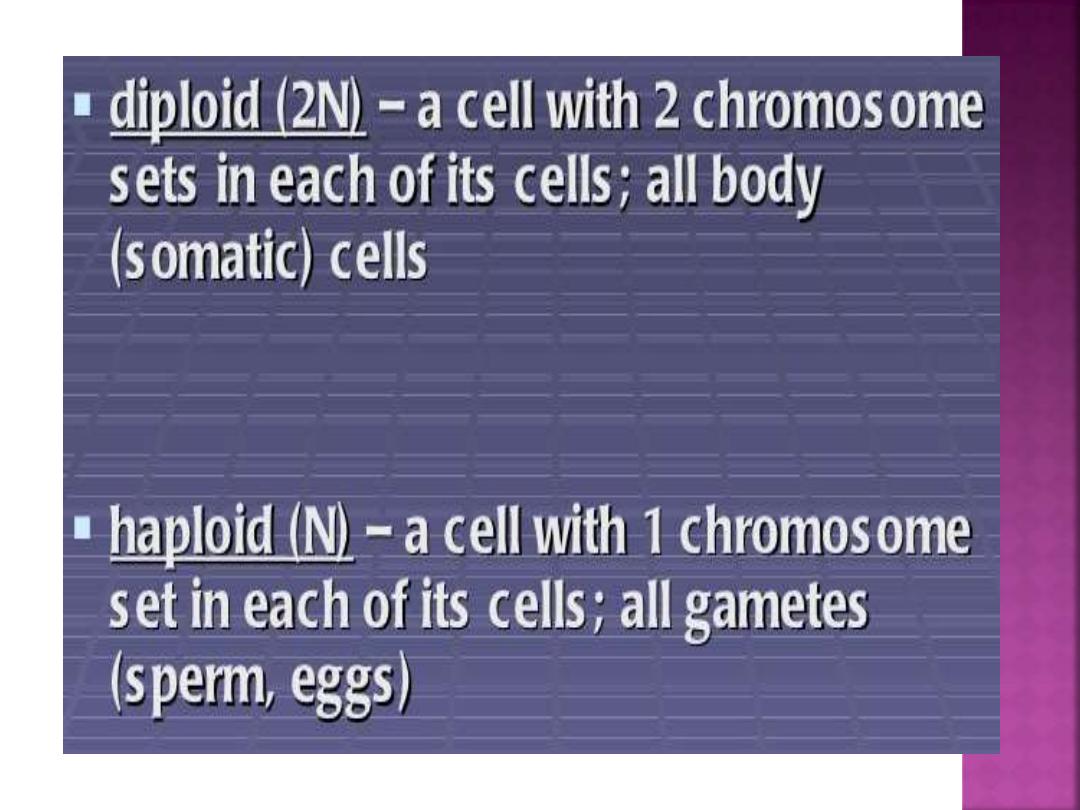

Human somatic cells contain 46 chromosome
(23 pairs=2N), but the germ cells contain 23
chromosomes (=N which are: sperm & eggs).
Homologous pairs come from:
- father.
- mother.
Human Karyotype:
pictures of homologous chromosomes lined up
together during Metaphaes in which the
chomosomes are arranged in order of decreasing
length.

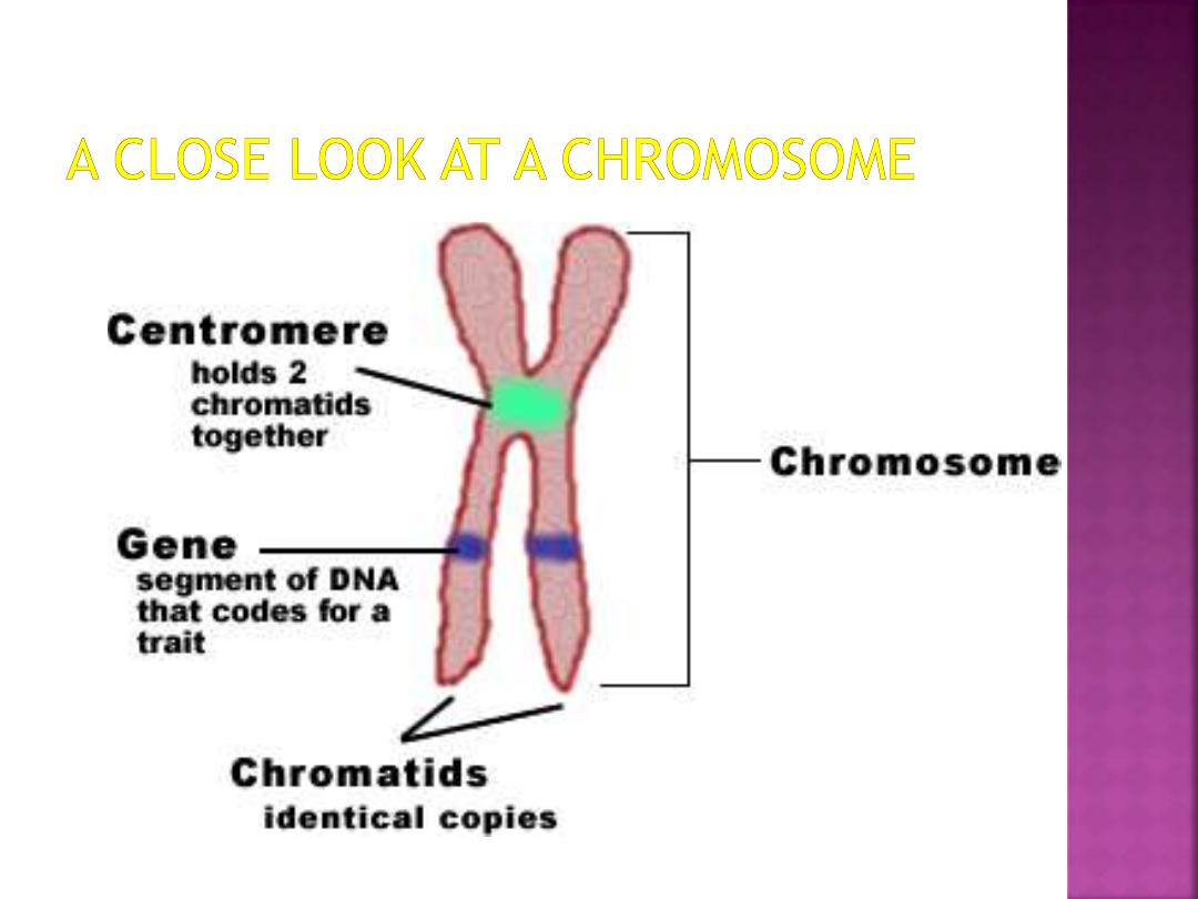
1. Chromosome - a thread-like structure carrying
the genetic material (DNA) and associated
proteins.
2. Homologous chromosomes - chromosomes from
male and female which have the same length,
shape, and genes
3. Chromatid - one of the thread-like structures of a
replicated chromosome
4. Sister chromatids - replicates of the chromatid
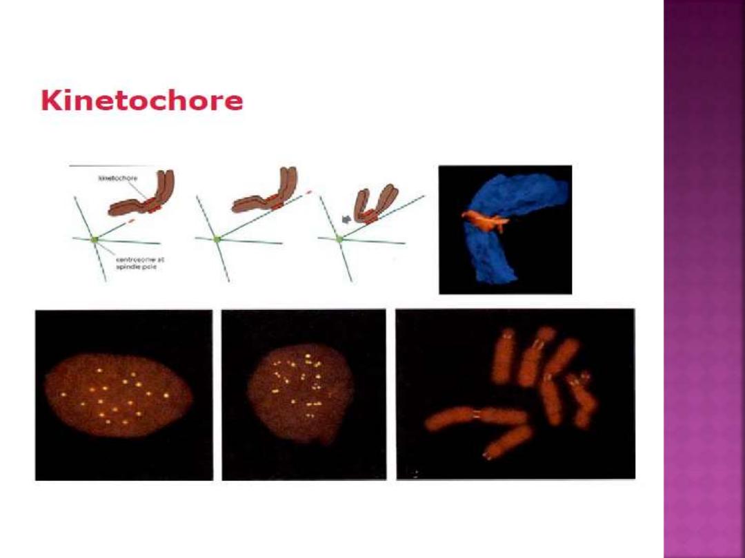
5. Centromere - unduplicated area between
chromatids where kinetochore is found
6. Gene - a section of DNA encoding a particular
trait.
7. Allele - an alternative form of a gene
Body Cell Types :
are about 210 types.
Life Span of cell : includes
- Born( Mitosis).
- Differentiate.
- Function.
- Die.
For example:
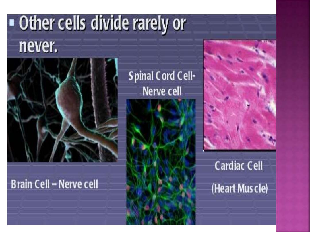
1- Neutrophil : 6-7 days circulating & 4 days in
tissue.
2- Red Blood Cell : 120 days.
3- Brain neuron, heart: 50-100 years.
Birth :
Mitosis (Except germ cells (divided by
Meiosis)).
Death :
1- Apoptosis: programmed cell death.
2- Necrosis.
The orderly program of events in the lifetime
of a cell is known as the cell cycle. One cell
cycle describes the period between a cell's
creation by mitosis, and its subsequent
division into two daughter cells.
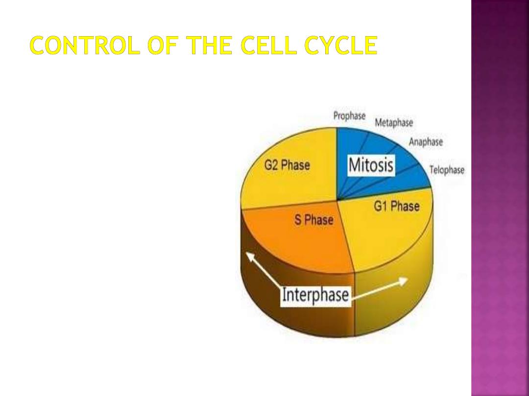
The typical cell cycle is divided into two
phases: a brief mitotic phase in which the cell
divides it`s nuclear and cytoplasmic contents,
and a longer period between divisions called
interphase.

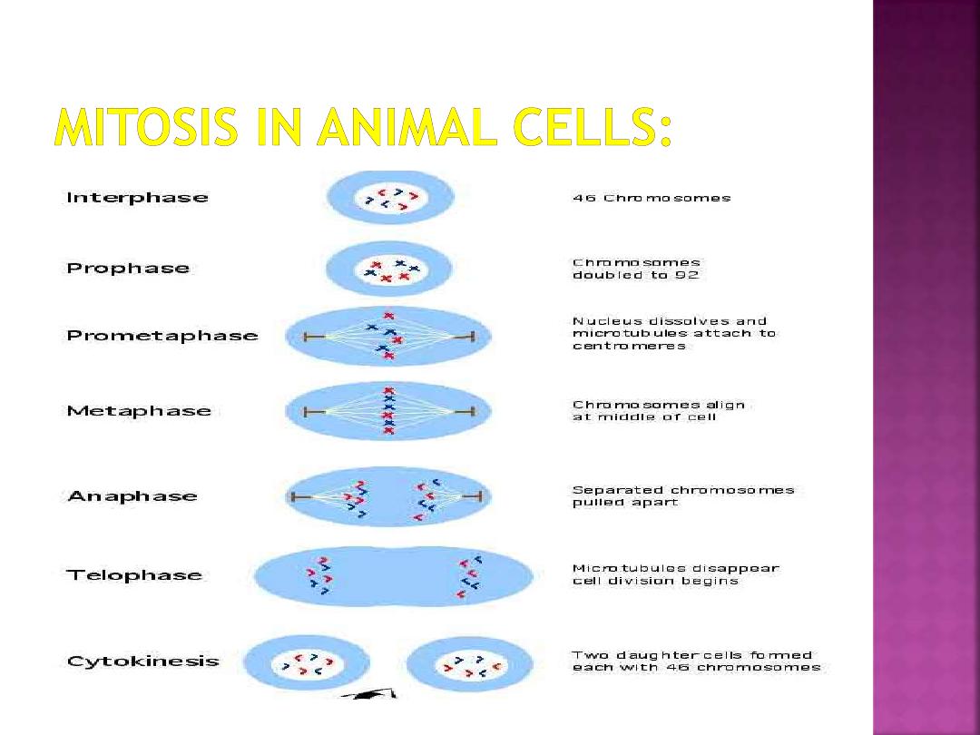
In eukaryotic (plant, animal & fungus) cells, the
division of chromosomes and cytoplasm into two
cells is known as the mitotic phase. During
mitosis and cytokinesis, each of the two
daughter cells will receive an exact copy of the
parent cell's chromosomes and roughly half of
the cytoplasm.
It is important to know that the parent cell's
chromosomes are replicated during the S
(synthesis) phase of the cell cycle before mitosis
can begin. The replicates (called sister
chromatids) remain attached to each other
through early mitosis.
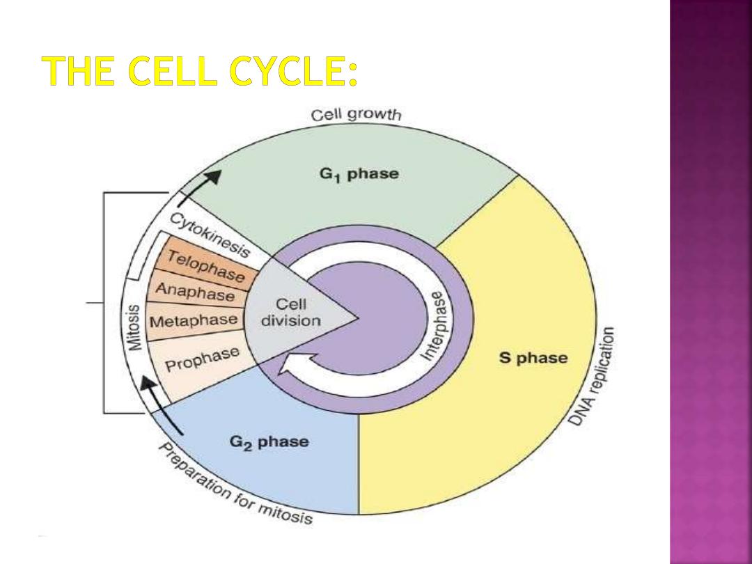
G0 phase:
Indefinite period. Specialized cell
functions , a cell that is in this phase is not
preparing to divide, but instead is carrying out
specialized functions .
G1 phase:
Metabolic changes prepare the cell for
division. At a certain point - the restriction point -
the cell is committed to division and moves into the
S phase.
S phase:
DNA synthesis replicates the genetic
material. Each chromosome now consists of two
sister chromatids.
G2 phase:
Metabolic changes assemble the
cytoplasmic materials necessary for mitosis and
cytokinesis.
M phase:
A nuclear division (mitosis) followed by a
cell division (cytokinesis).
The period between mitotic divisions - that is, G1, S
and G2 - is known as
interphase.
Series of events that takes place in a cell leading to
it`s division and duplication (replication).
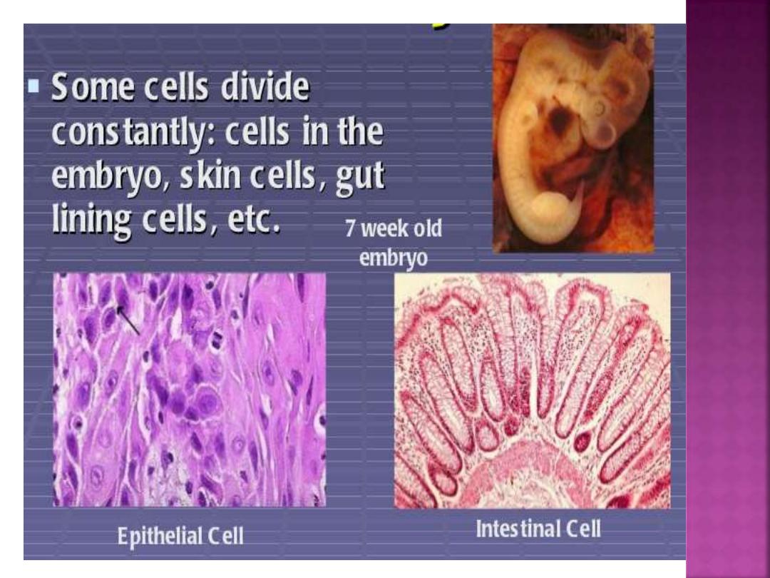
Producing new cells allow multicellular organisms to
grow and replace dead cells.
In an adult, cells reproduce at the same rate at
which they die.
Cell division occurs at least 10 million times every
second in an adult human body.
The life cycle of a cell is known as the
cell cycle
.
The cell cycle begins when a cell is formed and
when the cell divides & forms two new “daughter”
cells.
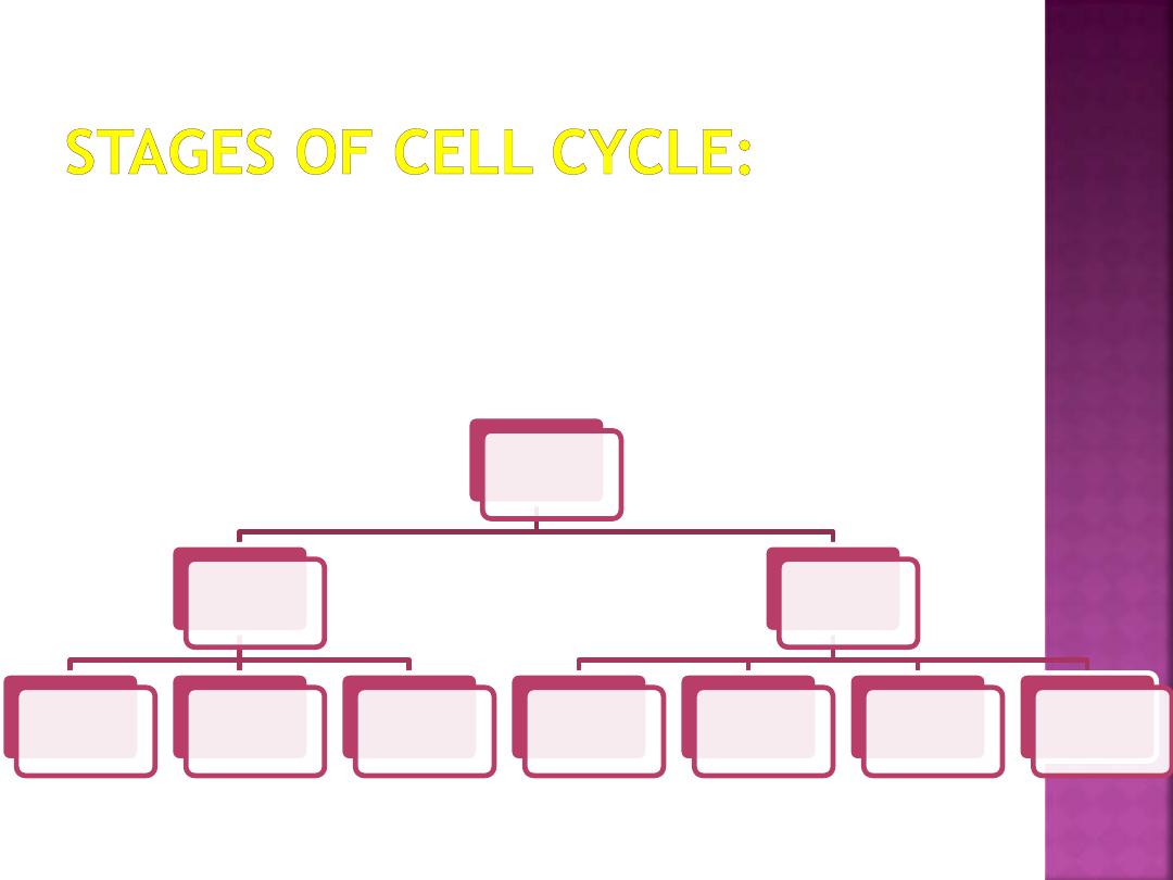
There are three stages:
1- Interphase.
2- Mitosis.
3-Cytokinesis
The cell
cycle
Interphase
G1 phase
S phase
G2 phase
Mitosis
prophase
metaphase
anaphase
telophase
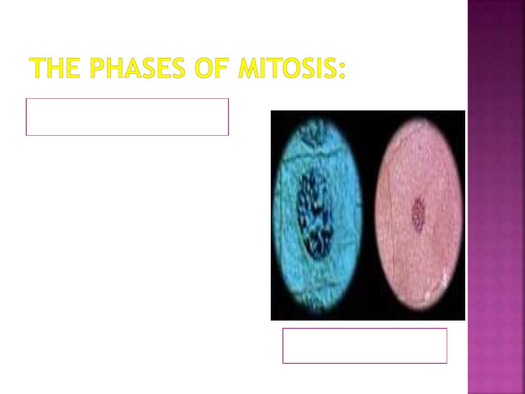
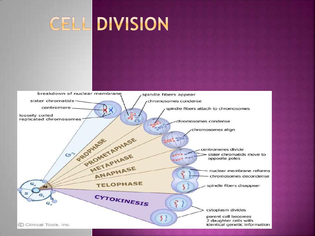
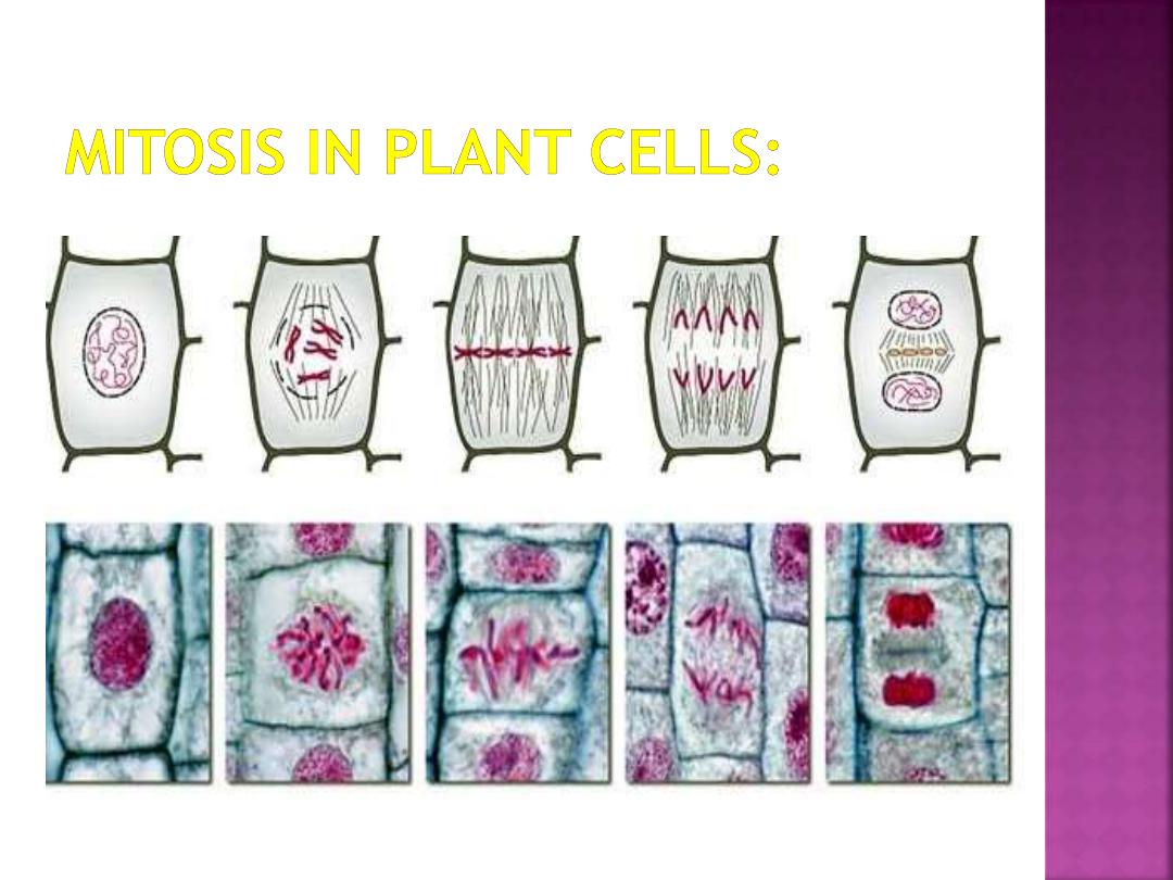
1-Prophase:
Plant & Animal cells
The beginning of mitosis is
called prophase. In early
prophase, the centrosomes
move toward opposite poles
of the cell, organizing the
spindle microtubules
between them.
The sister chromatids
become visible in the
nucleus as they condense.
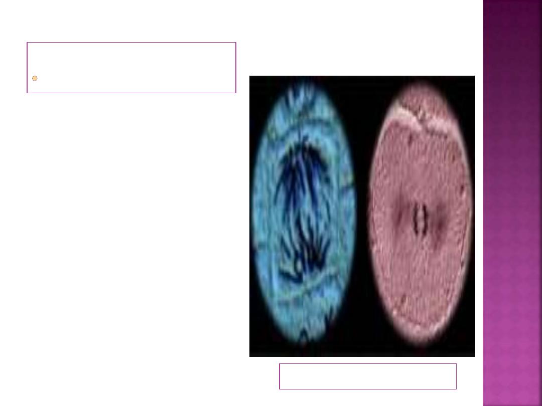
2-Metaphase:
Plant & Animal cells
The chromatids
remain lined up
between the poles of
the cell
during metaphase.
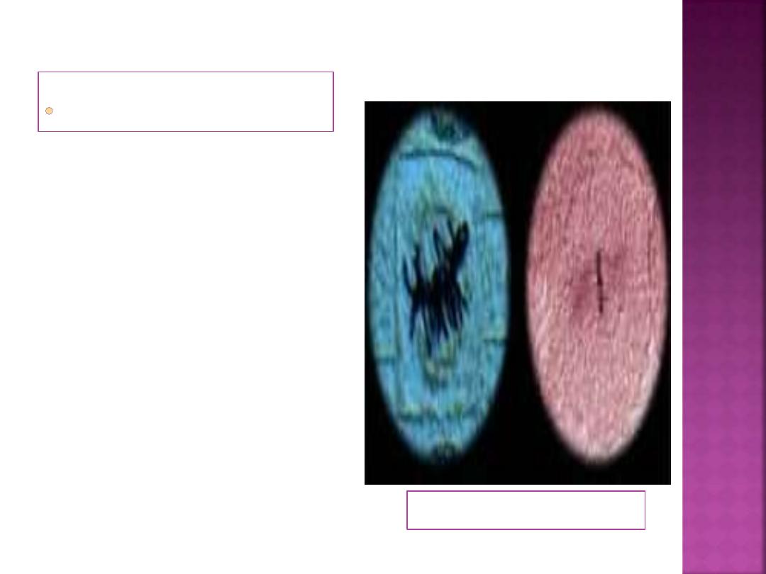
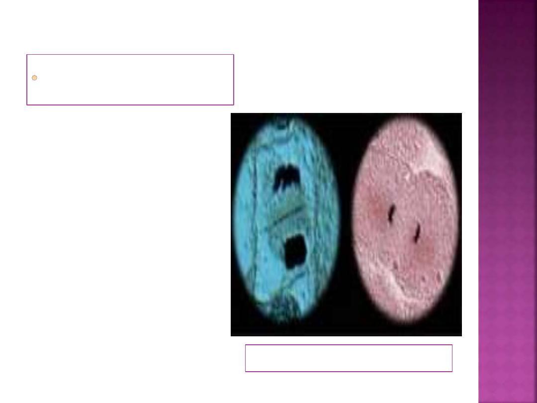
3-Anaphase:
Plant & Animal cells
Begins when the
pairs of sister
chromatids separate.
The separated
chromatids are now
called chromosomes,
and move toward the
poles of the cell.
4- Telophase:
Plant & Animal cells
The chromosomes
arrive at the poles
in telophase, and
new nuclear
membranes form
around them.
.
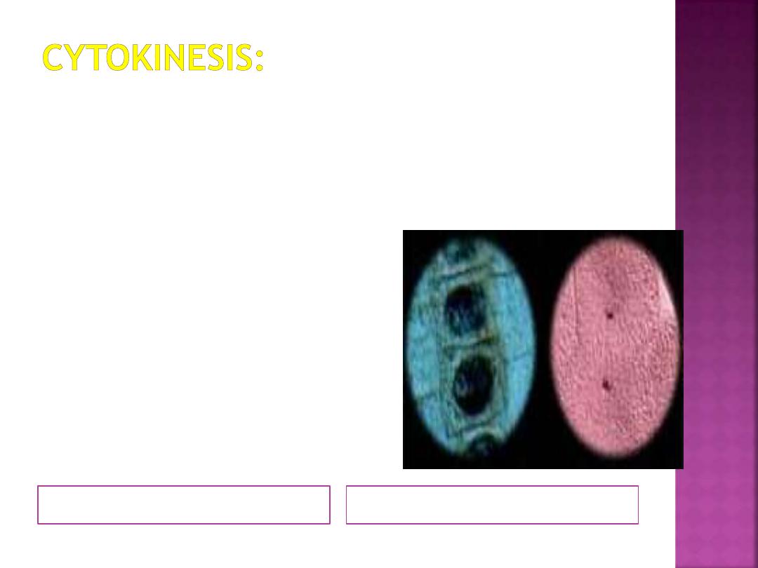
Plant & Animal cells
Division of the cytoplasmic
components is
calledcytokinesis.
In animal cells, cytokinesis
occurs when a ring of actin and
myosin filaments constricts the
plasma membrane at the
equator. Eventually, the parent
cell is divided into two cells.
In plant cells, a number of
small vesicles fuse at the
metaphase plate to form the
cell plate. Over time, the cell
plate reaches across the cell
and joins with the plasma
membrane.
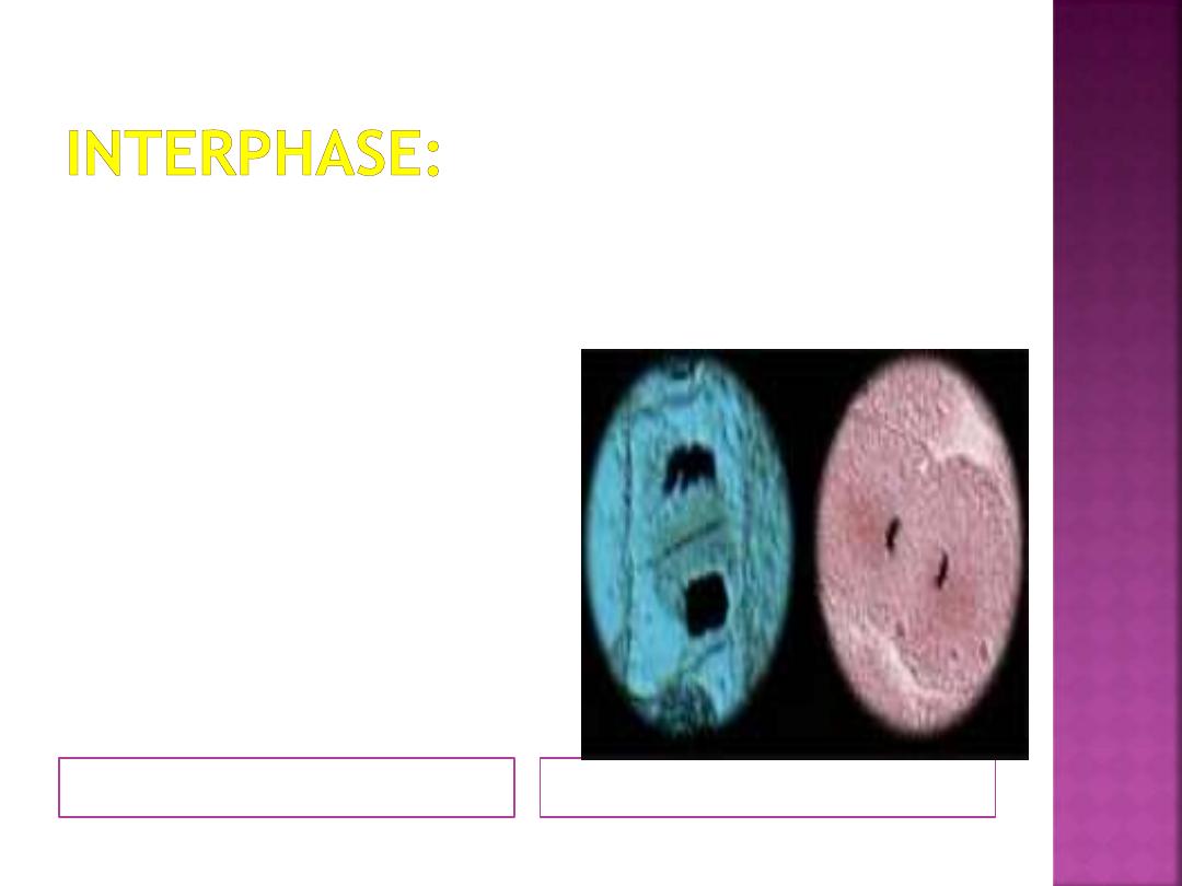
.
Plant & Animal cells
The process of mitosis and
cytokinesis creates two
separate cells, each with an
identical set of
chromosomes. After
cytokinesis, the daughter
cells will enter interphase.
For additional information on
mitosis in plant and animal
cells, or prokaryotic cell
division
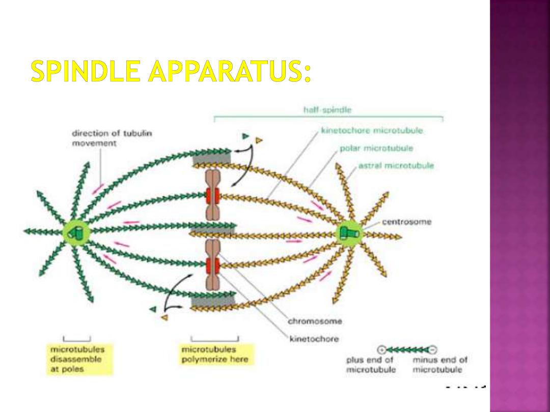
The cell has to create
a cytoskeleton
structure for the
chromsomes to attach
to during mitosis.
This structure is
called the
Spindle
Apparatus
and it is
made from
microtubules that
project from the
centrioles.
refers to the sub-cellular structure that
segregates chromosomes between daughter cells
during cell division.
It is also referred to as the mitotic
spindle during mitosis or the meiotic
spindle during meiosis.
It is composed of hundreds upon hundreds of
proteins, the fundamental machinery are the
spindle microtubules. Attachment of microtubules to
chromosomes is mediated by kinetochores, which
actively monitor spindle formation and prevent
premature anaphase onset.
bipolar attachment of sister kinetochores to
microtubules from opposite cell poles couples opposing
tension forces, aligning chromosomes at the cell equator
and poising them for segregation to daughter cells.
G1 Checkpoint
- Check
to see if DNA is
damaged.
G2 Checkpoint
- Check
to see if DNA is
replicated properly.
M Checkpoint
- spindle
assembly checkpoint,
check for alignment of
chromosomes
Apoptosis -
programmed cell death,
if any of the checks fail
neoplasm: abnormal growth of cells |
benign: non-cancerous | malignant:
cancerous : Tumor.
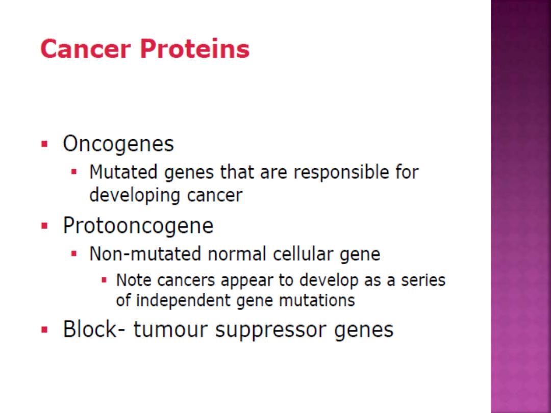
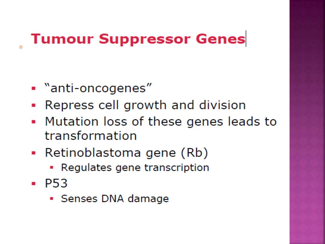
Cancer: cellular growth disorder that results
from the mutation of genes that regulate the
cell cycle, cells are abnormal and do not
perform their intended function.
When the rate of cell division (mitotic rate)
is greater than that of cell death in a tissue.

Cell-Cycle regulation by:
1- CDKs and cyclins.
2- CDK inhibitory proteins.
3-The Rb/E2F pathway.
4-Positive and negative phosphorylation of
CDKs.
5-Protein degradation.
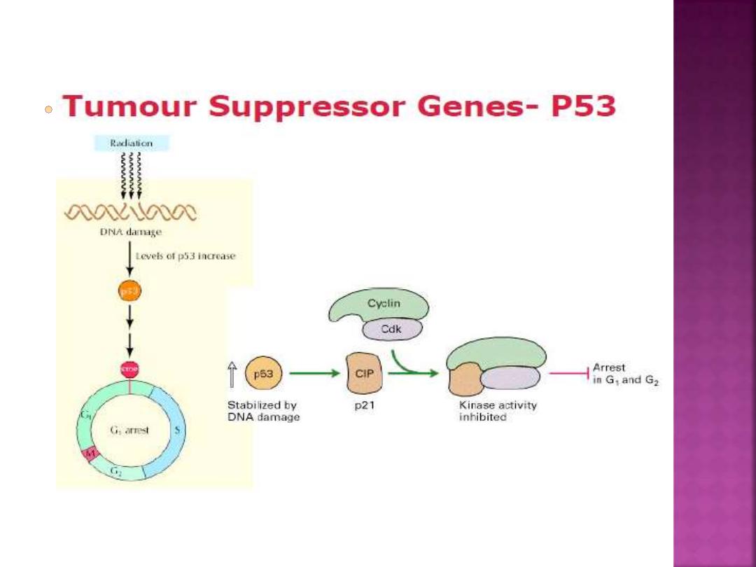
For example: p53
The p53 gene is a gene that codes for a
protein that inhibits the development and
growth of tumors (in addition to other
functions). It is known as a tumor suppressor
gene.
If this gene is mutated –- that is, altered in
some way by either the environment or
inheritance, cancers can develop and grow
out of control.
THANK YOU