.

At the end of this lecture the student will be able
to:
Define the cell membrane.
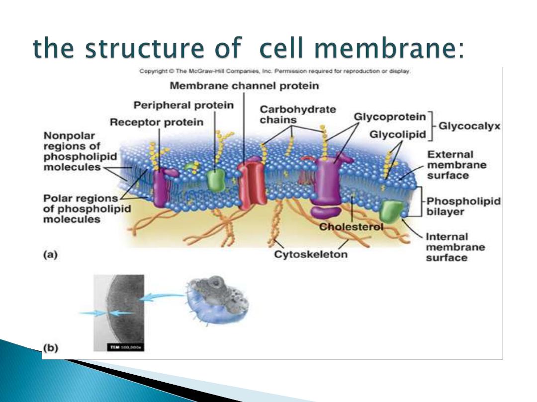
Describe the structure of cell membrane.
Determine the functions of cell membrane.
Recognize to the mechanisms of transport.
Compare between the
Exocytosis &
Endocytosis
Which is called also
plasma Membrane
or
Cytoplasmic Membrane
.
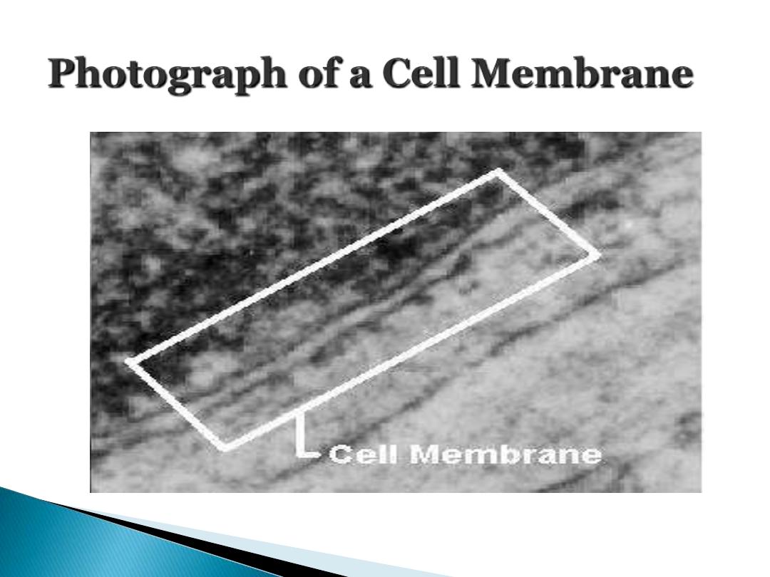
It is a
biological membrane, it is
surrounding and separating the interior
components (which are alive)of all cells from
the outside environment (which is nonliving).
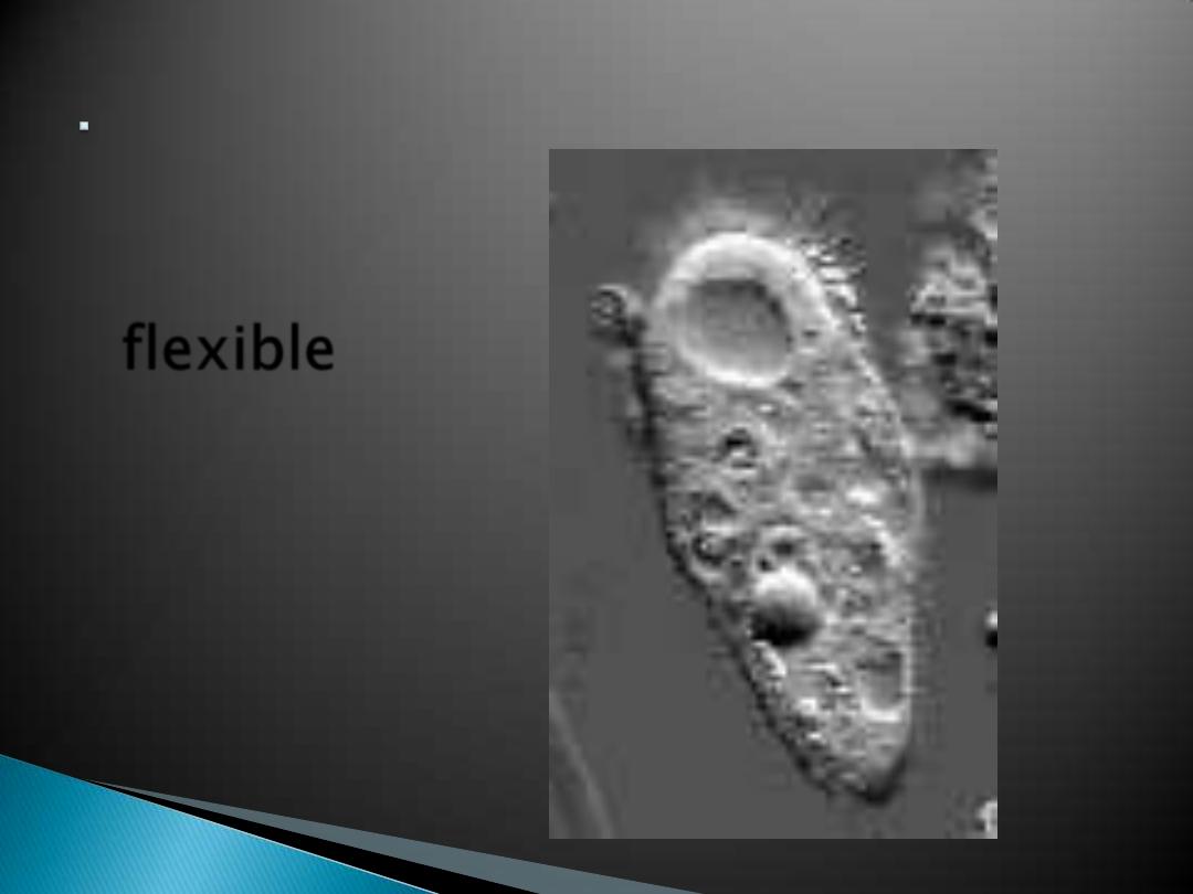
The cell
membrane is
flexible
and
allows a
unicellular
organism to
move.
Balanced internal condition of cells
Also called
equilibrium
Maintained by plasma membrane controlling
what enters & leaves
the cell
Protective barrier
Regulate transport in & out of cell
(selectively permeable)
Allow cell recognition.
Provide anchor sites for filaments of
cytoskeleton.
Provide a binding site for enzymes
Interlocking surfaces bind cells together
(junctions)
Contains the cytoplasm (fluid in cell)
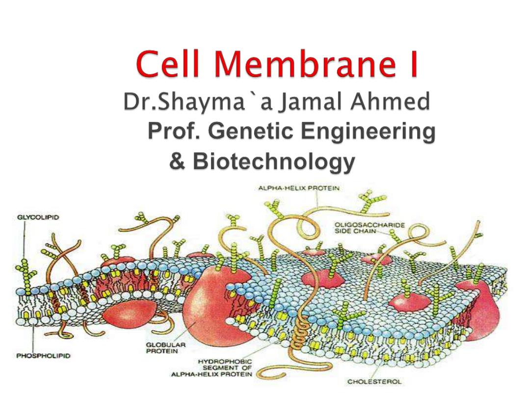
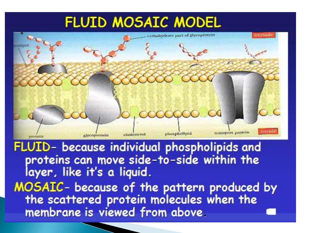
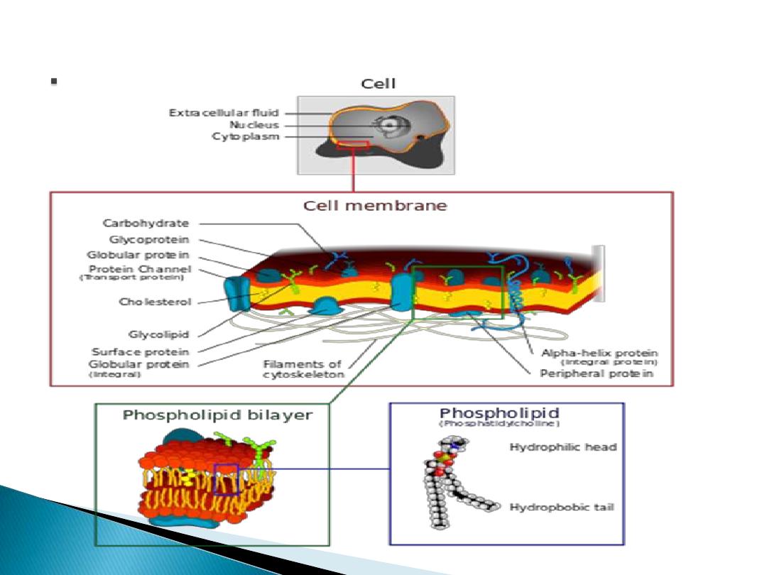
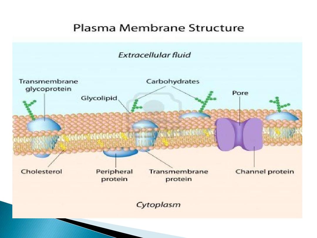
The plasma membrane (cell membrane) is
made of two layers of phospholipids. The
membrane has many proteins embedded in it.
The arrangement of protein & lipid molecules
with in the membrane may suggest different
models. The most accepted model is the
Fluid-Mosaic model
.
In 1972, Singer & Nicolson revised the model
in simple way:
They proposed that:
the globular protein
are inserted of the lipid bilayer, with their
nonpolar segments in contact with the
nonpolar interrior of the bilayer & their polar
portions stick out from the membrane
surface.
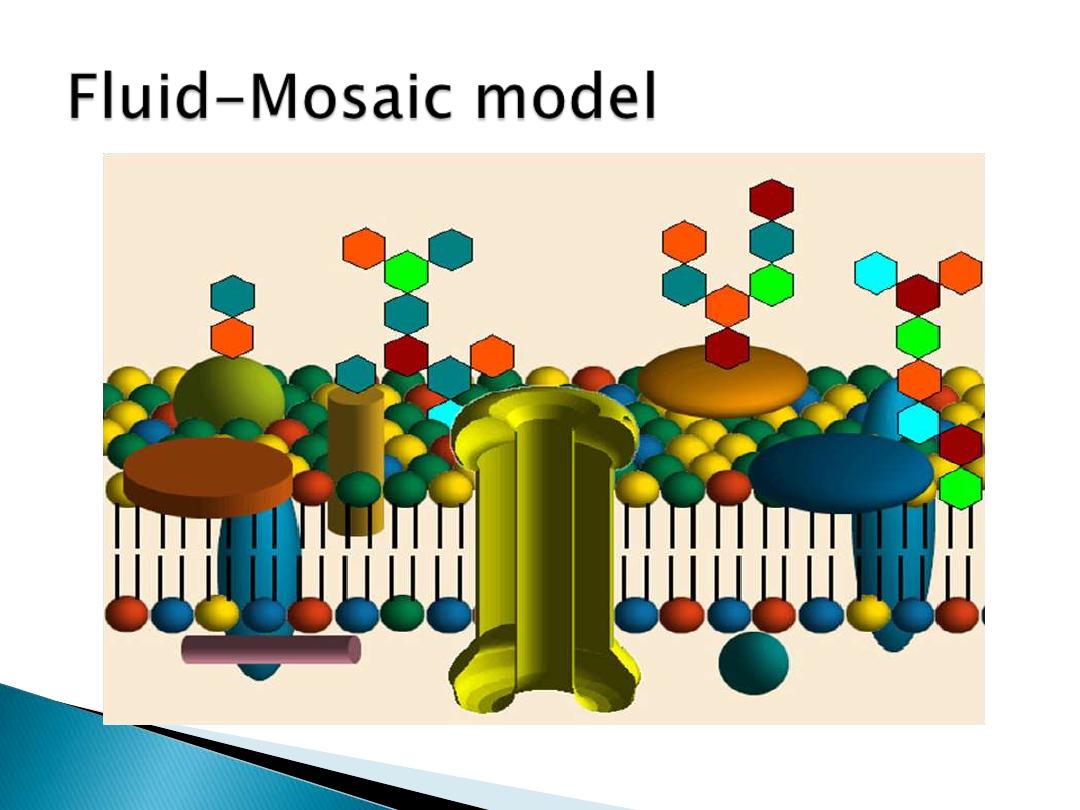
A. Mosaic:
an object comprised of bits and pieces
embedded in a supporting structure
1. membrane lipids form the supporting structure
2. membrane proteins provide the bits and pieces
3. both lipids and proteins may be mobile or 'fluid'
B. Membrane lipids:
the supporting structure
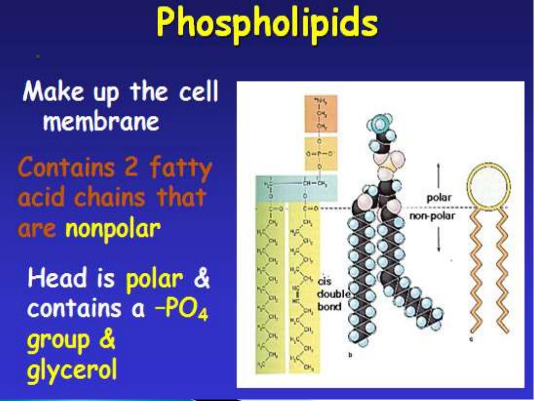
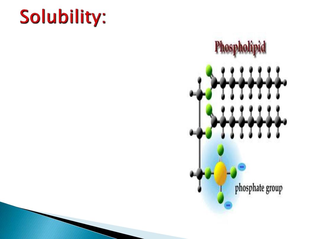
1. phospholipids
2. glycolipids
3. cholesterol
C. Membrane proteins:
the bits and pieces
1. integral (intrinsic) proteins
2. peripheral (extrinsic) proteins
1.
Lipids
: bilayer of phospholipids
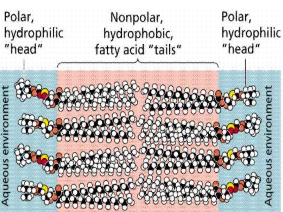
a.
Hydrophilic heads
- P0
4
end "water"
"loving" attracted to water on inner/outer
parts of cell
b.
Hydrophobic tails
- fatty acids "water"
"fearing" attracted to each other on inside
of bilayer
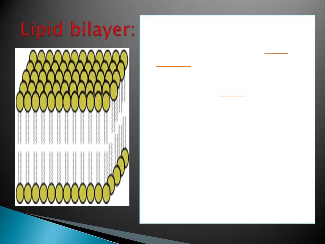
Diagram of the arrangement
of amphipathic lipid
molecules to form a
.
The yellow
groups separate the grey
hydrophobic tails from the
aqueous cytosolic and
extracellular environments.

c.
Glycolipids
- some carbohydrates
attached
to outer lipids (involved in cell to cell
recognition)
d.
Cholesterol
- regulates fluidity of
membrane
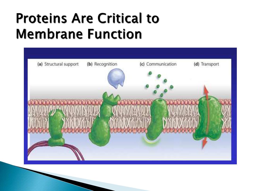
2.
proteins:
interspersed throughout the
membrane.
1)
Receptors:- hormones ,neurotransmitters.
2)
Enzymes: - reactions in & out of cell.
3)
Transport :- ions and molecules.
4)
Intercellular junctions.
5)
Cell-cell recognition.
6)
Cytoskeletal and extracellular matrix
attachment.
1- Integral proteins:
- inserted into the bilayer
(transmembrane - across
entire bilayer).
2- Peripheral proteins:
- on inner & outer surface.
3- Glycoproteins:
- carbohydrates on outer
surface.
glycocalyx - outer carbohydrate coat (cell
recognition and identification).
.
1
1- lipids can pass
through the cell
membrane easily.
2- Small molecules and larger
hydrophobic molecules move
through easily.
e.g. O
2
, CO
2
, H
2
O
3- Ions, hydrophilic molecules
larger than water, and large
molecules such as proteins
do not move through the
membrane on their own