Scapular region

OBJECTIVES
…
To list scapular muscles
To describe axillary gateways
To describe scapular anastomosis
To follow main nerves in the region
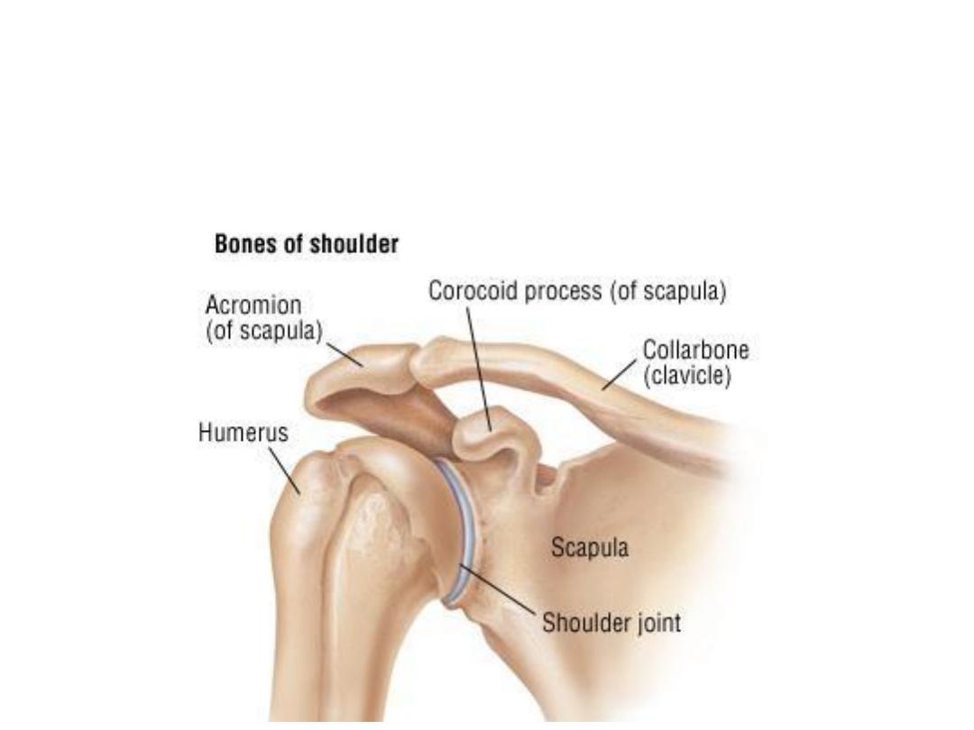
The scapula articulates:
-By its glenoid with the humeral head
-By its acromion with the clavicle
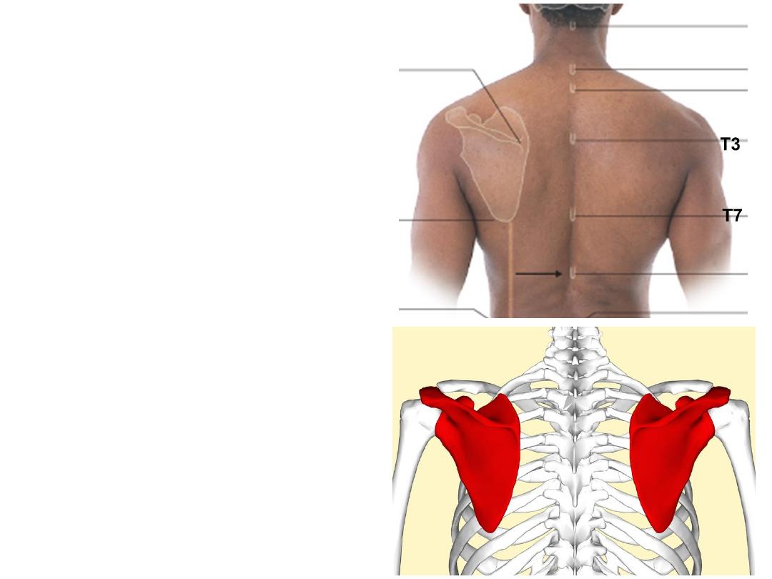
Surface localization:
-Scapula extends from T2-T7 vertebral
level
-Costal level is the same as the vertebral
level
-Scapular spine: T3
-Inferior scapular angle: T7
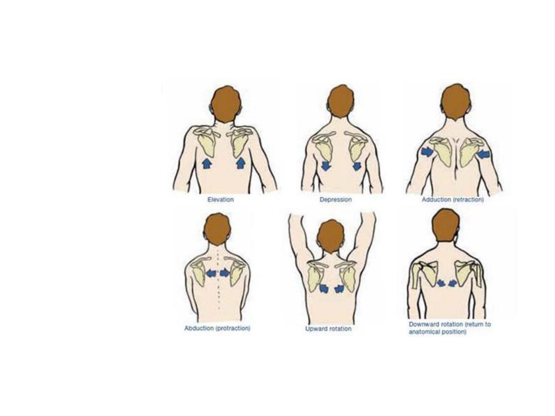
Scapular movements:
Elevation
Depression
Protraction
Retraction
Medial rotation
Lateral rotation
Muscles connecting the scapula to the axial skeleton
Muscle
Origin
Insertion
Innervation
Function
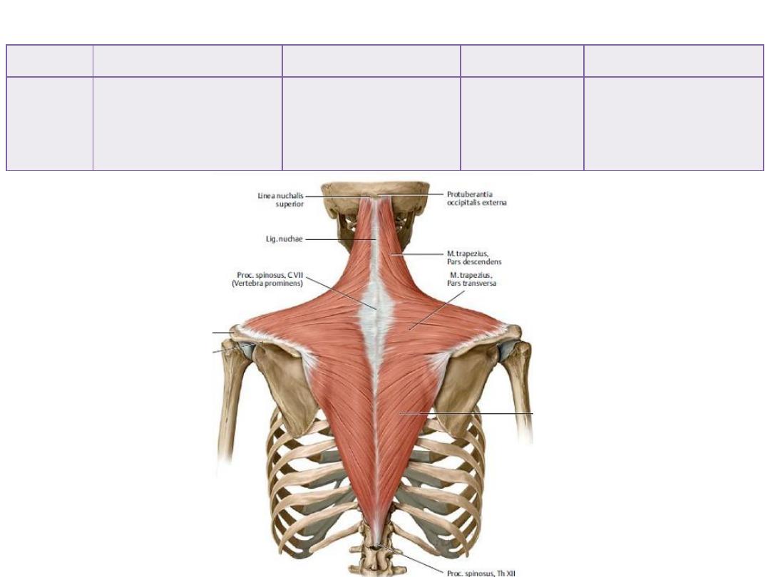
Trapezius
• Back of skull
• Ligamentum nuchae
• Spines of C7-T12
• spine of the scapula
• Lateral 1/3 of
clavicle
Spinal
accessory n
• Scapular elevator
• Lateral rotator
Muscle
Origin
Insertion
Innervation
Function
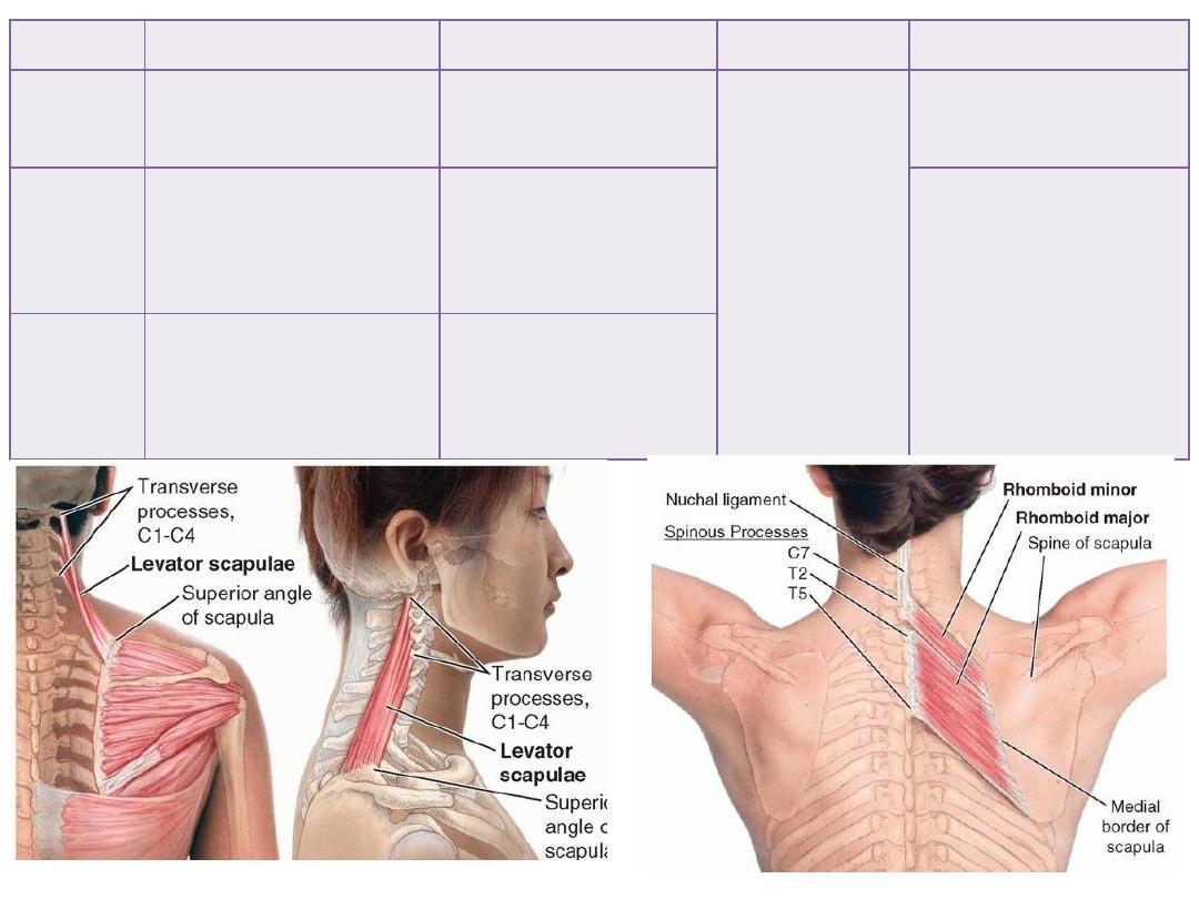
Levator
scapulae
Transverse processes of
C3-C6 vertebrae
superior angle to root
of spine of the scapula
Dorsal scapular
nerve
Elevator of scapula
Rhomboid
minor
Spines of C7 & T1
Medial border of
scapula at the spine
root
• Scapular elevator
• Retractor
Rhomboid
major
Spines T2-T5
Medial border of
scapula below the
spine root
Muscles connecting the scapula to the humerus
Muscle
Origin
Insertion
Innervation
Function
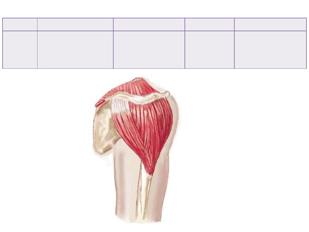
Deltoid
• Scapular spine
• Acromion
• Lateral 1/3 clavicle
Deltoid tuberosity of
humerus
Axillary nerve
• Shoulder abductor
• Flexor (anterior F)
• Extensor (post F)
Muscle
Origin
Insertion
Innervation
Function
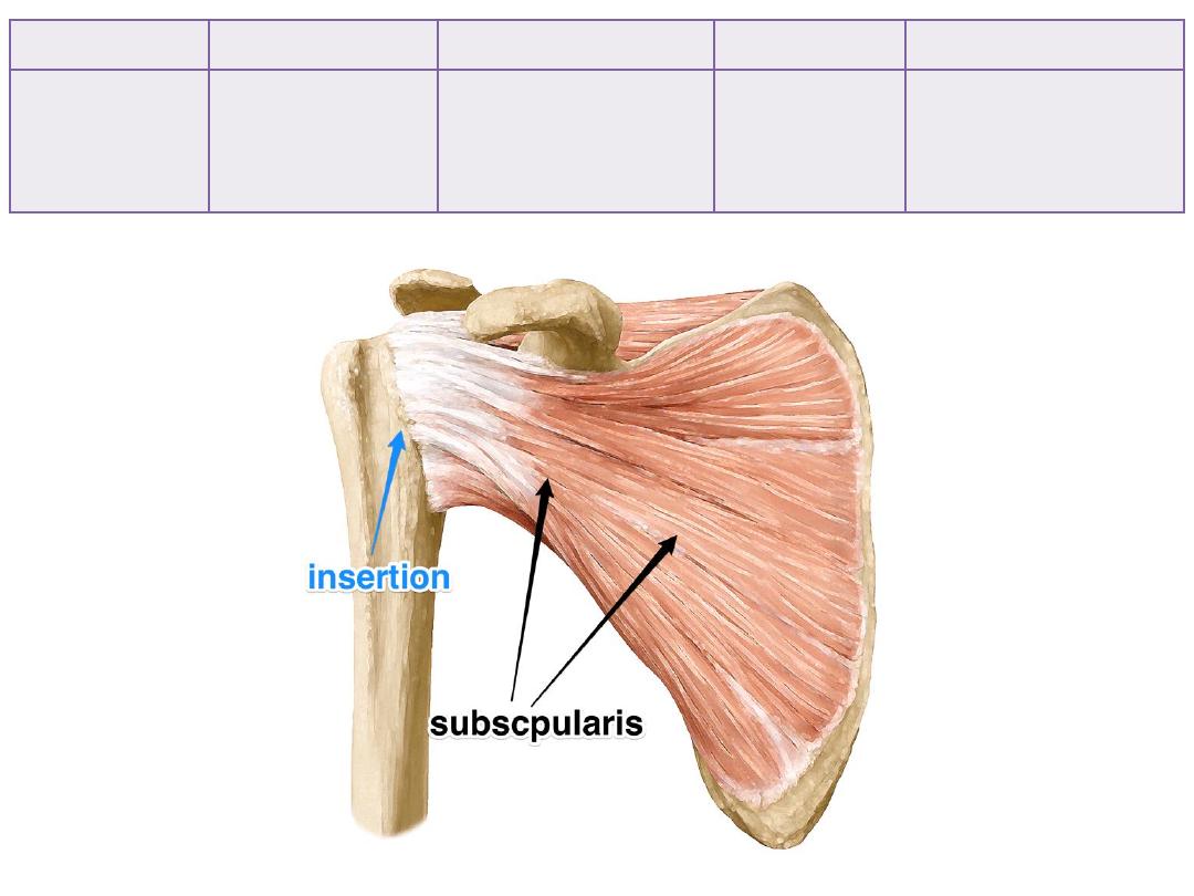
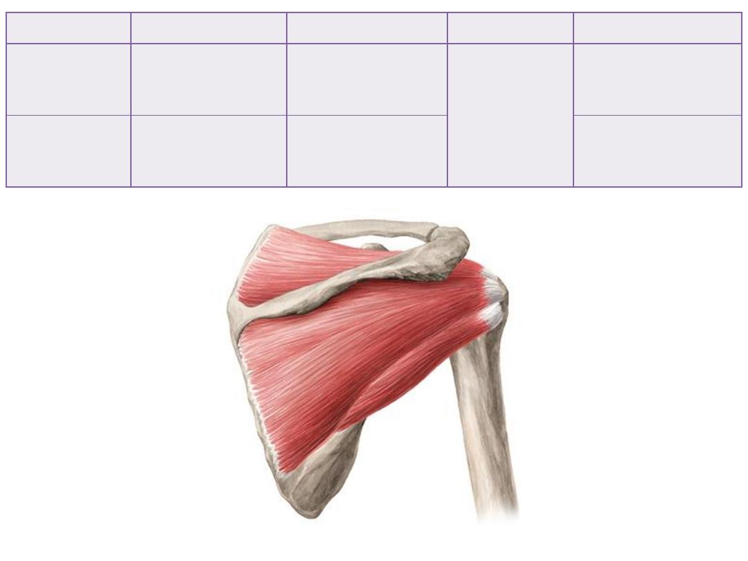
Subscapularis
Subscapular fossa
Lesser tuberosity of
humerus
Upper & lower
subscapular n
• Medial rotator
• Rotator cuff
Muscle
Origin
Insertion
Innervation
Function
Supraspinatus
Supraspinous fossa
Greater tuberosity
(upper facet)
Suprascapular n
• Initiates abduction
• Rotator cuff
Infraspinatus
Infraspinous fossa
Greater tuberosity
(middle facet)
• Lateral rotator
• Rotator cuff
Muscle
Origin
Insertion
Innervation
Function
Teres minor
Axillary border of
scapula
Greater tuberosity
(lower facet)
Axillary n
• Lateral rotator
• Rotator cuff
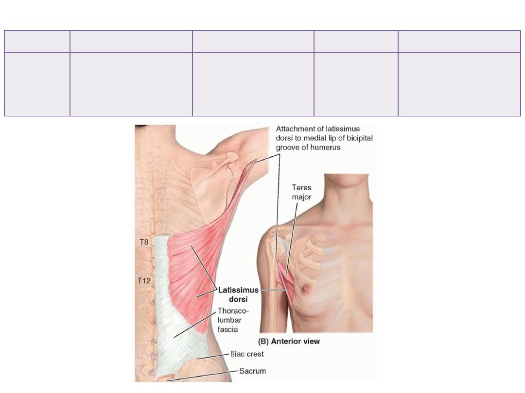
Teres Major
Oval area beside
infraspinous fossa
Medial lip of bicepital
groove
Lower
subscapular n
• Medial rotator
• Extensor
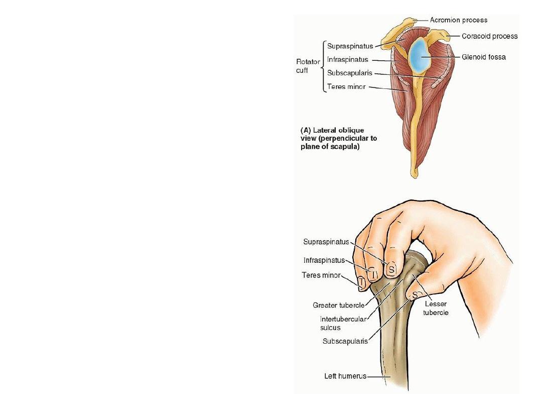
Rotator cuff:
These four muscles play important role
in stabilizing the shoulder joint in
position by:
1- Fusing with its capsule
2- Approximating the two articular
surfaces
Muscles connecting the trunk to the humerus
Muscle
Origin
Insertion
Innervation
Function
Latissimus
dorsi
• Spines of T7-12
• Iliac crest
• Lower 4 ribs
Intertubercular groove
Thoracodorsal
n
• Shoulder adductor
• Extensor
• Medial rotator
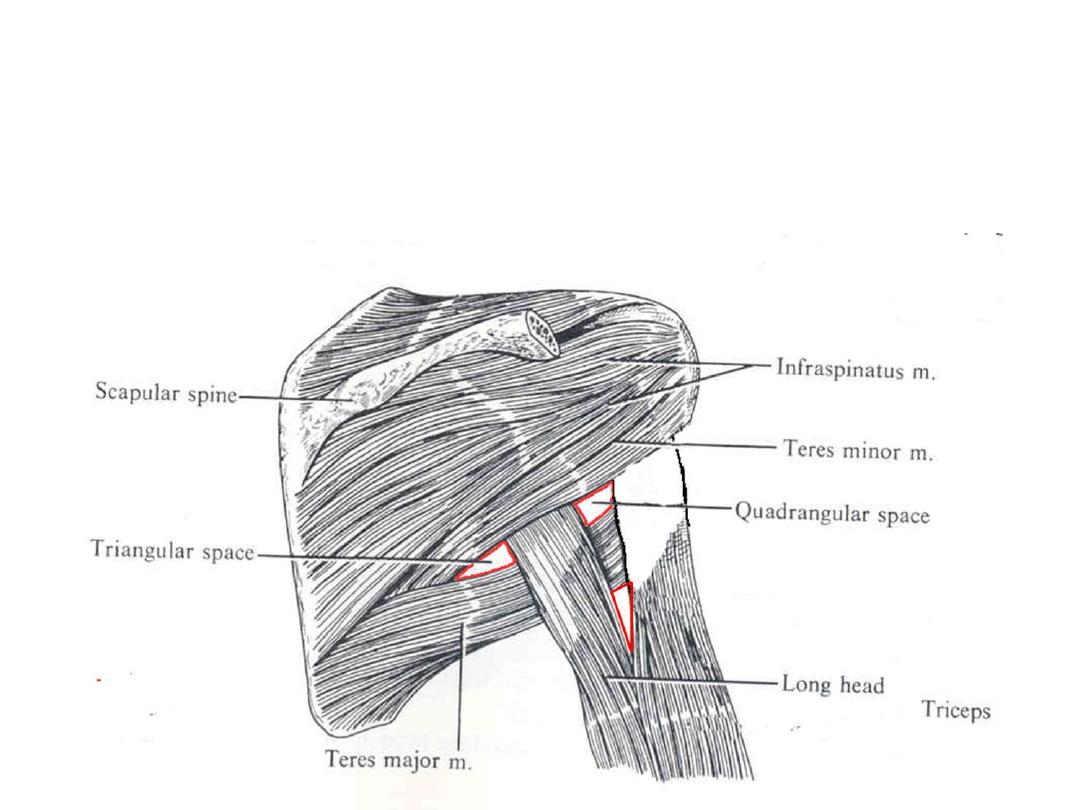
Communication with the axilla (axillary gateways):
1- Quadrangular space
2- Triangular space
3- Triangular interval
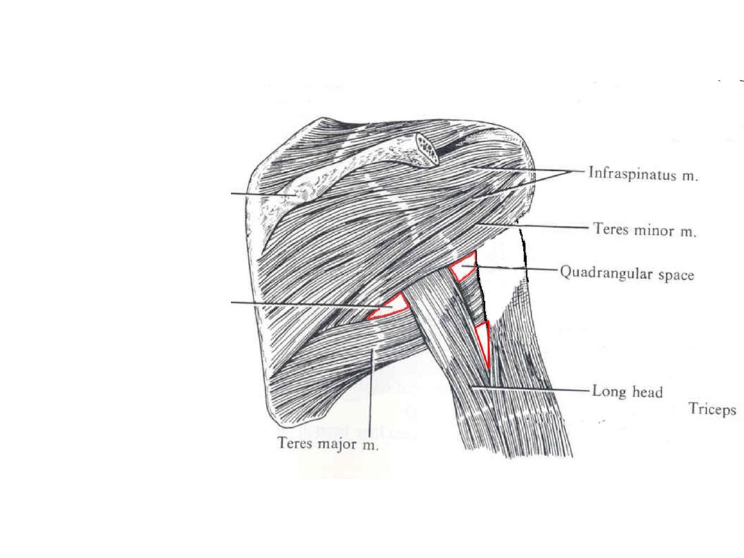
Quadrangular space:
Boundaries:
1- Teres major
2- Teres minor
3- Triceps long head
4- Humerus
Structures:
1- Axillary nerve
2- Posterior CHA
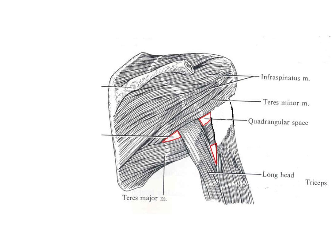
Triangular space:
Boundaries:
1- Teres major
2- Teres minor
3- Triceps long head
Structures:
Circumflex scapular n
Triangular interval:
Boundaries:
1- Teres major
2- Humerus
3- Triceps long head
Structures:
1- Radial n
2- Profundal brachii artery
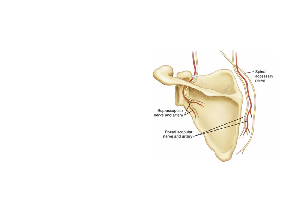
Nerves:
1- Suprascapular n (C5
& 6
):
- Branch of upper trunk of BP
- Descends to the region to pass in the
scapular
notch
underneath
the
transverse scapular ligament
- Enters supraspinatus
- Hooks around scapular spine &
reaches the infraspinatus to supply it
2- Dorsal scapular n (C5):
- Arises from BP roots
- Descends deep to levator scapulae &
rhomboids supplying them
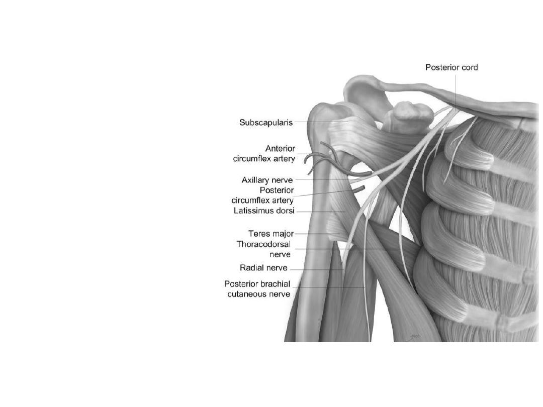
3- Thoracodorsal n (C5, 6 &7):
-
Branch of posterior cord of BP
-
Descends on subscapularis
accompanied
by
the
subscapular artery
-
Enters latissimus dorsi near
its insertion to supply it
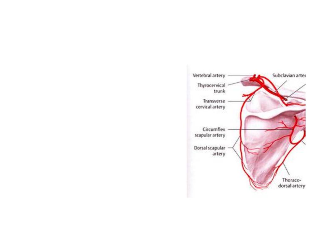
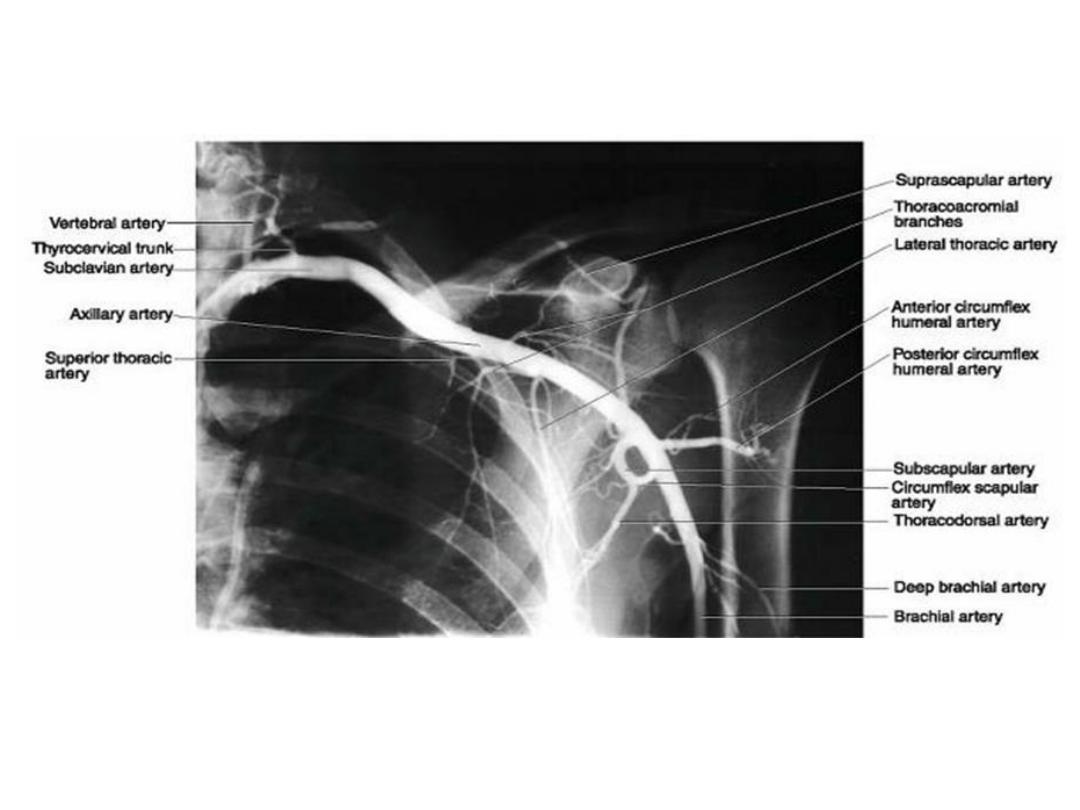
Arteries:
1- Suprascapular artery:
- Branch of 1
st
part of subclavian a
- Passes in the neck then descends
with its accompanying nerve
- Shares the same course of the nerve
but it passes above the ligament
- Shares in scapular anastomosis
2- Dorsal scapular artery:
- Arises from subclavian artery or from
its transverse cervical branch
- Descends deep to levator scapulae &
rhomboids supplying them
- Shares in scapular anastomosis
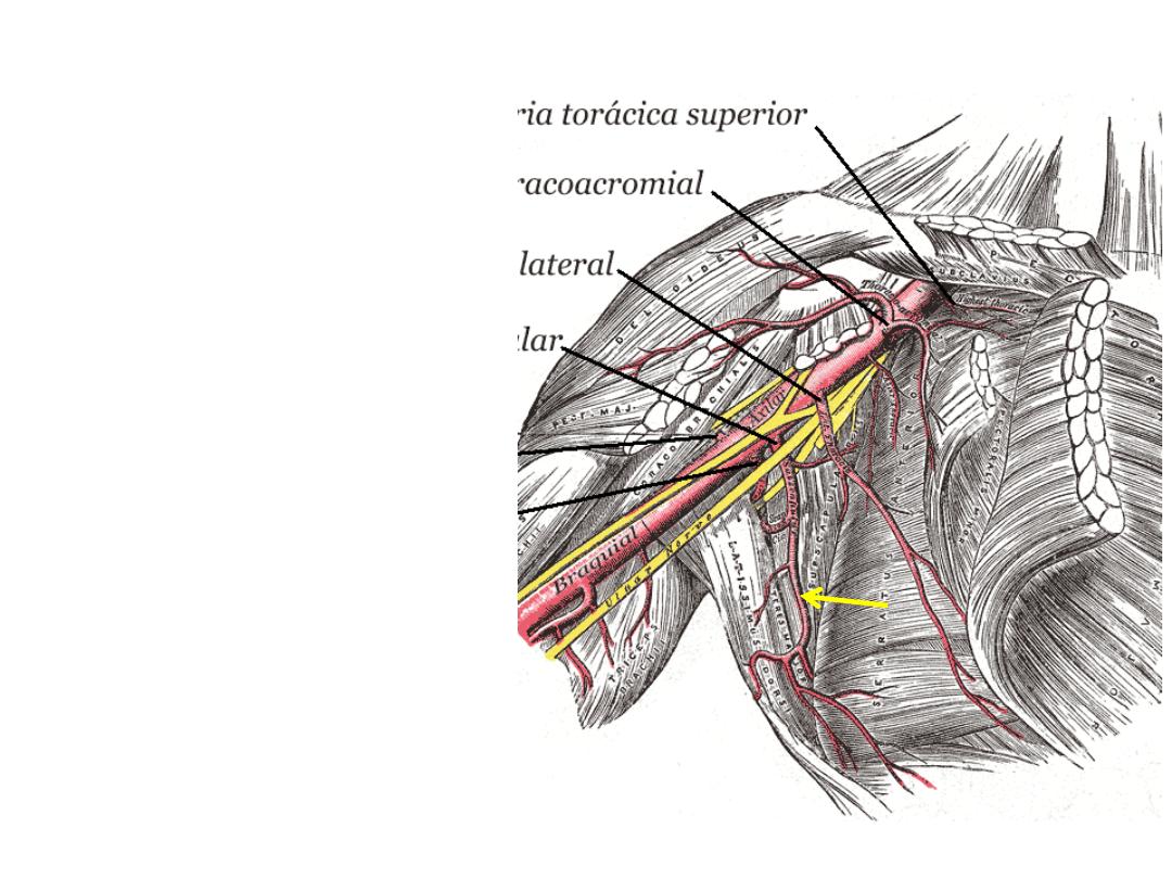
3- Subscapular artery:
-
Branch of 3
rd
part of axillary
artery
-
Descends
on
subscapularis
accompanied by thoracodorsal
nerve
-
Gives the circumflex scapular
branch to the back muscles
-
Shares in scapular anastomosis
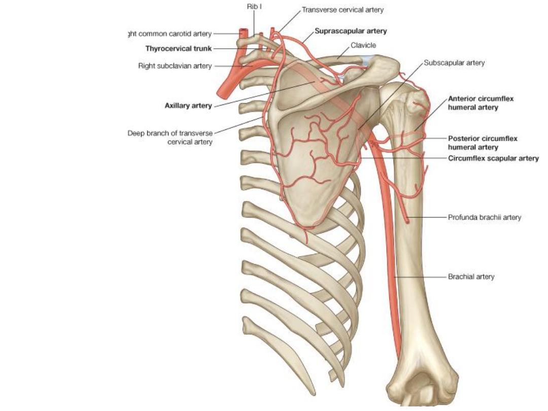
Scapular anastomosis
Connects the 1
st
part of
subclavian to the 3
rd
part
of axillary arteries