1
Lecture 08 Pathology D. Hameed
Syphilis
Definition:
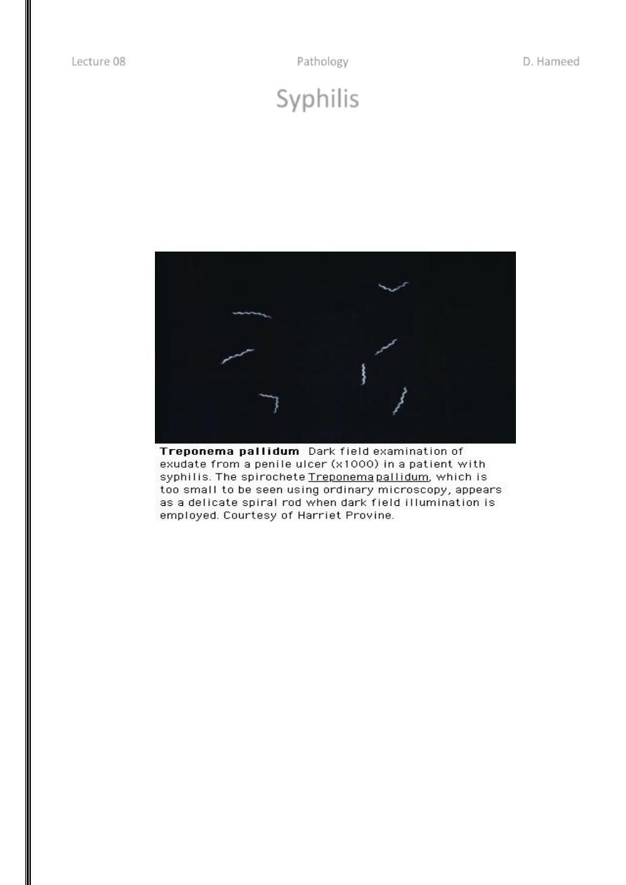
Syphilis is a systemic infection caused by the spirochete Treponema pallidium, which is
transmitted mainly by direct sexual intercourse (venereal syphilis) and less commonly via
placenta (congenital syphilis) or by accidental inoculation from the infectious Materials T.
Pallidum spirochetes cannot be cultured but are detected by silver stains, dark field examination
and immunofluorescence technique
Pathogenesis:
The organism is delicate and susceptible to drying and does not survive long outside the
body.
The organism invades mucosa directly possibly aided by surface abrasions following
intercourse with an infected person, a primary lesion, an ulcer known as the chancre,
develops at the site of infection usually on the external genetalia but also lips and
anorectal region. Within hours, the T. pallidum pass to regional lymph nodes and gain
access to systemic circulations. Thereafter, the disease is unpredictable. Its incubation
period is about 3 weeks.
Whatever the stage of the disease and location of the lesions the histologic hallmarks of
syphilis are Obliterative endarteritis Plasma cell rich mononuclear cell infiltrates.
The endarteritis is secondary to the binding of spirochetes to endothelial cells mediated by
fibronectin molecules bound to the surface of the spirochetes. The mononuclear infiltrates
are immunologic response.

2
Host humeral and cellular immune responses may prevent the formation of chancre on
subsequent infections with T. pallidum but are insufficient to clear the spirochetes.
STAGES OF SYPHILIS
Primary
Secondary
Latent
- Early latent
- Late latent
Late or tertiary
May involve any organ, but main parts are:
- Neurosyphilis
- Cardiovascular syphilis
- Late benign (gumma)
PRIMARY SYPHILIS (The Chancre)
Incubation period 9-90 days, usually ~21 days.
Develops at site of contact/inoculation.
Classically: single, painless, clean-based, indurated ulcer, with firm, raised borders. Atypical
presentations may occur.
Mostly anogenital, but may occur at any site (tongue, pharynx, lips, fingers, nipples, etc...)
Non-tender regional adenopathy Very infectious.
May be darkfield positive but serologically negative. Untreated, heals in several weeks, leaving
a faint scar.
SECONDARY SYPHILIS
Seen 6 wks to 6 mos after primary chancre
Usually w diffuse non-pruritic, indurated rash, including palms & soles.
May also cause:
- Fever, malaise, headache, sore throat, myalgia, arthralgia, generalized
lymphadenopathy
- Hepatitis (10%)
- Renal: an immune complex type of nephropathy with transient nephrotic
syndrome
- Iritis or an anterior uveitis
- Bone: periostitis
- CSF pleocytosis in 10 - 30% (but, symptomatic meningitis is seen in <1%)

3
SECONDARY SYPHILIS (Cont.)
The skin rash:
- Diffuse,
- Often with a superficial scale (papulosquamous).
- May leave residual pigmentation or depigmentation.
Condylomata Lata:
- Formed by coalescence of large, pale, flat-topped papules.
- Occur in warm, moist areas such as the perineum.
- Highly infectious.
Mucosal lesions:
- ~ 30% of secondary syphilis patients develop mucous patch (slightly raised, oval
area covered by a grayish white membrane, with a pink base that does not bleed).
- Highly infectious
Lesions of syphilis resolve without treatment although person remains infected
LATENT SYPHILIS
Positive syphilis serology without clinical signs of syphilis (& has normal CSF).
It begins with the end of secondary syphilis and may last for a lifetime.
Pt may or may not have a h/o primary or secondary syphilis.
Is divided into early and late latency.
LATENT SYPHILIS (cont.)
1. Early latent:
- The first year after the resolution of primary or secondary lesions, or
- A reactive serologic test for syphilis in an asymptomatic individual who has had a
negative serologic test within the preceding year.
- Infectious.
2. Late latent:
- Usually not infectious, except for the pregnant woman, who may transmit infection
to her fetus.
LATE SYPHILIS ‘Tertiary Syphilis’
- Is the destructive stage of the disease.
- Lesions develop in skin, bone, & visceral organs (any organ).
- The main types are:
Late benign (gummatous)
Cardiovascular &

4
Neurosyphilis
- Can be crippling and life threatening
- Blindness, deafness, deformity, lack of coordination, paralysis, dementia may occur
- It is usually very slowly progressive, barring certain neurologic syndromes which may
develop suddenly due to endarteritis and thrombosis in the CNS
- Late syphilis is noninfectious.
NEUROSYPHILIS
Divided into 5 groups, which may overlap:
Asymptomatic neurosyphilis
Syphilitic meningitis
Meningovascular syphilis
General paresis
Tabes dorsalis
TABES DORSALIS
Occurs 20-30 years after the initial infection.
It is uncommon.
More common in whites and in men.
It’s a slowly progressive, degenerative disease involving the posterior columns and
posterior roots of the spinal cord.
Results in progressive loss of peripheral reflexes, impairment of vibration and position
sense, and progressive ataxia.
Bladder incontinence & impotence are common.
Chronic destructive changes of the large joints of the affected limbs may be seen in
advanced cases (i.e., Charcot's joints).
CARDIOVASCULAR SYPHILIS
May not manifest clinically until 20-30 years after infection, but usually begins within 5-10 years
after initial infection.
Primarily aortic insufficiency and aortic aneurysm of the ascending aorta. Other large arteries
may sometimes be involved, and rarely the coronary ostia may be involved.
Caused by obliterative endarteritis of the vasa vasorum with resultant damage to the intima &
media of the great vessels, causing dilatation of the ascending aorta and eventually results in
stretching of the ring of the aortic valve, producing aortic insufficiency. The valve cusps remain
normal.
Asymptomatic aortitis is best diagnosed by visualizing linear calcifications in the wall of the
ascending aorta.
More common in men than in women and possibly in blacks than in whites.

5
Syphilitic gummas
There are grey white rubbery masses of variable sizes. They occur in most organs but in skin,
subcutaneous tissue, bone, Joints and testis. In the liver, scarring as a result of gummas may
cause a distinctive hepatic lesion known as hepar lobatum.
Collapse of the bridge of the nose and palate can occur with perforation
Osteitis and periosteitis may lead to thickening and deformity of long bones such as the sabre
tibia
Histologically, gummas look like a central coagulative necrosis characterized by peripheral
granumatous responses. TheTrepanosomas are scanty in these gummas and difficult to
demonstrate
Congenital syphilis
This infection is most severe when the mother's infection is recent. Treponemas do not invade
the placental tissue or the fetus until the fifth month of gestation (since immunologic
competence only commences then) syphilis causes late abortion, still birth or death soon after
delivery or It may persist in latent forms to become apparent only during childhood or adult life.
The out come of congenital syphilis depends on stage of maternal infection (i.e. the degree of
maternal spirochataemia). In primary and secondary stages, the fetus is heavily infected and
may die of hydrops in utero or shortly after birth. Liver and pancrease show diffuse fibrosis. The
placenta is heavy, and pale with plasmacytic villitis. After maternal second stage, the effects of
congenital syphilis are progressively less severe.
Less dramatic visceral disease, papular lesions on skin and mucosae such as the nose snuffles,
may be seen with Huchinson's teeth, and interstial keratitis.
Children infected in utero who are sero -positive show no lesions until two or more years after
birth are classified as having late congenital syphilis. The late congenital syphilis is distinctive for
the triads: Interstial keratitis; Hutchinson teeth and Eight nerve deafness
TESTS FOR SYPHILIS
Dark field Microscopy
VDRL, RPR
FTA-ABS, MHA-TP
Direct Fluorescent Antibody (DFA)
