1
Lecture 07 Pathology D. Hameed
Schistosomiasis
Schistosomiasis, AKA Bilharzia
Parasitic disease caused by several species of flatworm
Affects many in developing countries
Can contract it by wading or swimming in lakes, ponds and other bodies of water
infested with the parasite’s snail host.
A Brief History...
First described by German pathologist
Theodore Maximilian Bilharz
Bilharz performed autopsies on Egyptian
patients who had died from the disease:
found male & female parasite eggs in the liver portal system, bladder.
Later seen in Japan, called Katayama fever
o Symptoms: rash on legs, fever, diarrhoea, bloody stools
emaciation, edema
death.
Classification
Genus: Schistosoma
Species: S. mansoni
S. japonicum
S. haematobium
S. indicum
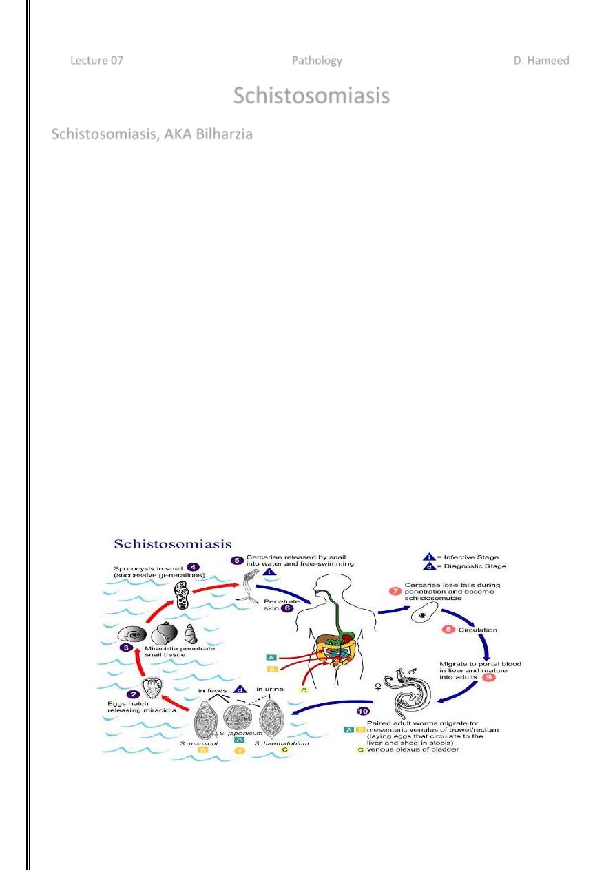
Life Cycle (Basic)
2
Life Cycle (Eggs larvae into snail)
1. Parasite eggs released into freshwater (from human
urine, feces)
2. Eggs hatch
ciliated miracidia, free swimming
3. Miracidia find & infect snail host (different species
prefer diff’t snail sp.)
4. Each miracidia transforms into many fork-tailed, free
swimming forms called cercariae within 4-6 weeks of
entering snail.
5. Cercariae leave snail and move into water at a rate of
1500/day for up to 18 days.
Life Cycle (Into human lymphatics lungs liver)
6. Cercariae find a human host, penetrate skin, and
differentiate into larval forms called schistosomulae.
7. Migrate through the host’s skin, gain access to the
lymphatic system.
8. Travel to the lungs (stay 3-8 days and ~70% are
eliminated)
9. Migrate to liver portal system, mature into male &
female adults
Life Cycle
(maturation movement to target organs egg production)
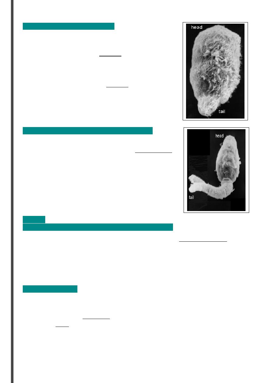
10. In liver, m & f pair up
female inserts herself into the gynecophoral canal of male
they are now ‘paired’.
11. Migrate to favoured sites:
i. S. mansoni – mesenteric venules of large bowel & rectum
ii. S. japonicum – mesenteric veins of the small intestine
iii. S. haematobium – perivesical venous plexus surrounding the bladder
Life Cycle (Egg release)
12. Females release eggs.
i. Egg characteristics
- Covered in microbarbs
cling to vascular endothelium
- Pores, which allow the release of
1. Antigens
2. Enzymes (aid in passage of eggs through host tissues)
3
13. Eggs enter lumen of excretory organs
50%
passed out of body
50%
trapped in tissues, carried away by blood circulation, lymph.
Acute Infection (Early)
Cercariae penetrate skin
rash
- called schistosome or swimmer’s itch.
Eggs laid in target organs release antigens
cause Katayama fever
- fever
- urticarial
- malaise
- diarrhea
Chronic Infection (Late)
Symptoms of chronic infection caused by eggs that travel to various parts of body
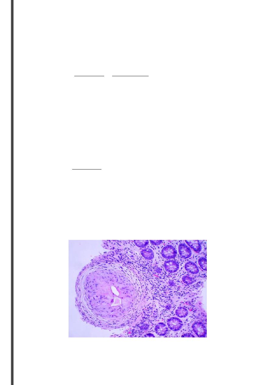
Eggs remain trapped in host tissues secrete Ags granulomatous inflammatory
immune response
Granulomas: macrophages surrounded by lymphocytes (CD4, CD8 Tcells),
which aggregate at site of infection.
Fibroblast cells also at site of infection.
During late stage of chronic infection, they replace the granulomas. Their
prolif. is stim. by factors produced by the schistosome egg, & by cytokines
from macrophages & CD4 Tcells.
Fibroblasts mediate collagen deposition in the granuloma, leading to fibrosis (=fibrous
connective tissues development
Granuloma

4
Chronic Infection (When eggs meet the GI tract)
- In S. mansoni infections
- Wall of colon is damaged as eggs pass through
- Inflamm. response
ulcers, inflammatory polyps
Can lead to fibrosis
- Clinically: diarrhea, abdominal pain
- Eggs can also accumulate in the appendix
Can lead to appendicitis (inflammation of the appendix)
Chronic Infection (When eggs meet the meet the liver/spleen)
- Hepatosplenic schistosomiasis
- Eggs carried by portal circulation
liver
- Granulomatous response
- Granulomas are walled off with fibrous tissue
fibrosis obstructs portal veins
portal
hypertension
Esophageal varices (dilated esophageal veins
bursting can cause
bleeding to death. Caused directly by portal hypertension.)
Splenomegaly (enlarged spleen)
Chronic Infection (When eggs meet the meet the heart)
- In those with severe hepatosplenic schistosomiasis
- Blood gets shunted directly back to the heart (doesn’t pass through liver).
- Eggs accumulate in heart, sometimes lodged in pulmonary arterioles.
- Form granulomas
block pulmonary circulation
pulmonary hypertension.
Can lead to right ventricular strain, and eventually cardiovascular collapse.
Chronic Infection
(When eggs meet the meet the genitourinary areas & CNS)
Genitourinary complications
Eggs lodge themselves in wall of bladder & can develop into polyps
Polyps can erode, ulcerate & cause hematuria (blood cells in urine)
Eggs lodge in ureters and urethra, cause lumps and lesions kidney failure
Eggs lodge into ovaries, the uterus, cervix, fallopian tubes lumps complications
incl. infertility
(For the men: eggs can also lodge into the testes and the prostate )
CNS complications
S. haematobium and S. mansoni can migrate to the spine
S. japonicum found in the brain and causes encephalopathy (general brain dysfunction)
5
Diagnosis
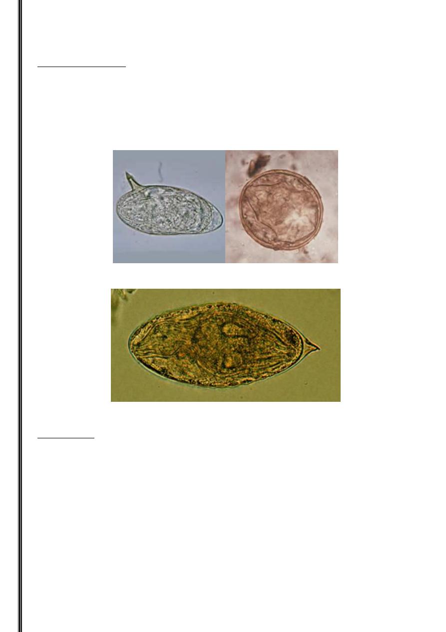
Microscopic Detection
Take stool or urine sample to detect eggs
S. haematobium eggs are oval and have a spike at the tip
S. japonicum eggs small and almost spherical with tiny spine
S. mansoni eggs have a spike on the side (spine)
S. mansoni S. japonicum
S. haematobium
Antibody tests
An earlier and more sensitive form of detection
Some complications
Cross-reactivity with other helminthic infections (other flatworm
parasites)
Can’t tell the difference between current and old infections as antibodies
stay long after infection is over.
Can’t tell you anything about overall worm burden so we can’t tell how
serious the infection is
6
Antigen tests:
Detect antigens in blood with immunoelectrophoresis
2 types are detected though share similar complications with antibody tests
Molecular detection:
20-25% of schistosomiasis genome has been sequenced can use 2 probes to detect S.
mansoni DNA in human blood
Genome sequencing has the potential to yield DNA vaccines