
1
Hormonal Control of Calcium & Phosphate Metabolism
ﺩ.ﺑﺎﻥ ﺟﺎﺑﺭ
Objectives
■
Understand the importance of maintaining homeostasis of body calcium and
phosphate concentrations, and how this is accomplished.
■
Describe the body pools of calcium, their rates of turnover, and the organs that
play central roles in regulating movement of calcium between stores.
■
Identify the major hormones and other factors that regulate calcium and
phosphate homeostasis and their sites of synthesis as well as targets of their action.
Extracellular fluid calcium concentration is normally regulated precisely,
the normal value of about 9.4 mg/dl, which is equivalent to 2.4 mmol /l.
This precise control is essential because calcium plays a key role in many
physiologic processes, including contraction of muscles; blood clotting;
and transmission of nerve impulses(Excitable cells, such as neurons, are
sensitive to changes in calcium ion concentrations, hypercalcemia cause
progressive depression of the nervous system; conversely, hypocalcemia
cause the nervous system to become more excited.
About 0.1 % of the total body calcium is in the extracellular fluid,
About 1 % is in the cells and its organelles,
The rest is stored in bones.
The calcium in the plasma is present in three forms:
40 % of the calcium is combined with the plasma proteins .
10 % of the calcium is combined with anionic substances of the
plasma and interstitial fluids (citrate and phosphate, for instance)
in such a manner that it is not ionized;
the remaining 50 % of the calcium in the plasma is ionized. This
ionic calcium is the form that is important for most functions of
calcium in the body, including the effect of calcium on the heart,
the nervous system, and bone formation.
Approximately 85 % of the body's phosphate is stored in bones,14 to 15%
is in the cells, and less than 1 % is in the extracellular fluid. Although
extracellular fluid phosphate concentration is not nearly as well regulated
as calcium concentration, phosphate serves several important functions
and is controlled by many of the same factors that regulate calcium.

2
Fluxes of Calcium and Phosphate
Maintaining constant concentrations of calcium in blood requires frequent
adjustments, which can be described as fluxes of calcium between blood and
other body compartments. Three organs participate in this process when
necessary:
The small intestine is the site where dietary calcium is absorbed.
Importantly, efficient absorption of calcium in the small intestine is
dependent on expression of a calcium‐binding protein in epithelial
cells.
Bone serves as a vast reservoir of calcium. Bone is composed of
organic matrix that is greatly strengthened by deposits of calcium
salts ; known as hydroxyapatite, which contain Ca/P ratio on a
weight basis varying between 1.3 and 2.0. Stimulating net
resorption of bone mineral , releases calcium and phosphate into
blood, and suppressing this effect allows calcium to be deposited in
bone.
The kidney is critcally important in calcium homeostasis. Under
normal blood calcium concentrations, almost all of the calcium that
enters glomerular filtrate is reabsorbed from the tubular system
back into blood
Parathyroid hormone (PTH)
Parathyroid hormone is protein hormone secreted from cells of the
parathyroid glands and finds its major target cells in bone and kidney.
Normally there are four parathyroid glands in humans; they are located
immediately behind the thyroid gland.nIt contains mainly chief cells
(secrete PTH) and a small number of oxyphil cells( not certain, but they are
believed to be modified or depleted chief cells that no longer secrete
hormone).

3
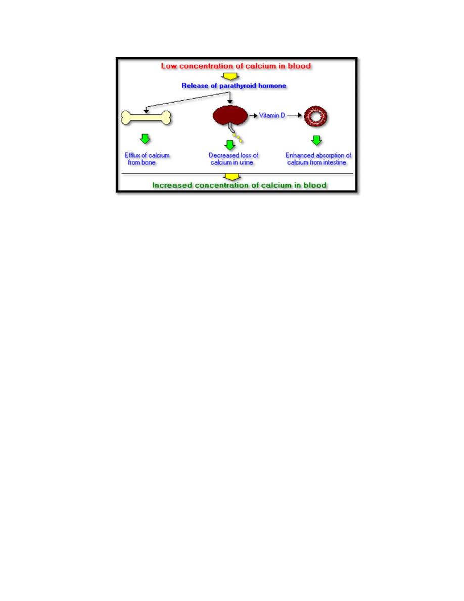
Physiologic Effects of Parathyroid Hormone
The main function of parathyroid hormone is bring calcium ion
concentrations in extracellular fluid back within the normal range if it fall
below normal. Parathyroid hormone accomplishes its job by stimulating
three processes (figure 1):
Mobilization of calcium from bone: PTH stimulates osteoclast
activity and bone resorption, but this occurs through an indirect
mechanism.
PTH binds to receptors on the adjacent osteoblasts, causing them
to release cytokines, which activates receptors on preosteoclast cells, causing
them to differentiate into mature osteoclasts. The mature osteoclasts then
release enzymes and acids that promote bone resorption.
Enhancing absorption of calcium from the small intestine: PTH
stimulates this process, but indirectly by stimulating production of
the active form of
vitamin D
in the kidney.
Vitamin D induces synthesis of
a calcium‐binding protein in intestinal epithelial cells that facilitates efficient
absorption of calcium into blood.
Suppression of calcium loss in urine: PTH stimulates tubular
reabsorption of calcium. Another effect of PTH on the kidney is to
stimulate loss of phosphate ions in urine.
Control of Parathyroid Hormone Secretion
PTH is released in response to low extracellular concentrations of free
calcium .The parathyroid cell monitors extracellular free calcium
concentration via an integral membrane protein that functions as a
calcium‐sensing receptor
. When calcium concentrations fall below the
normal range, there is a steep increase in secretion of parathyroid
hormone. Low levels of the hormone are secreted even when blood
calcium levels are high.
4
Disease States
Excessive secretion of parathyroid hormone is seen in two forms:
Primary hyperparathyroidism is the result of parathyroid gland
disease, most commonly due to a parathyroid tumor which secretes
the hormone without proper regulation. Common manifestations of
this disorder are hypercalcemia, kidney stones and decalcification of
bone.
Secondary hyperparathyroidism is the situation where disease
outside of the parathyroid gland leads to excessive secretion of
parathyroid hormone. A common cause of this disorder is kidney
disease ‐
if the kidneys are unable to reabsorb calcium, blood calcium levels
will fall, stimulating continual secretion of parathyroid hormone to maintain
normal calcium levels in blood.
Secondary hyperparathyroidism can also
result from inadequate nutrition. A prominent effect of secondary
hyperparathyroidism is decalcification of bone, leading to pathologic
fractures
Inadequate production of parathyroid hormone ‐ hypoparathyroidism ‐
typically results in decreased concentrations of calcium and increased
concentrations of phosphorus in blood. Common causes of this disorder
include surgical removal of the parathyroid glands and disease processes
that lead to destruction of parathyroid glands. The resulting hypocalcemia
often leads to tetany and convulsions, and can be acutely life‐threatening.

5
Treatment focuses on restoring normal blood calcium concentrations by
calcium infusions, oral calcium supplements and vitamin D therapy.
Vitamin D (Calcitriol)
vitamin D or calcitriol is a steroid hormone . Cholecalciferol D3 is
generated in the skin of animals when light energy is absorbed by a
precursor molecule 7‐dehydrocholesterol. Vitamin D3, does not have
significant biological activity. Rather, it must be metabolized within the
body
to
the
hormonally‐active
form
known
as
1,25‐
dihydroxycholecalciferol. This transformation occurs in two steps,
Within the liver, cholecalciferal is hydroxylated to 25‐
hydroxycholecalciferol by the enzyme 25‐hydroxylase.
Within the kidney, 1‐alpha‐hydroxylase act on 25‐
hydroxycholecalciferol, yielding 1,25‐dihydroxycholecalciferol, the
biologically active form.
1‐alpha‐hydroxylase in the kidney is tightly regulated and serves
as the major control point in production of the active hormone.
The major inducer of 1‐alpha‐hydroxylase is
parathyroid
hormone
; it is also induced by low blood levels of phosphate.Each
of the forms of vitamin D is hydrophobic, and is transported in
blood bound to carrier proteins.
Physiological Effects of Vitamin D
Vitamin D is well known as a hormone involved in mineral metabolism
and bone growth.

6
Its most dramatic effect is to facilitate intestinal absorption of
calcium, although it also stimulates absorption of phosphate and
magnesium ions. In the absence of vitamin D, dietary calcium is
not absorbed at all efficiently. Vitamin D stimulates the
expression of a number of proteins involved in transporting
calcium from the lumen of the intestine, across the epithelial cells
and into blood (calcium transporters ‐ calbindin).
The crutial effect of vitamin D on bone is to provide the proper
balance of calcium and phosphorus to support mineralization.
vitamin D has potent effects on the growth and differentiation of
many types of cells.
Disease States
Vitamin D deficiency: The classical manifestations of vitamin D
deficiency is rickets, which is seen in children and results in bony
deformaties including bowed long bones. Deficiency in adults leads to
the disease osteomalacia. Both rickets and osteomalacia reflect
impaired mineralization of newly synthesized bone matrix, and usually
result from a combination of inadequate exposure to sunlight and
decreased dietary intake of vitamin D. Vitamin D deficiency or
insufficiency occurs in several other situations, which you might predict
based on the synthetic pathway described above: like Genetic defects in
the vitamin D receptor and Severe liver or kidney disease.
Vitamin D toxicity: Excessive exposure to sunlight does not lead to
overproduction of vitamin D. Vitamin D toxicity is inevitably the result of
overdosing on vitamin D supplements. However, ingestion of excessive
(milligram) quantities of vitamin D over periods of weeks of months can
be severely toxic to humans.

7
Calcitonin
hormone
Calcitonin is a peptide hormone known to participate in calcium and
phosphorus metabolism. It is from the parafollicular or C cells in the thyroid
gland
Physiologic Effects of Calcitonin
Calcitonin plays a role in calcium and phosphorus metabolism. In particular,
calcitonin has the ability to decrease blood calcium levels at least in part by
effects on two well‐studied target organs:
Bone: Calcitonin suppresses resorption of bone by inhibiting the activity
of osteoclasts, a cell type that "digests" bone matrix, releasing calcium
and phosphorus into blood.
Kidney: Calcitonin inhibits tubular reabsorption of these two ions,
leading to increased rates of their loss in urine.
Control of Calcitonin Secretion
The most prominent factor controlling calcitonin secretion is the extracellular
concentration of ionized calcium. Elevated blood calcium levels strongly
stimulate calcitonin secretion, and secretion is suppressed when calcium
concentration falls below normal
Disease States
A large number of diseases are associated with abnormally increased or
decreased levels of calcitonin, but pathologic effects of abnormal calcitonin
secretion are not generally recognized.
• There are several therapeutic uses for calcitonin. It is used to treat
hypercalcemia resulting from a number of causes. Calcitonin also
appears to be a valuable aid in the management of osteoporosis
(excessive bone resorption and decreased bone formation)

8
Summary of Hormonal Control Systems
Maintaining normal blood calcium and phosphorus concentrations is managed
through the concerted action of three hormones that control fluxes of calcium
in and out of blood and extracellular fluid:
Parathyroid hormone
serves to increase blood concentrations of calcium.
Mechanistically, parathyroid hormone preserves blood calcium by several
major effects:
Stimulates production of the biologically-active form of vitamin D
within the kidney.
Facilitates mobilization of calcium and phosphate from bone. To prevent
detrimental increases in phosphate, parathyroid hormone also has a
potent effect on the kidney to eliminate phosphate (phosphaturic effect).
Maximizes tubular reabsorption of calcium within the kidney. This
activity results in minimal losses of calcium in urine.
Vitamin D
acts also to increase blood concentrations of calcium. It is
generated through the activity of parathyroid hormone within the kidney. The
most important effect of vitamin D is to facilitate absorption of calcium from
the small intestine. In concert with PTH, vitamin D also enhances fluxes of
calcium out of bone.
Calcitonin
is a hormone that functions to reduce blood calcium levels. It is
secreted in response to hypercalcemia and has at least two effects:
Suppression of renal tubular reabsorption of calcium. In other words,
calcitonin enhances excretion of calcium into urine.
Inhibition of bone resorption, which would minimize fluxes of calcium
from bone into blood.
Although calcitonin has significant calcium-lowing effects in some species, it
appears to have a minimal influence on blood calcium levels in humans.

9

10