Lec. No.10 Centrosome
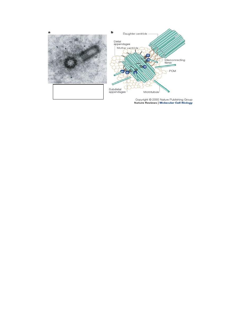
In cell biology, the centrosome is an organelle that serves as
the main microtubules organizing center ( MTOC) of the
animal cell ,it is duplicated during S phase of the cell cycle .
Centerioles , found only in animal cells, these paired
organelles are located together near the nucleus. Each
centerioles is made of nine bundles of microtubules (three per
bundle) arranged in a ring.
Just before mitosis, the two centrosomes move part until they
are on opposite side of the nucleus and organized into a
spindle-shaped formation that called spindle fibers.These
spindle fibers act as indicator for the alignment of the
chromosomes as they separate later during the process of cell
division.
Functions of centrioles.
In nondividing cells, the mother centriole can attach to the
inner side of the plasma membrane forming a basal body.
In almost all types of cell, the basal body forms a
nonmotile
In cells with a flagellum, e.g. sperm, the flagellum
develops from a single basal body. (While sperm cells
have a basal body, eggs have none. So the sperm's basal
body is absolutely essential for forming
which will form a spindle enabling the first
division of the zygote to take place.
In ciliated cells such as
o
the columnar epithelial cells of the lungs
o
6

Cilia & Flagella
Eukaryotic cilia and flagella are hair
‐like cellular appendages
composed of specialized microtubules and covered by a
specialized extension of the cellular membrane, found in
most microorganisms and animals, cilia function to move a
cell or to help transport fluid or materials past them. The
respiratory tract in humans is lined with cilia that keep
inhaled dust, and harmful microorganisms from entering the
lungs. Cilia are usually shorter and occur together in much
greater numbers than flagella.
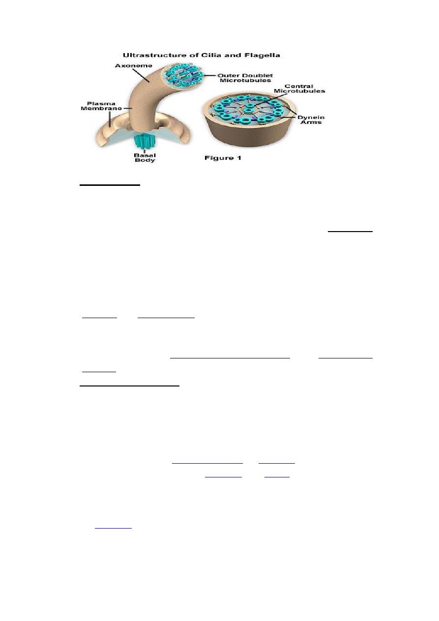
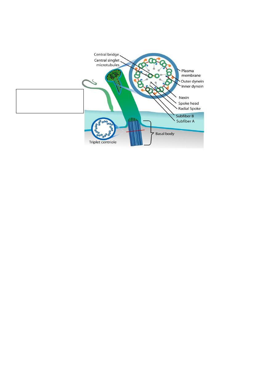
In eukaryotic cells, cilia and flagella contain the motor
protein (dynein) and (microtubles), the core of each of the
structures is termed the (axoneme) and contains two central
microtubules that are surrounded by an outer ring of nine
doublet microtubules. Dynein molecules are located around
the axoneme. A plasma membrane surrounds the entire
axoneme complex, which is attached to the cell at a structure
termed the basal body.
7
Centrosomes

Basal Body:
Basal bodies are modified centrioles that give rise to cilia and
flagella. Basal body (also known as a kinetosome). Basal
bodies maintain the basic outer ring structure of the axoneme
of the cilia and The basal body is structurally identical to the
centrioles.
Inclusions bodies
Inclusion bodies, sometimes called elementary bodies, are
nuclear or cytoplasmic aggregates of stainable substances,
usually proteins. Inclusion bodies can also be hallmarks of
genetic diseases, as in the case of Neuronal Inclusion bodies
in disorders like frontotemporal dementia and Parkinson's
disease.
Inclusions bodies: are considered to be nonliving
components of the cell that neither possess metabolic activity
nor are bounded by membranes. The most common
inclusions are glycogen, lipid droplet, pigments, and crystals.
1-Glycogen:
form of energy storage in
The olysaccharide structure represents the main storage form
of glucose in the body.
, glycogen is made and stored primarily in the
8

cells of the
and the
, hydrated with three or four
parts of water. Glycogen functions as the secondary long-
term energy storage, with the primary energy stores being
fats held in
Muscle glycogen is converted into glucose by muscle cells,
and liver glycogen converts to glucose for use throughout the
body.
2-Lipids: is a storage forms of triglycerides, are stored in
specialized cells, adipocytes, also located as individual
droplets in various cell types. Especially those of the liver,
lipids are source of energy.
3- Pigments: there are deposits of colored substances
include:
A- Melanin: the most common pigment in the body, its
dark brown pigment present in the skin, hair, retina, and
some parts of the central nervous system (C.N.S). Melanin is
produced by the
, in a
specialized group of cells known as
B-Lipofuscin: its yellow to brown pigment found in long
lived cells, like neurons of the C.N.S and cardiac muscles.
Lipofuscin pigments are membrane-bound and represent the
indigestible remnants of lysosomal activity.
C- Hemosiderin: it’s a gold-yellow pigment. It’s the end
product of Hb degradation of old red blood cells. They are
present in the liver, spleen and bone marrow.
D- Crystals: Crystals are structures of crystalline forms of
certain proteins . They are not commonly found in cells, with
the exception of steroid cells, and interstitial cells of testes,
and occasionally in macrophages.
9

