Lec.4 Cell Biology
Tools of Cell Biology1. Microscopy
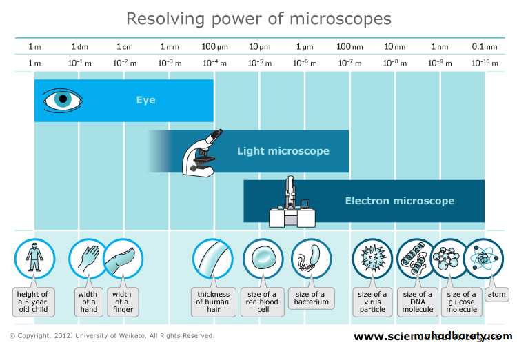
The microscope is one of the cell biologists most important tools. The vast majority of living organisms are too small to be seen in any detail with the human eye, and cells and their organelles can only be seen with the aid of a microscope. The most important features of a microscope are Magnification and Resolution.Magnification is how much bigger a sample appears to be under the microscope than it is in real life .
Resolution is the ability to distinguish between two points on an image i.e. the amount of detail.
The resolution of an image is limited by the wavelength of radiation used to view the sample. This is because when objects in the specimen are much smaller than the wavelength of the radiation being used, they do not interrupt the waves, and so are not detected. The wavelength of light is much larger than the wavelength of electrons, so the resolution of the light microscope is a lot lower. Using a microscope with a more powerful magnification will not increase this resolution any further. It will increase the size of the image, but objects closer than 200nm will still only be seen as one point.
Light Microscope :
In which light ray passing through a specimen are brought into focus by a set of glass lenses and resulting image is then viewed by the human eye . Light microscopes are able to magnify objects up to about a thousand times. Since most cells are between 1 and 100 μm in diameter, they can be observed by light microscopy, as can some of the larger subcellular organelles, such as nuclei, chloroplasts, and mitochondria. However, the light microscope is not sufficiently powerful to reveal fine details of cell structure, The limit of resolution of the light microscope is approximately 0.2 μm; two objects separated by less than this distance appear as a single image, rather than being distinguished from one another.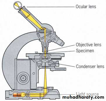
Light microscope
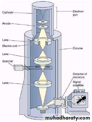
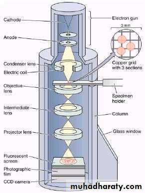
Electron Microscope :
An electron microscope is a microscope that uses a beam of electrons as a source of illumination. The electron microscope can achieve a much greater resolution than that obtained with the light microscope because the wavelength of electrons is shorter than that of light. Thus, under optimal conditions, the resolving power of the electron microscope is approximately 0.2 nm. The (E.M.) can magnify 250,000 times or more.
Two types of electron microscopy—transmission and scanning—are widely used to study cells
Transmission electron microscope (TEM):
The specimen is embedded in plastic and then cut into extraordinarily thin section.
A section placed on small metal grid .The electron beam passes through the specimen and then falls through the specimen and then falls onto photographic plate or a fluorescent screen .The result is a two dimensional image.
Scanning electron microscopy (SEM):
The specimen is coated with thin film of gold or some other metal.
Here the electron beam is projected on the sample. The electrons do not go through the sample but bounce off. This way it is possible to visualize the surface structure of the specimen. The S.E.M. image is viewed on a type of television. The result is a three dimensional image.
(TEM) (SEM)
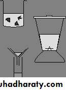
3- Cell fractionation
This means separating different parts and organelles of a cell, so that they can be studied in detail. All the processes of cell metabolism (such as respiration or photosynthesis) have been studied in this way. The most common method of fractionating cells is to use differential centrifugation:1. Cut tissue (e.g. liver, heart, leaf, etc) in ice-cold isotonic buffer. Cold to stop enzyme reactions, isotonic to stop osmosis, and buffer to stop pH changes.
2. Grind tissue in a blender to break open cells.
3. Filter. This removes insoluble tissue (e.g. fat, connective tissue, plant cell walls, etc). This filtrate is now called a cell-free extract, and is capable of carrying out most of the normal cell reactions.
4. Centrifuge filtrate at low speed
(1 000 x g for 10 min)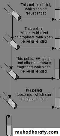
5. Centrifuge supernatant at medium speed
(10 000 x g for 30 min)6. Centrifuge supernatant at high speed
(100 000 x g for 1 hour)7. Centrifuge supernatant at very high speed
(300 000 x g for 3 hours)8. Supernatant is now organelle-free cytoplasm
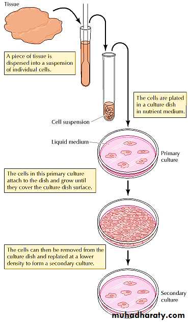
4-Growth of Animal Cells in Culture
Cell culture is the process by which cells are grown under controlled conditions, generally outside of their natural environment.
Animal cell culture are initiated by the dispersion of a piece of tissue into suspension of its components cells , which is then added to a culture dish containing nutrient media . In addition to slats and glucose , the media used for animal cell cultures contain various amino acids and vitamins , which the cells can not make for themselves .
Most animal cell types attach and grow on the plastic surface of dishes used for cell culture .The initial cell cultures established from a tissue are called primary cultures .The cells in a primary culture usually grow until they cover the culture dish surface. They can then be removed from the dish and replaced at a lower density to form secondary cultures.
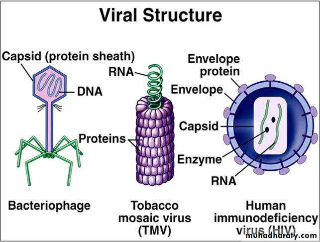
5. Viruses
Viruses are intracellular parasites that cannot replicate on their own. They reproduce by infecting host cells and usurping the cellular machinery to produce more virus particles. In their simplest forms, viruses consist only of genomic nucleic acid (either DNA or RNA) surrounded by a protein coat. Viruses are important in molecular and cellular biology because they provide simple systems that can be used to investigate the functions of cells. Because virus replication depends on the metabolism of the infected cells.