
1
General examination
Important steps in general examination of a child:
Setting.
Position.
Consciousness and orientation.
Respiration.
State of nutrition and hydration.
Body examination.
Vital signs.
Anthropotric measures.
Special things if present.
1-Setting:
Introduce yourself.
Gel.
Good light.
Right side.
Patient lie flat and central.
Exposure from nipple to mid-thigh.
2-Position:
Flexed with fisted hands as the patient we saw in the ward.
Extended posture frog like.
Opisthotonous his head and his heels on the bed while his body is arched
commonly seen in kernicterus "bilirubin encephalopathy".
Tetanus neonatorum.
Decerberate and decorticate posture.
3-Consciousness and orientation:
Conscious, lethargic, comatose.
Lethargic with crying on examination only (implying serious problem).
Orientation: in child of more than 4-5 years old >> ask about (place, time, person).
Ibnlatef
Notes
Pediatrics

2
4-Respiration:
Regular or not, seek about sign of respiratory distress if present.
Chyne stokes breathing hyperpnea + pause, indicates severe brain insult,
respiratory failure or heart failure, type 1 hypoxia is imminent PO2 <92%, type 2 PCO2
>45mmHg + hypoxia.
Chyne stokes breathing pattern is normal in premature or in neonate (1 week) old age.
5-State of nutrition and dehydration:
CHO deficiency.
Protein deficiency edema.
Fat deficiency medial aspect upper thigh & buttock "sites of storage of fat"
Vitamins and minerals.
Water and electrolytes (signs of dehydration).
Note: wrinkling of skin in the area around the thigh is sign of wasting look for
abdominal distension + eversion of umbilicus.
6-Body examination:
General:
o
Age and sex.
o
Any external corrections (cannula, IV fluid, oxygen mask).
o
Built (average build, thin, emaciated, obese).
Shape of the head:
o
Normal.
o
Bracheocephaly.
o
Scelocephaly.
Hair:
o
Distribution.
o
Fragile or not.
o
Thick or silky.
o
Discoloration reddish color of the hair in malnutrition, failure to thrive.
o
Alopecia (loss of hair) localized as in skin disease or generalized as SLE.
Fontanels:
o
Examine it when baby is sitting and not crying.
o
Size normal 2.5 cm if large decrease of bone hypothyroidism.
o
Depressed or sunken dehydration.
o
Bulging increased intra-cranial pressure ICP – hypernatremia – fluid therapy.
o
Anterior fontanel diamond shape – close in 6-18 months.
o
Posterior fontanel triangular shape – close in 3 months.

3
Face Skin color:
o
Pallor anemia (pallor check), nephrotic syndrome (off colored), hypopituitarism,
shock.
o
Jaundice increased serum bilirubin ((jaundice appears clinically when increase
more than 3.5 mg/dl in child and 5 mg/dl in neonate)).
o
Plethoric face (red color face) polycythemia, vasodilation, vascular overload.
o
Pinkish color face polycythemia, chronic hypoxia.
o
Earthy pale complexion uremia.
o
Pigmentation racial, actinic, in disease like Addison's.
o
Malar flush in mitral stenosis.
Eye:
o
Anemia look at palpebral conjunctiva.
o
Polycythemia congested conjunctiva.
o
Jaundice look at sclera.
o
Puffiness (edema of the eyelids) in renal disease and myxedema and allergic.
o
Xanthelasma yellowish plaques around the eye.
o
Sub-conjunctival hemorrhage in bleeding tendency, conjunctivitis, severe cough.
o
Sunken eye dehydration.
o
Tears on crying or not.
o
Any discharge (like pus).
o
White spots in the iris Vit. A deficiency.
o
Signs of dehydration sunken eye + dryness (tears and glistening).
Ear:
o
Discharge.
o
Large or small ears.
o
Low set ears.
o
Boat ear (congenital).
Nose:
o
Nasal discharge.
o
Look inside for any polyps.
o
Bleeding.
o
Flaring of ala nasi (sign of respiratory distress).
Lips:
o
Cyanosis.
o
Ulcer.
o
Herpes labialis.
o
Angular stomatitis and cheilosis Iron deficiency anemia & vitamin deficiency.
Gums:
o
Red + swollen + suppuration gingivitis.
o
Gingival hypertrophy in scurvy, leukemia, drugs like phenytoin.
o
Bleeding gums inflammation, Vit. C deficiency.

4
o
Chelosis vitamin deficiency.
Teeth:
o
Number of teeth.
o
Dental caries.
o
Teeth loss.
Tongue:
o
Color red in glossitis, pale in severe anemia, yellow in jaundice, blue in central
cyanosis.
o
Moisture dry tongue in dehydration and air and drugs like anticholinergic.
o
Fur in air breathers.
o
Smooth tongue in anemia.
Buccal mucosa:
o
Thrush candida infection.
o
Aphthus ulcer.
o
Petechial hemorrhage bleeding tendency and infection.
o
Pigmentation Addison's disease.
o
Pallor anemia.
o
Dryness of the mouth sign of dehydration.
Congenital anomalies:
o
Cleft lip and cleft palate and Cleft uvula.
Neck:
o
Lymphadenopathy ((L.N in neck + axillary + inguinal + epi-trochlear L.N near elbow
enlargement of two L.N in non-adjacent site called generalized
lymphadenopathy)).
o
Neck mass and Thyroid.
o
Swelling midline or lateral.
o
Using of accessory muscle in respiration sign of respiratory distress.
Chest:
o
Abnormal shape.
o
Rachitic rosary beaded ribs in rickets.
o
Signs of dyspnea flaring of ala nasi – cyanosis – dusky – suprasternal, intercostal,
subcostal rescission.
Abdomen:
o
Abdominal distention distention (5F) – flat – scaphoid.
o
Skin rash allergy, contact dermatitis, candidiasis.
o
Sings of wasting loss of muscle + loss of subcutaneous fat + look at thigh,
buttock, arm and pectoralis major muscle.
o
Sings of dehydration skin turgor – elasticity.
Groin:
o
Wasting loss of muscle bulk.
o
Thinning loss of subcutaneous fat (exam thickness of skin fold).

5
o
L.N.
o
Hernia in pediatric (indirect inguinal hernia = swelling of the scrotum).
Lower limbs:
o
Joint swelling and deformities (knee joint swelling) and Muscle wasting.
o
Edema (on the shaft of the tibia – dorsum of foot pressure at least for 1 min).
o
Bowing of leg in rickets.
o
Ankle joint widening in rickets.
o
Color jaundice, pallor, cyanosis.
o
Nails pallor – koilonychias (chronic iron deficiency anemia) – leukonychia (in liver
disease and hypo-proteinemia).
o
Fungal infection of the foot.
Back:
o
Sacral edema.
o
Pigmentation and Rash.
o
Meningocele and myelomeningeocele.
o
Vertebral column pass your finger along the vertebral column.
Upper limbs:
o
Abnormal movements and Joint swelling and deformities.
o
Muscle wasting (wasting of thinner or hypo-thinner muscles).
o
Skin color anemia, cyanosis, jaundice, pigmentations.
o
Skin lesions purpura, petechiae, purpupic spots, ecchymosis, hematoma.
o
Palmer erythema, spider navei, central pallor of the palm.
o
Nails clubbing, koilonychias, onycholysis ((GIT causes of clubbing in pediatric
are: celiac disease, cystic fibrosis, liver cirrhosis, IBD)).
o
Hand moisture.
o
Skin retraction.
o
Creases indicate Hg less than 7 – pallor indicate Hg less than 12.
o
Widening of wrist joint on rickets.
7-Vital signs:
(all of them calculated by chart or using the following method)
Blood pressure:
o
5 methods:
Auscultation: cuff = 2/3 of arm circumference.
Palpitory method: only systolic.
Flushing pale red.
Osmometry.
Doppler.
o
There is special chart for blood pressure:
Example: 4 years child BP = 4+90/4+60 = 94/64 mmHg
Age in years + 90
Age in years + 60
6
Temperature:
o
From Tympanic membrane (more common), Oral, Axillary (+0.5), Rectal (-0.5).
o
One degree increase lead to 10 beat increase in the heart rate.
o
36.5 – 37.5 = normal.
o
< 36.5 = sub-normal.
o
< 35 = hypothermia.
o
> 37.5 = febrile.
o
Less than 38 = Low grade fever.
o
More than 38 = High grade fever.
o
> 39 = hyperthermia.
o
> 41 = hyperpyrexia.
Respiratory rate:
o
2 months age 60/min.
o
2 months – 1 year 50/min.
o
1 year – 5 years 40/min.
o
5 yeas – 10 years 30/min.
o
More than 10 years 20/min.
o
Periodic breathing: occurs when the breath pause for up to 10 seconds at time,
there may be several such pauses close together, followed by series of rapid
shallow breaths, then breathing returns to normal. This is common condition in
premature babies in first few weeks of life. Even healthy full term babies sometimes
spells periodic breathing, usually after sleeping deeply. Home care: supine position,
avoid soft pillows and smoking, never snake your baby to breath brain injury.
Periodic breathing
Apnea
Breathing stops up to 10 seconds
Stops more than 20 seconds
No
Infant may become limp
No cyanosis
Cyanosis
No change in heart rate
Decrease heart rate
Pulse rate:
o
Measures:
Newborn (< 1 month) 120-160 bpm.
Infant (1-12 month) 80-140 bpm.
Toddler (1-3 year) 80-130 bpm.
Preschooler (3-5 year) 80-120 bpm.
School age (6-12 year) 70-100 bpm.
'Adolescent (> 13 year) 60-100 bpm.
o
Rate:
Tachycardia: Fever, shock, drugs (salbutamol), sinus tachycardia, anemia,
thyrotoxicosis.

7
Bradycardia: sick sinus syndrome, athletes, cretinism, drugs (propanol), sleeping,
heart block, heart failure.
o
Rhythm:
Regular – regular.
Regular – irregular (ectopic).
Completely irregular.
Radio-femoral delay: post ductal coarctation of aorta.
Radio-radial delay: pre ductal coarctation of aorta.
Brachio-femoral delay.
o
Character:
Jet of pulse: e.g. big and thrusting pulse.
Watson's water hammer pulse.
Gallop rhythm: can be assessed by palpation, we find S1, S2, S3, tachycardia.
DDx: heart failure and valvular heart disease.
o
Volume: small volume, normal volume, large volume.
o
Pulsus paradoxus: decrease in systolic blood pressure >15 mmHg with inspiration,
occur in asthma and acute pericarditis.
o
Non-cardiac causes of large volume pulse Thyrotoxicosis, Severe anemia, Stress.
o
Cardiac causes of small volume Aortic stenosis, Coarctation of aorta, Pericardial
effusion, Cardiac tamponade.
o
Causes of radial pulse absence Arteriovenous fistula, TAR: Thrombocytopenia-
absent radius syndrome (Thrombocytopenia, absence of radial artery, congenital
absence of radius bone)
o
Tachycardia + small volume in shock or diarrhea.
o
Water hummer (collapsing pulse) large volume, dorsum of hand.
o
Differential cyanosis: cyanosis present in foot, but not hand coarctation of aorta.
o
By ending of pulse examination: 80 bpm, regular, normal character, good volume,
no radio-femoral delay, normal peripheral pulsation.
Capillary refill.
Pulse oximetry.
Blood glucose.
8-Anthropotric measures:
Weight
:
o
Normal Birth weight 2.5 - 4.5 kg.
o
<2.5 kg low birth weight.
o
<1.5 kg very low birth weight.
o
<1 kg extremely low birth weight.
o
Baby double his weight at 6 months.
Post ductal coarctation:
Bluish discoloration of
the lower limbs but not
the upper limbs & head

8
o
Triple at 1 year.
o
Quadruple at 2 year.
o
Every year 3.5 kg increase (10 g/day).
Height:
o
Normal birth 50 cm.
o
First year 75 cm.
o
Second year 85 cm.
o
Forth year 100 cm.
o
After that 6 cm/year.
OFC = occiputo-frontal circumference:
o
At birth 35 cm.
o
2 cm per month in the first 3 months.
o
1 cm per month in 3-6 months.
o
0.5 cm per months in 6 months – 1 year.
o
12 cm in one year.
o
10 cm in the rest of life.
o
At birth = 35 cm.
o
At 6 months = 44 cm.
o
At 1 year = 47 cm.
Notes:
Indication for measuring blood pressure below 3 years:
o
Cardiac case.
o
Renal case.
o
CNS case.
OFC in chart:
o
95-5 normal.
o
Below 5 microcephaly.
o
Above 95 macrocephaly – megalocephaly – hydrocephaly.
Height in chart:
o
95-5 normal.
o
Below 5 short stature.
o
Above 95 long stature.
o
Measure length (lying) if baby less than 2 years.
o
Measure height (stand) if baby more than 2 years.
Weight in chart:
o
95-5 normal.
o
Below 5 marasmus – kwashiorkor – marasmus on kwashiorkor.
o
Above 95 obese.
9
Other notes:
o
Causes of macrocephaly: Familiar, big ventricles, fluid (hydrocephalus), big bone
(rickets or thalassemia major).
o
In acute illness weight is most affected anthropometric measure.
o
In chronic illness length is most affected anthropometric measure.
o
TB and bronchiectasis decrease weight.
o
Asthma increase weight (due to steroids use) and cause short stature.
Tiny child vs. stunted growth:
o
In tiny child the height and weight both decreased in a similar manner and often
there is a history of tinny child in family (seek about similar condition in family).
o
While in stunted growth the height and weight are severely decreased and may be
not the same and there is no similar condition in the family and often associated
with other diseases.
9-Special things if present:
Hydrocephalus, clubbing, cyanosis.
Sown syndrome features.
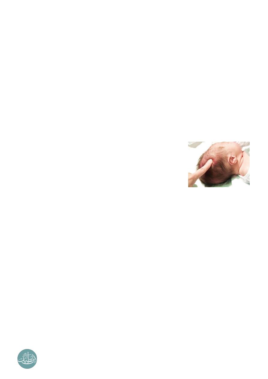
Craniotabes
o
It is a softening of the skull bones.
o
Can be a normal finding in infants, especially premature infants.
o
It may occur in up to one third of all newborn infants.
o
It is harmless in the newborn, unless it is associated with other problems these
can include rickets and osteogenesis imperfecta (brittle bones).
o
Maneuver press the bone along the area where the bones of the skull come
together "posterior parietal". The bone often pops in and out, similar to pressing on
a ping-pong ball if the problem is present. No testing is done unless osteogenesis
imperfecta or rickets is suspected.
----------------------------------------------------------------------------------------------
www.facebook.com/ibnlatef
https://goo.gl/RpvNsl