Pathology
Lab 1&2
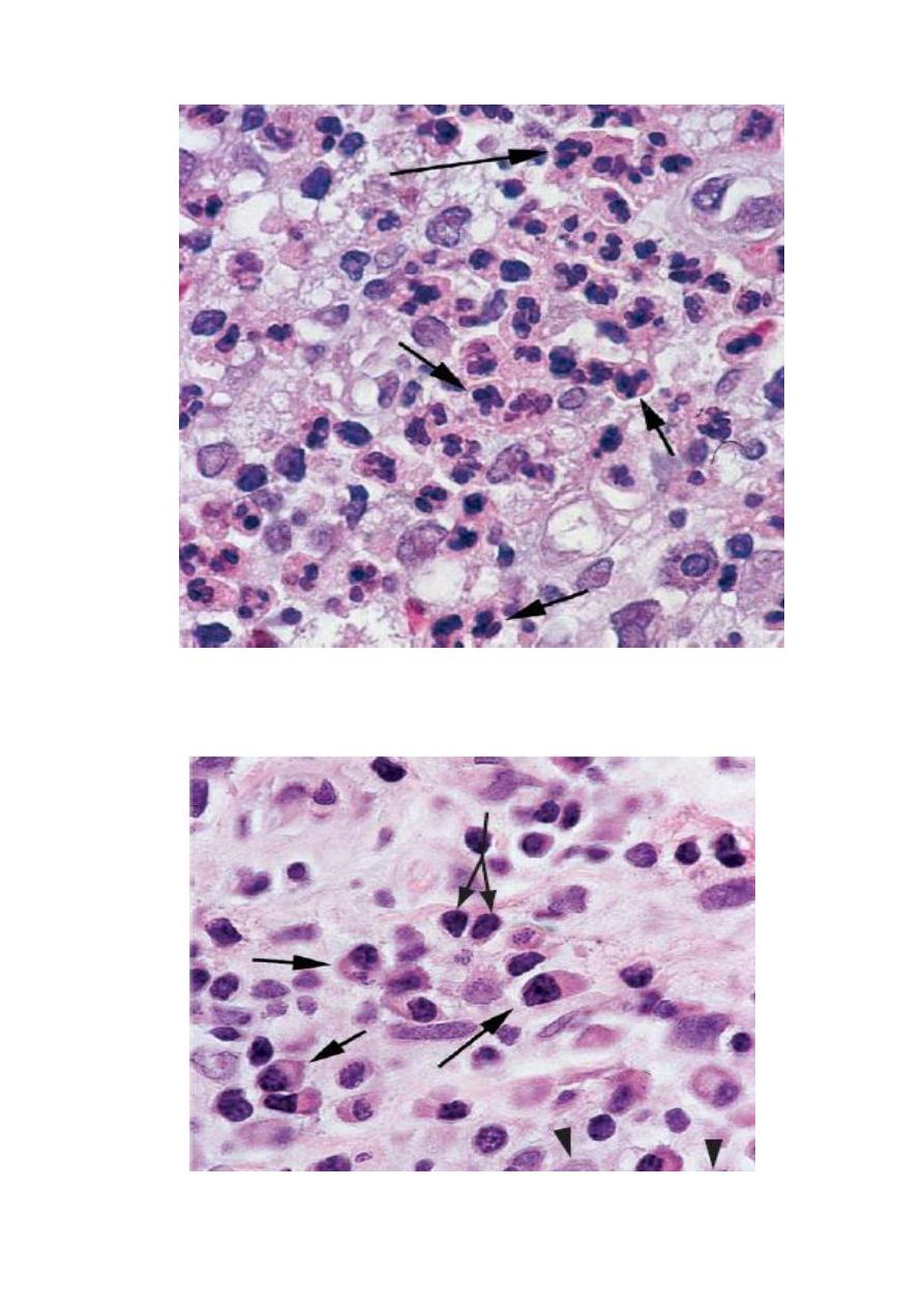
Chronic inflammation. Lymphocytes (double-headed arrow),
plasma cells (arrows) and a few macrophages (arrowheads) are
present.
Acute inflammation with densely packed polymorphonuclear
neutrophils (PMNs) with multilobed nuclei (arrows), With edema (red
arrws).
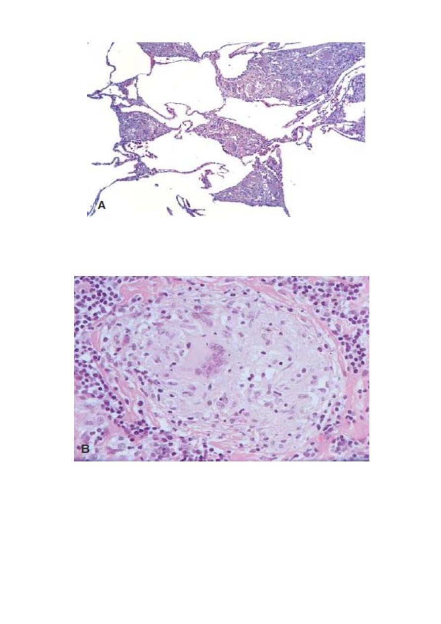
A. Section of lung from a patient with sarcoidosis reveals numerous discrete
granulomas
.
B. A higher-power photomicrograph of a single granuloma in a lymph node from
the same patient depicts a multinucleated giant cell amid numerous pale epithelioid
cells. A thin rim of fibrosis separates the granuloma from the lymphoid cells of the
node.
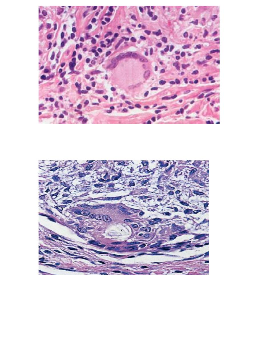
A foreign body giant cell has numerous nuclei randomly arranged in the
cytoplasm.
A Langhans giant cell shows nuclei arranged on the periphery of an abundant
cytoplasm.
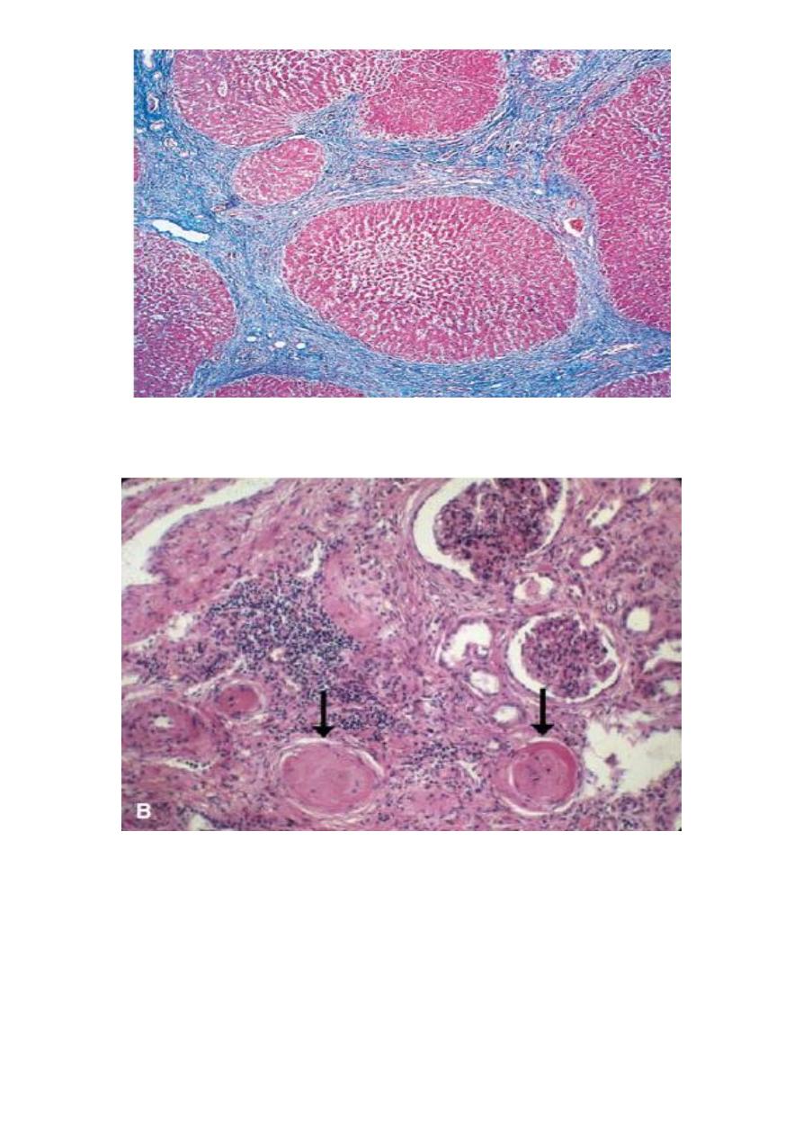
A microscopic section shows regenerating nodules (red ) surrounded by
bands of connective tissue (blue).
Many glomeruli have been destroyed and appear as circular scars
(arrows).
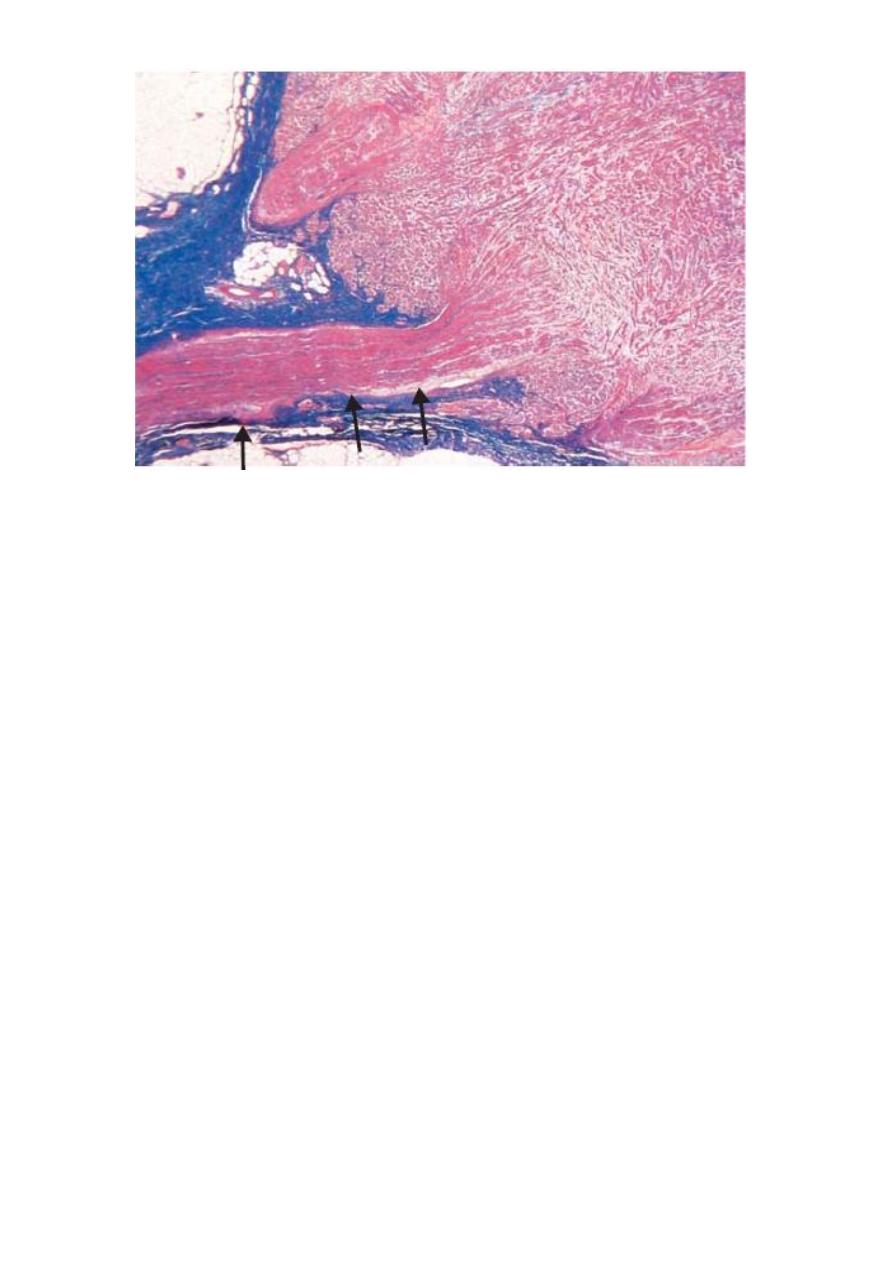
Traumatic neuroma. In this photomicrograph, the original nerve
(arrows) enters the neuroma. The nerve is surrounded by dense
collagenous tissue, which appears dark blue with this trichrome stain.
Excessive repair obstructs axonal reconnection.
Lab:-3
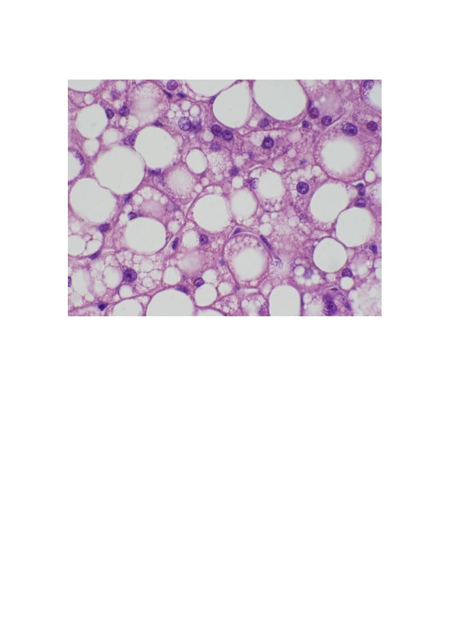
1- Fatty change:-
Organ:- Liver
Lesion:-
1-Enlargment of hepatocyte.
2-Acccumulation of fatty material inside the cytoplasm in a vacuoles with
different size and shapes.
3-Hepatocyte nucleus pushed to one side by a large droplet of fat make the cell
ring like shaped (sign cell).
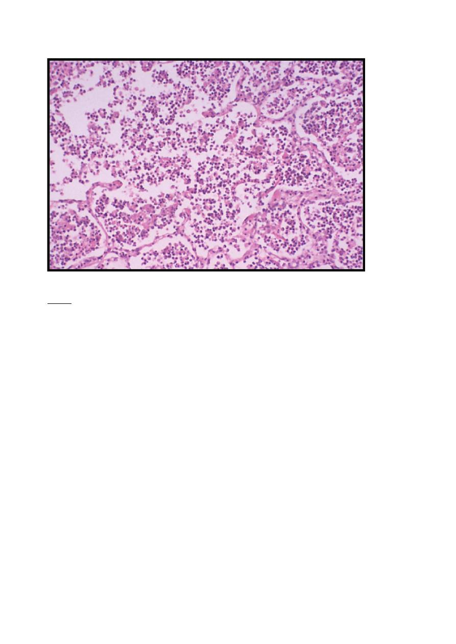
2-liquifactive necrosis:-
Organ:lung
Lesion:
1-loss of cellular and architectural details of tissue.
2-present of necrotic foci.
3-present of liquid material, which represent the pus.
4-present of neutrophils.
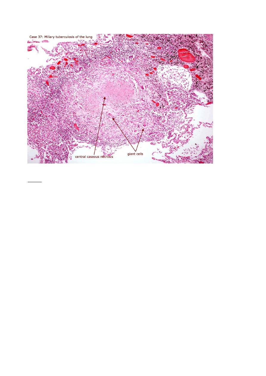
3-Caseous necrosis:
Organ:
Lesion:
1- loss of cellular and architectural details of tissue.
2- present of homogenous mass at the necrotic area.
3- present of neutrophils with giant cell (C shaped).
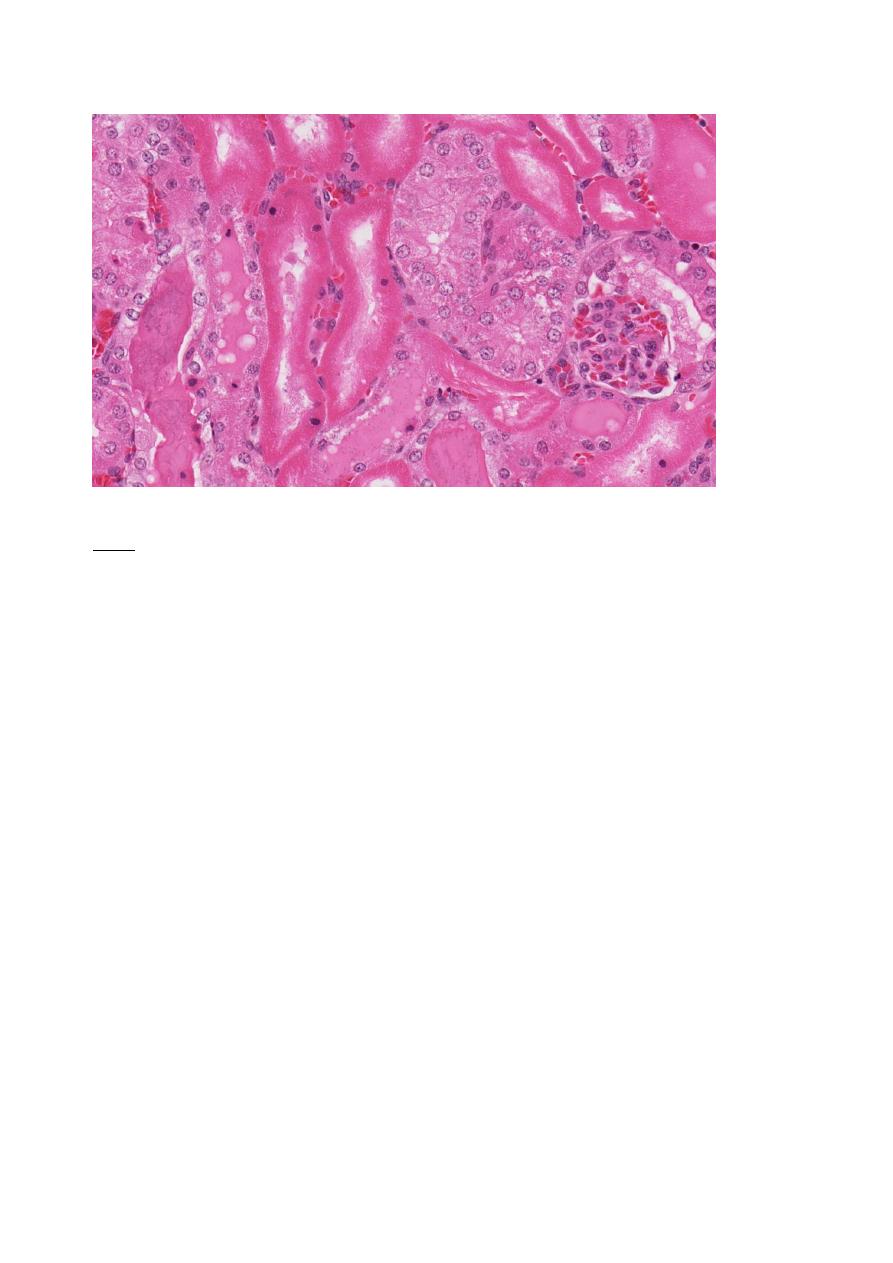
4-Coagulativenecrosis:
Organ:kidny
Lesion:
1- loss of cellular but architectural details of tissue remain.
2- Pyknosis
3- Karyonheis of nucleus
4- Karyolusis of nucleus
5- Ghost stage
6- hemorrhage
Lab 4:-
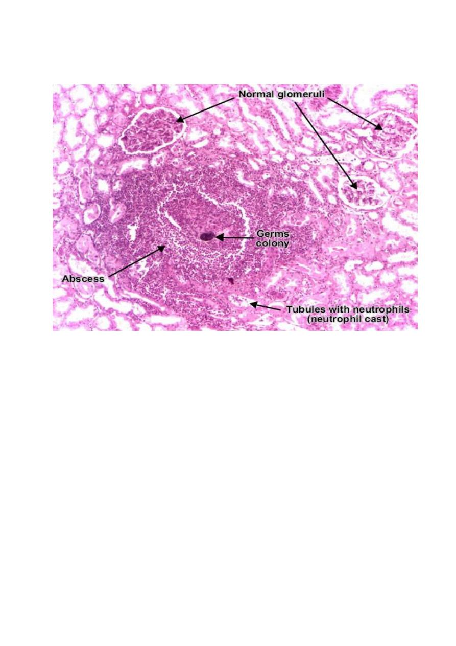
The renal interstitium presents abscesses (suppurative necrosis), consisting of
purulent exudate (pus) : neutrophils, fibrin, cell debris and central germ colonies
(hematoxylinophils). Tubules are damaged by exudate and may contain neutrophil
casts, which can be found in urine. In the early stages, glomeruli and vessels are
spared
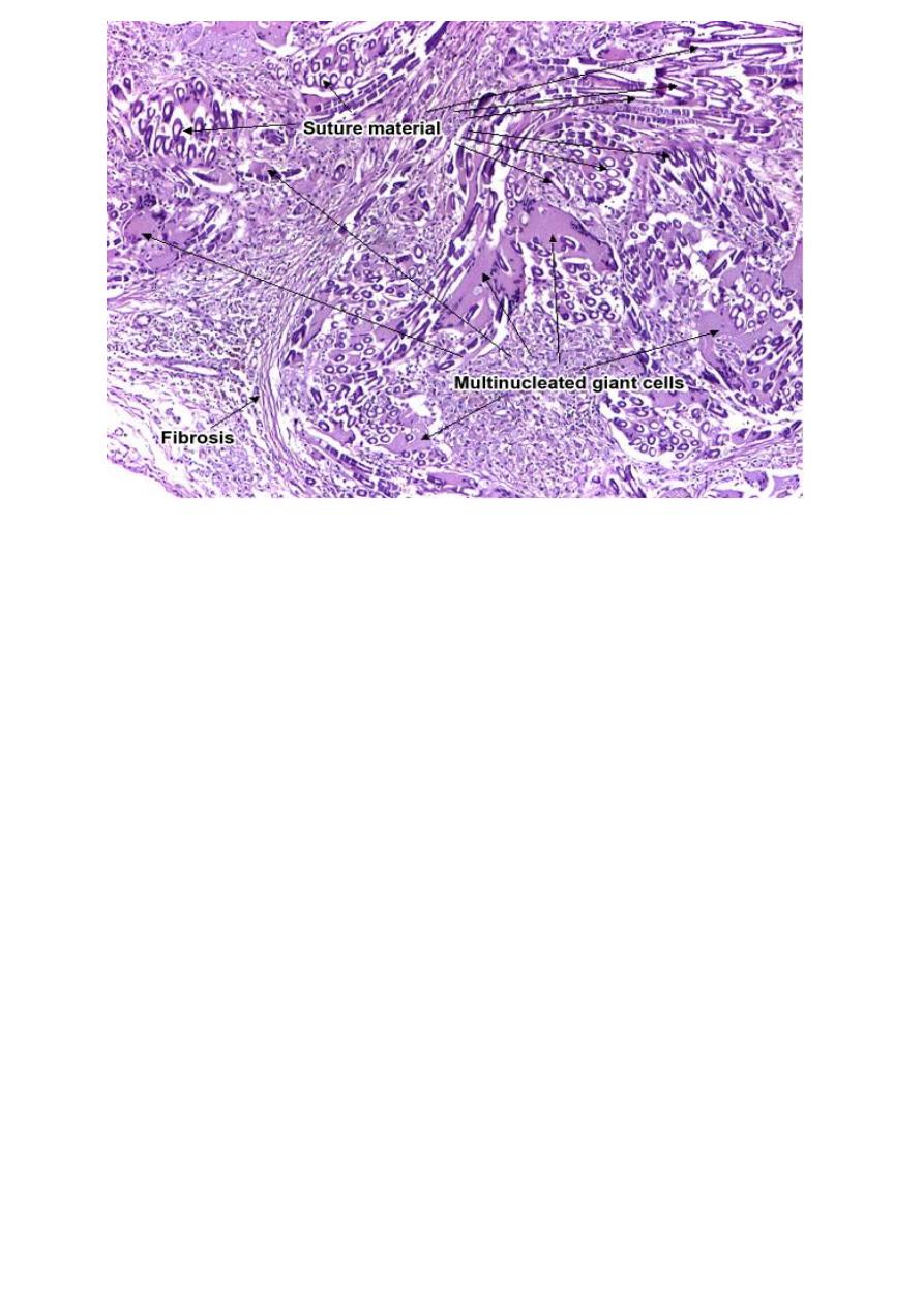
Microscopically, foreign body granuloma to suture material (nylon, silk) contains
multinucleated giant cells, with haphazardly arranged nuclei. These giant cells are
fused macrophages. The foreign body is birefringent, and sometimes may be
visible by polarized light in the middle of the granuloma or inside the giant cells.
These granulomas are non-necrotic. (HE, ob. x4).
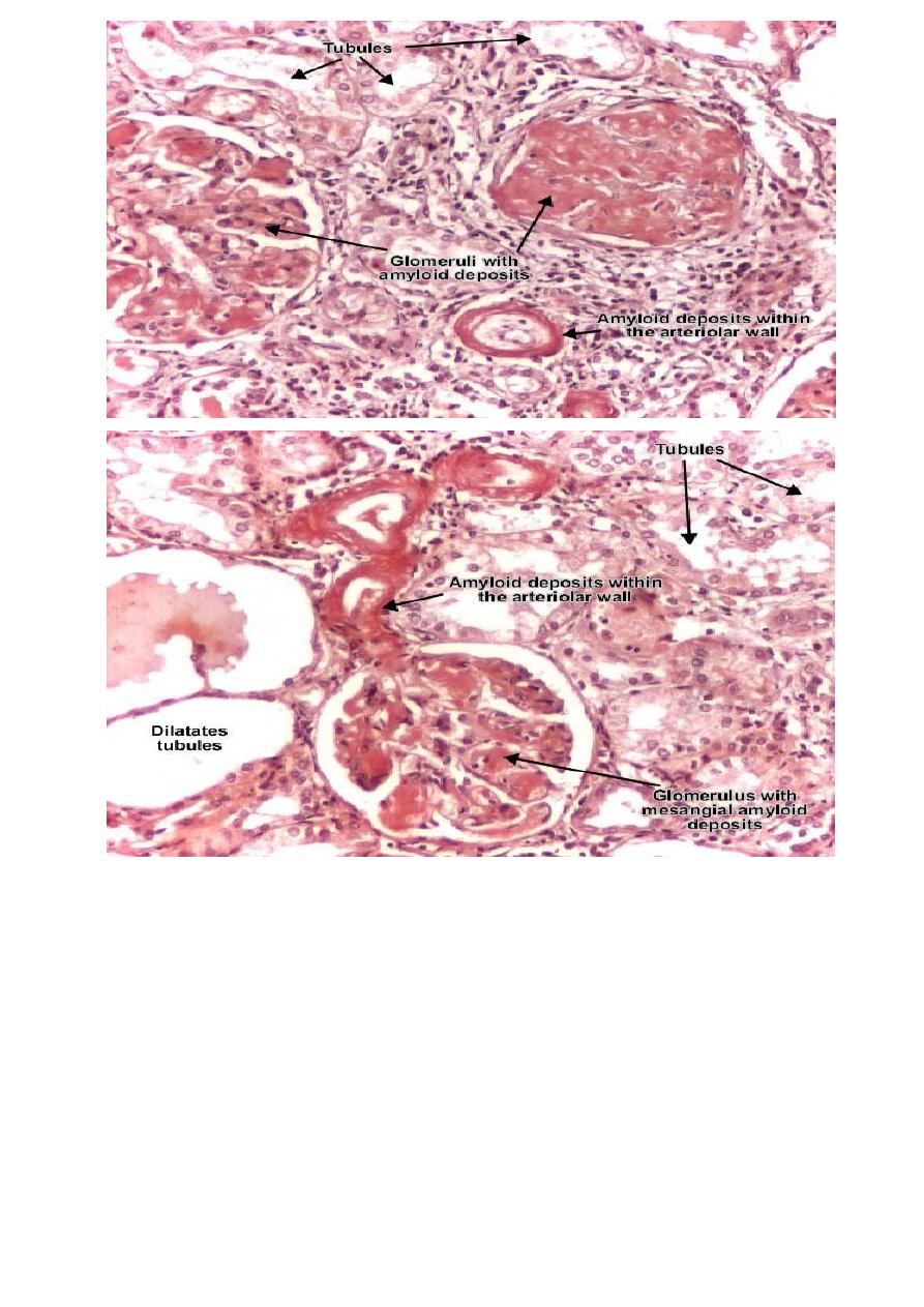
Amyloid (an abnormal protein) accumulates as extra-cellular deposits, nodular or
diffuse, as pink, amorphous material. Initially, the deposits appear in the glomeruli:
within the mesangial matrix and along the basement membranes of the capillary
loops. Continuous accumulation of the amyloid will compress and obliterate the
capillary tuft. With progression, amyloid deposits appear also peritubular and
within the arteriolar wall, narrowing them. Congo red is a special staining, elective
for amyloid. (Congo Red, ob. x20).
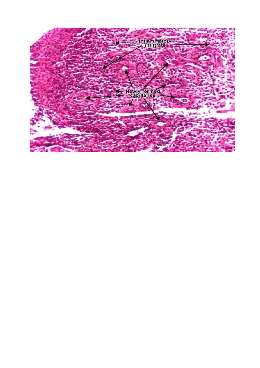
Healing (repair) by connective tissue has the granulation tissue as a hallmark. It
consists of new capillaries (result of proliferation of endothelial cells -
angiogenesis or neovascularization) in an edematous atmosphere of fibroblasts
(spindle shaped), myofibroblasts, mononuclear inflammatory cells, macrophages,
neutrophils, cellular debris. (Hematoxylin-eosin, ob. x10).
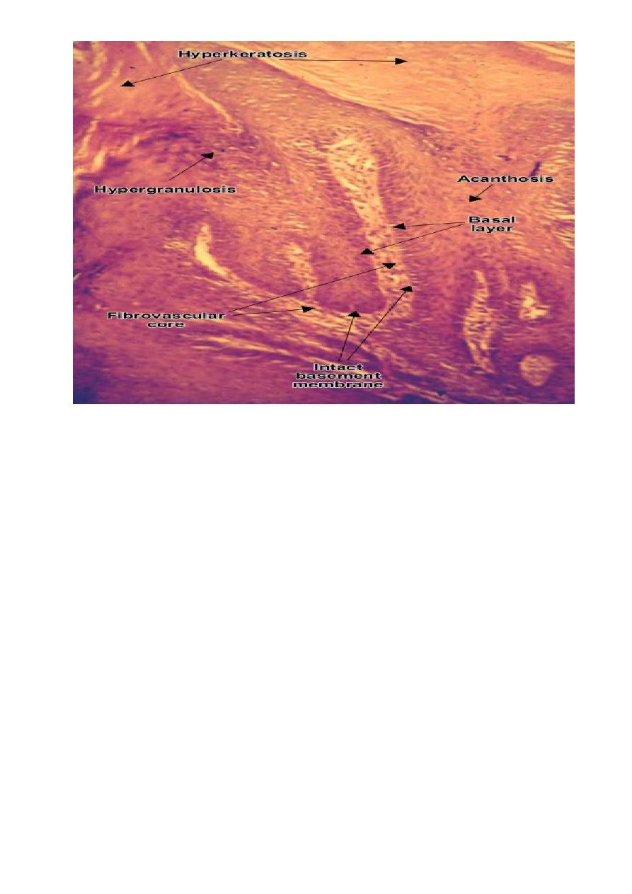
Squamous cell papilloma (verruca vulgaris) is a benign epithelial tumor. Tumor
cells proliferate and produce finger-like or warty projections; secondary, the
subjacent vessels and connective tissue (fibrovascular core) grows to sustain and
feed the tumor. The tumor cells resemble normal squamous cells, but there is an
increase of the layers number: acanthosis, hypergranulosis and hyperkeratosis. The
basement membrane is intact. (H&E, ob. X10).
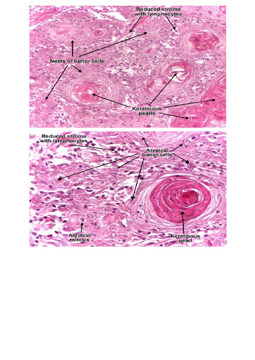
Tumor cells transform into keratinized squames and form round nodules with
concentric, laminated layers, called "cell nests" or "epithelial/keratinous pearls".
The surrounding stroma is reduced and contains inflammatory infiltrate
(lymphocytes). Poorly differentiated squamous carcinomas contain more
pleomorphic cells and no keratinization. (H&E, ob. x10).
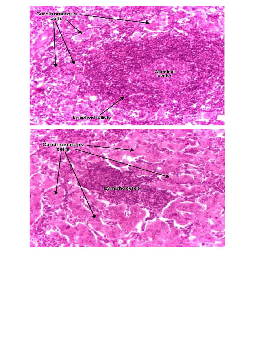
Lymph node with carcinoma metastasis : clusters of tumor cells, atypical, with
carcinomatous character. (H&E, ob. x20)
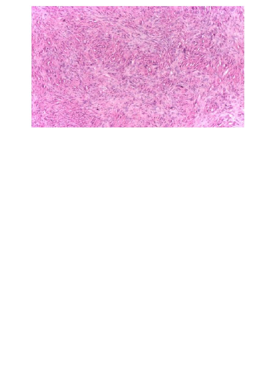
Fibrosarcoma (fibroblastic sarcoma) is a malignant connective (soft) tissue tumor
which originates from fibroblasts. The tumor may present different degrees of
differentiation : low grade (differentiated), intermediate malignancy and high
malignancy (anaplastic). The degree of differentiation is set according with:
resemblance of tumor cell with mature fibroblast (spindle-shaped), amount of
collagen secretion and mitotic rate. Tumor cells are arranged in short fascicles
which split and merge, giving the appearance of "fish bone". Poorly differentiated
tumors consist in more atypical cells, pleomorphic, giant cells, multinucleated,
numerous atypical mitoses and reduced collagen production. Presence of immature
blood vessels (sarcomatous vessels lacking endothelial cells) favors the
bloodstream metastasizing. (Hematoxylin-eosin, ob. X20)
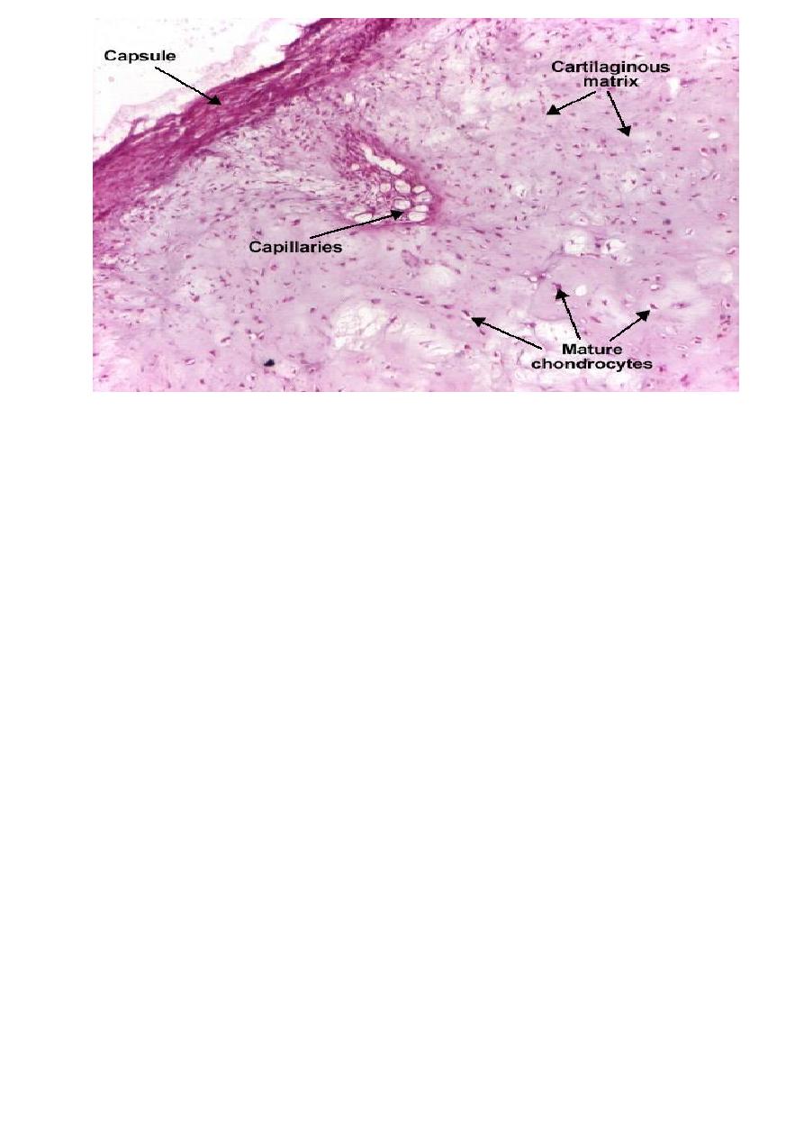
Chondroma is a benign cartilaginous tumor, encapsulated, with a lobular growing
pattern. Tumor cells (chondrocytes, cartilaginous cells) resemble normal cells and
produce the cartilaginous matrix (amorphous, basophilic material). Characteristic
are the vascular axes within the tumor, which make the distinction with normal
hyaline cartilage. (H&E, ob. x10)
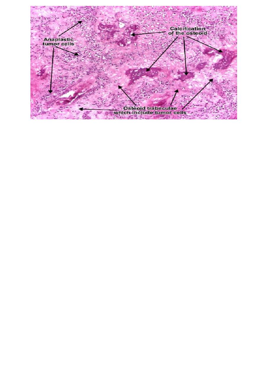
Osteosarcoma (osteogenic sarcoma) is a malignant tumor whose neoplastic cells
present osteoblastic differentiation and form tumor bone. Tumor cells are very
pleomorphic (anaplastic), some are giant and present numerous and atypical
mitotic figures. These cells produce osteoid describing irregular trabeculae
(amorphous,
eosinophilic/pink)
with
or
without
central
calcification
(hematoxylinophilic/blue, granular) - tumor bone. Tumor cells are included in the
osteoid matrix. Cartilage may be present. Presence of immature blood vessels
(sarcomatous vessels lacking endothelial cells) favors the bloodstream
metastasizing. (Hematoxylin-eosin, ob. x10)
Lab 5
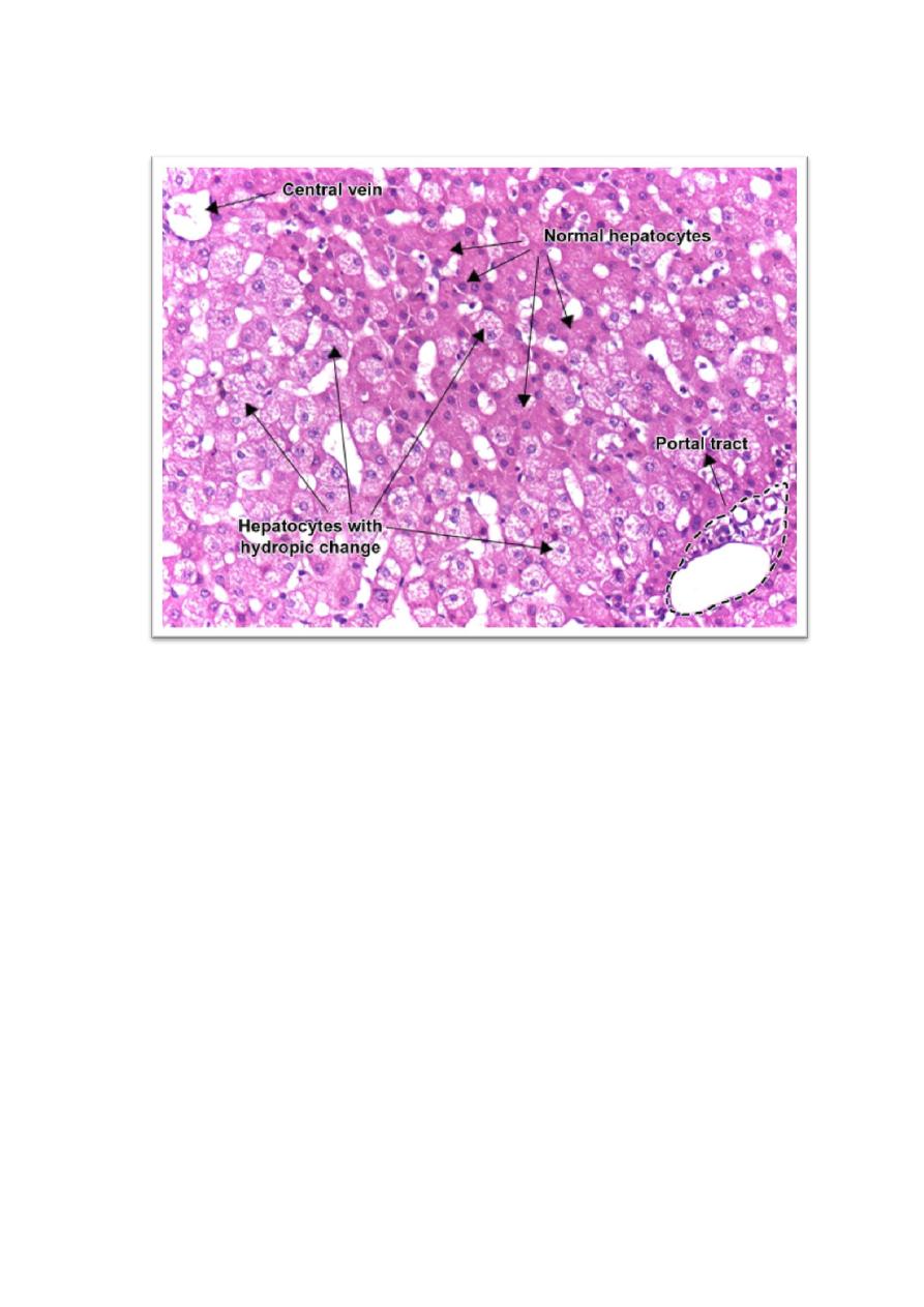
Organ:- liver.
Diagnosis:- Cellular swelling.
Lesion:- The cells are enlarged, with a clear cytoplasm and a normal nucleus in central
position.
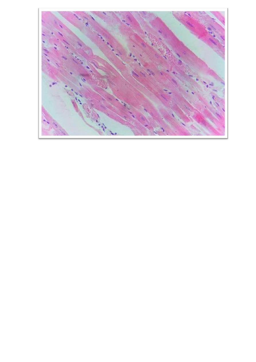
Organ:-
Skeletal muscles.
Diagnosis:-
Zenker's degeneration.
Lesion:-
The muscle fibers are swollen, have a loss of cross striations, and show a
hyaline appearance.

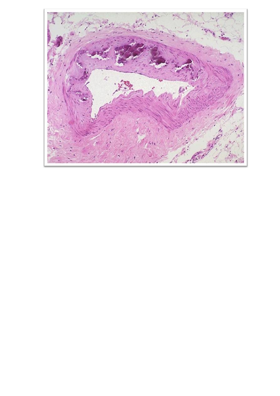
Organ:-Coronary artery.
Diagnosis:-Atherosclerosis (fibrous plaque).
Lesion:- The atheromatous fibrous plaque is localized in the intima of the artery, beneath
the endothelium, producing the thickening of the wall and, secondary, the narrowing of
the lumen and the atrophy of the muscular layer. The fibrous plaque contains collagen
fibres(eosinophilic), precipitates of calcium (hematoxylinophilic) and rare lipid-laden
cells. (H&E, ob. x4).
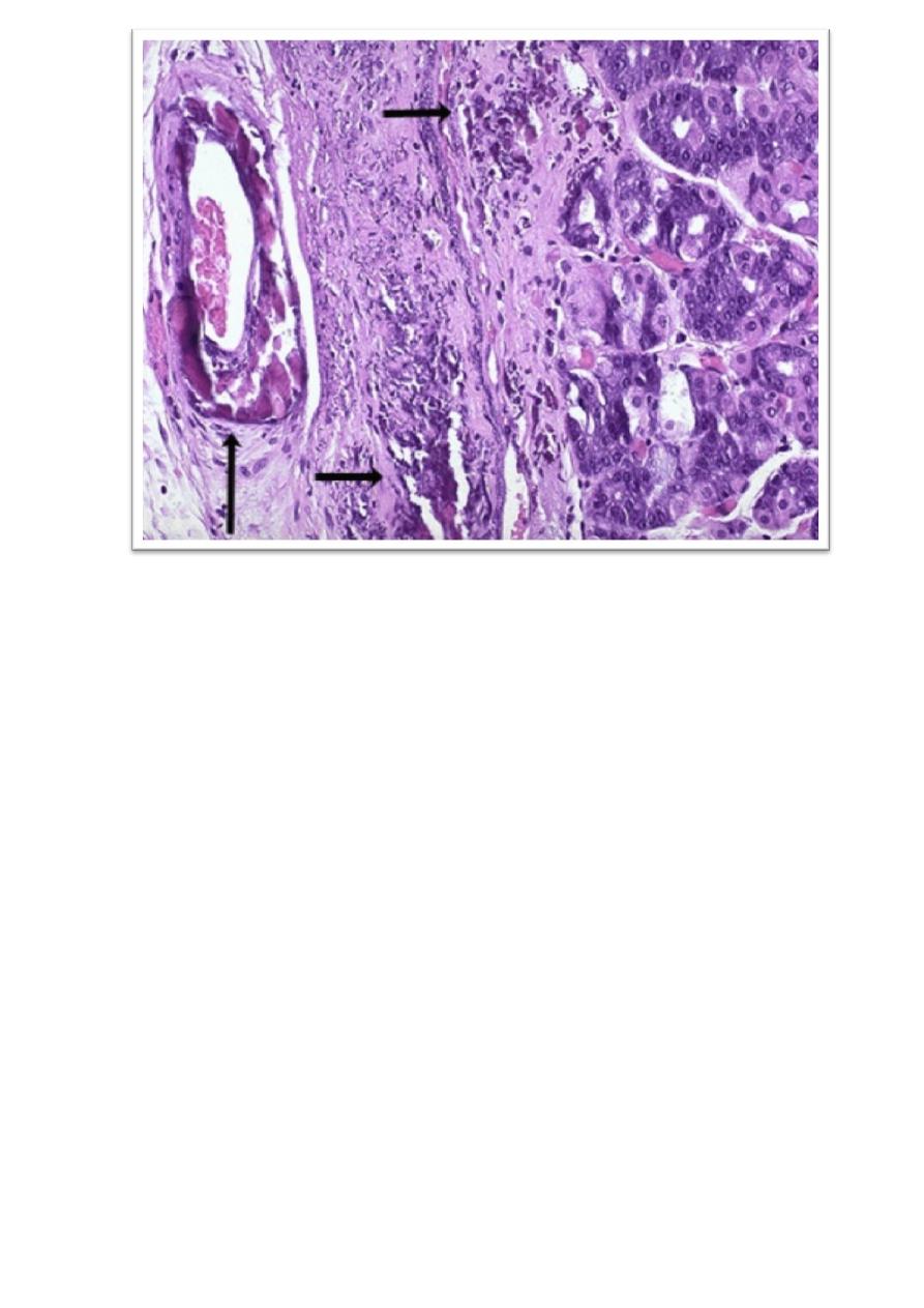
Organ:-Artery.
Diagnosis:- Dystrophic calcification (arteriosclerosis).
Lesion:- degenerated or necrotic tissue, This occurs as a reaction to tissue damage,
calcification can occur even if the amount of calcium in the blood is not elevated.
Organ:-Artery and damaged tissue.
Diagnosis:- Dystrophic calcification (wall of the stomach)
.
Lesion:- Artery with calcification in its wall, There are also irregular bluish-purple
deposits of calcium in the submucosa.