Dr. Ahmed M. Hussein
Spectrum of coronary artery disease-Silent ischemia
-Chronic stable angina-Acute coronary syndromes (ACS)
NSTE-ACS (Unstable angina , NSTEMI)
STEMI
-Heart failure
-Arrhythmia
-Sudden death
Chronic stable angina
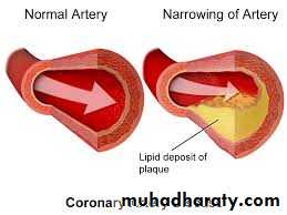
Angina pectoris is the clinical syndrome caused by transient myocardial ischaemia. It may occur whenever there is an imbalance between myocardial oxygen supply and demand. Coronary atheroma is by far the most common cause of angina.
Investigations
Resting ECG often normal.Exercise ECG.
Myocardial perfusion scanning.Stress echocardiography.
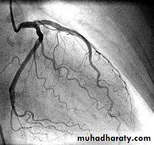
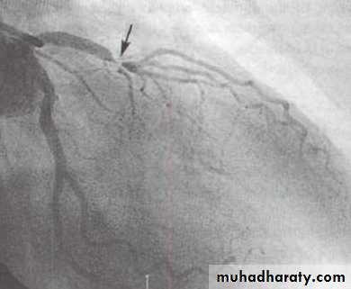
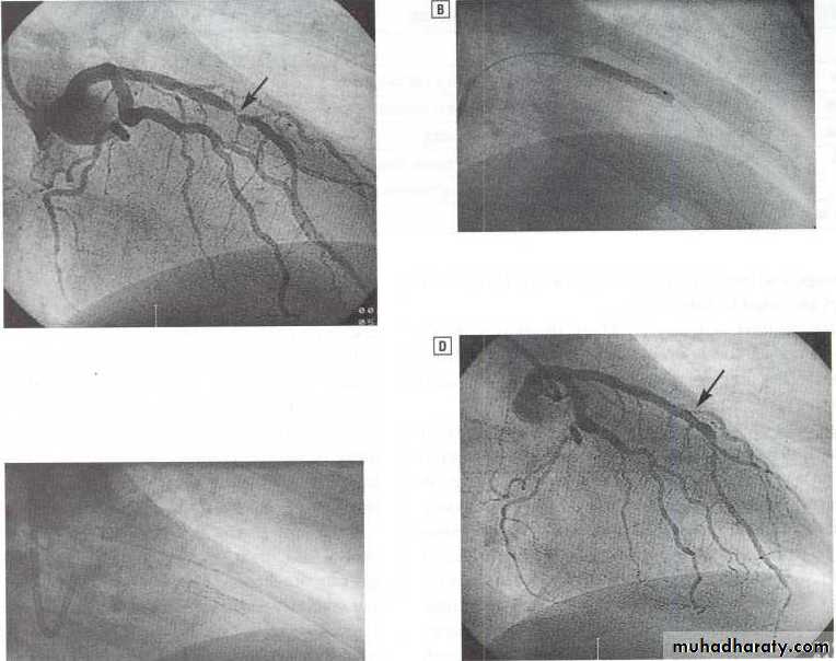
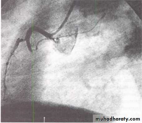
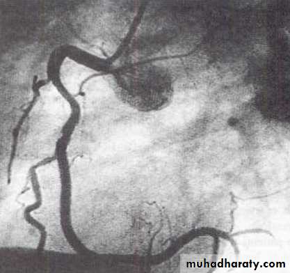
Coronary arteriography
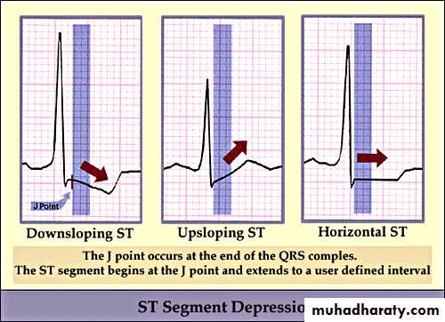
ST depression
ST Segment Depression
• Upward sloping depression of ST segment is not indicative of IHD• It is called J point depression or sagging ST seg
• Downward slopping or Horizontal depression of ST segment leading to T↓is significant of IHD
6
For more presentations www.medicalppt.blogspot.com
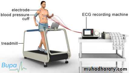
Exercise (stress) ECG
Coronary AngiographyFor more presentations www.medicalppt.blogspot.com
Management
Risk factors modification such as smoking, hypertension and hyperlipidaemia.Drugs
Antiplatelet therapy
Low-dose aspirin reduces the risk of adverse events such as MI and should be prescribed for all patients with coronary artery disease indefinitely .Clopidogrel (75 mg daily) is an equally effective.
Anti-anginal drug treatment
Nitrates
Beta-blockers
Calcium channel antagonists
Potassium channel activators
Invasive treatment
Percutaneous coronary intervention PCI.
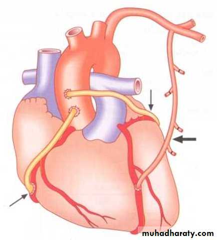
CABG
ACUTE CORONARY SYNDROMES
No ST ElevationST Elevation
Unstable Angina
NSTEMI STEMI
Myocardial Infarction
NSTEMI
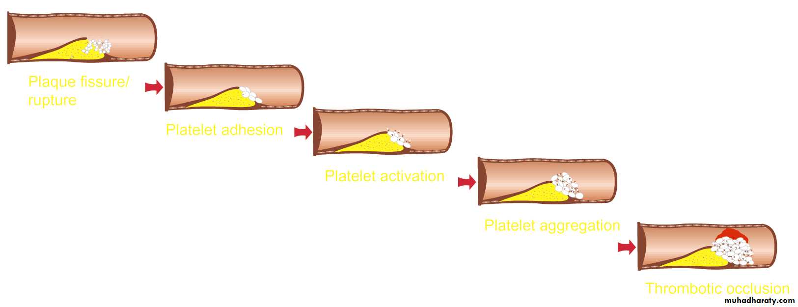
Pathogenesis of ACS
Sequence of eventsPlaque Rupture
Platelet Adhesion
Platelet Activation
Platelet Aggregation
Thrombotic Occlusion
Anti-platelet drugs
Platelet rupture
Platelet Adhesion
Platelet Activation
Platelet Aggregation
Thrombotic Occlusion
MI
Thrombus Formation and ACS
UA
NSTEMI
Plaque Disruption/Fissure/Erosion
Thrombus FormationNon-ST-Segment Elevation Acute Coronary Syndrome (ACS)
ST-Segment Elevation Acute Coronary Syndrome (ACS)Terminology:
High Serum TroponinDifferential diagnosis:
• Pericarditis• Pulmonary embolism
• Pneumothorax
• Aortic dissection
• Esophageal spasm
Ischemia and Infarction
18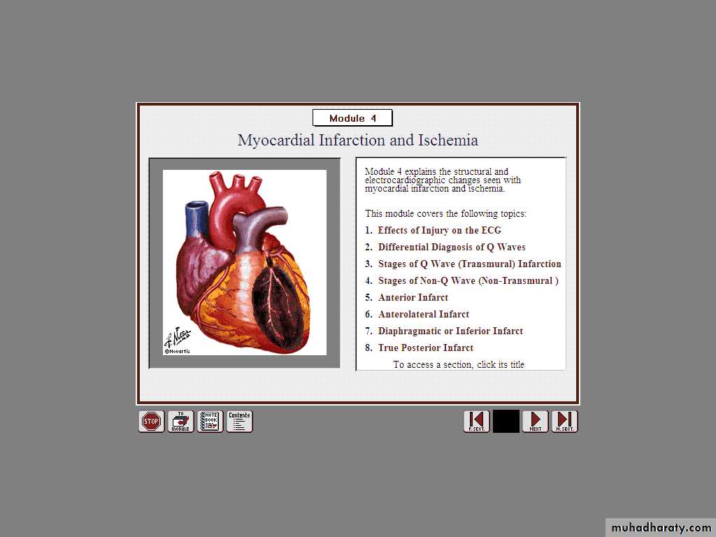
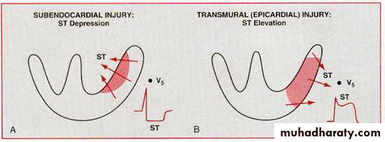
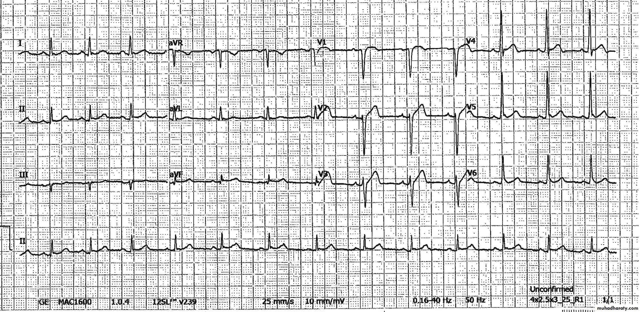
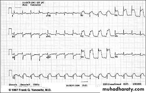
TRANSMURAL Injury ST Elevation
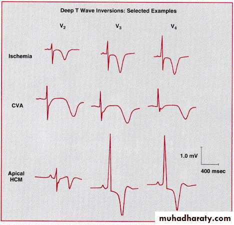
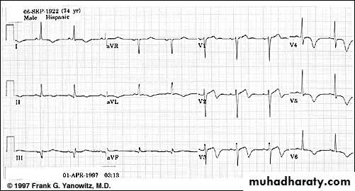
T Wave InversionDeep symmetric inverted T waves
In more than 2 precardial leads
85% of the patients with such T wave↓had > 75% stenosis of the coronary artery
T wave ↓are significantly associated with MI or death during follow up
Stages of STEMI
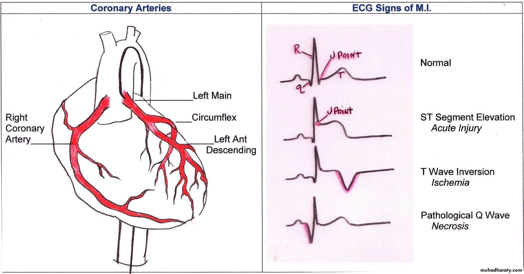
ST elevation
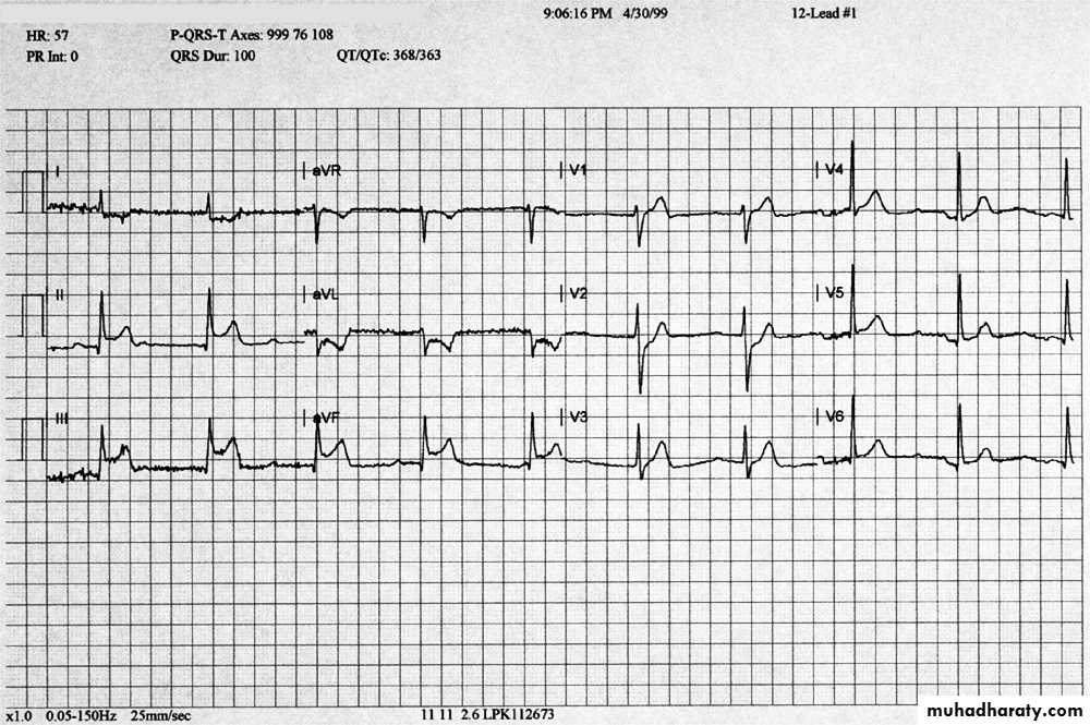
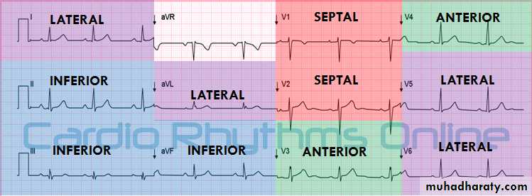
Arrangement of Leads on the EKG
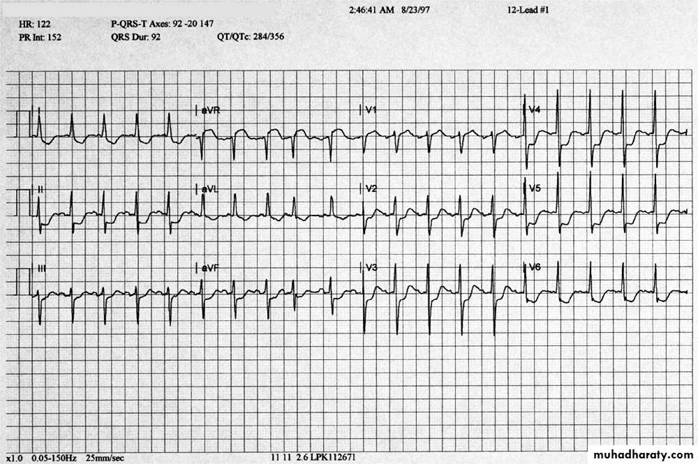
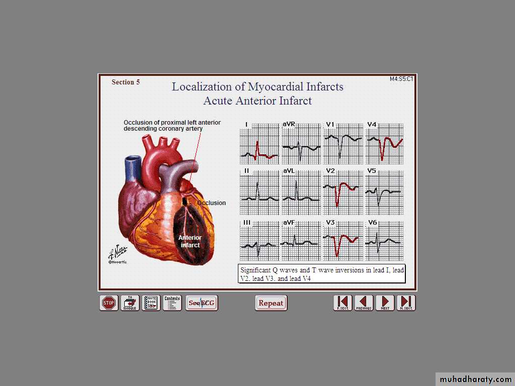
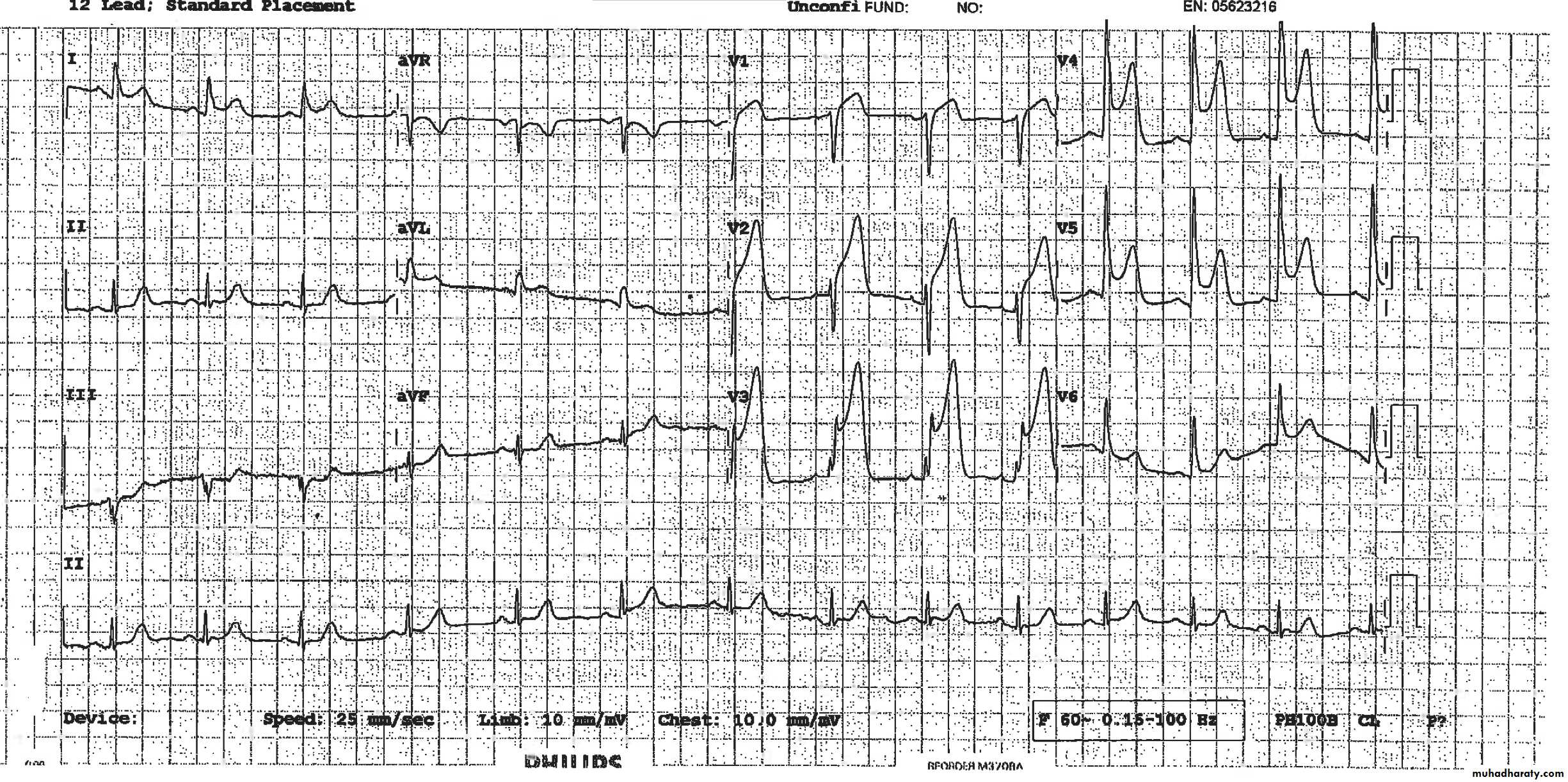
Acute Anterior MI
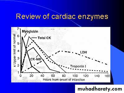
Cardiac Enzymes
Cardiac imaging
2D echocardiographyreveals regional wall motion abnormality also useful to identify mechanical complications of MI
Radionuclide imaging
used infrequently in the diagnosis of acute MI
mainly used to risk stratify patients with CHD
Very Striking
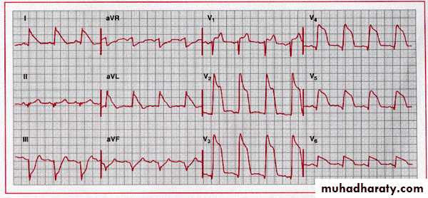
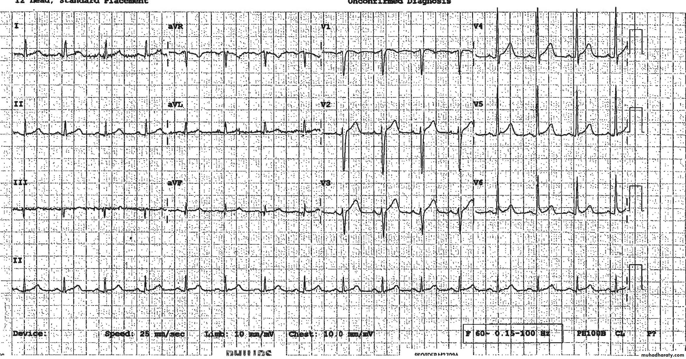
Acute Antero-Lateral MI
27
28
Severe Chest Pain – Why ?
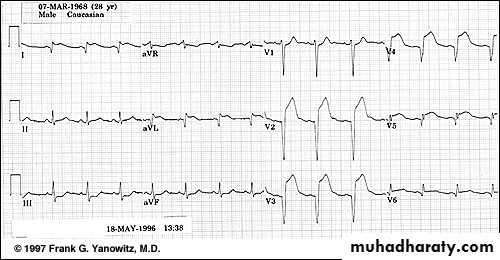
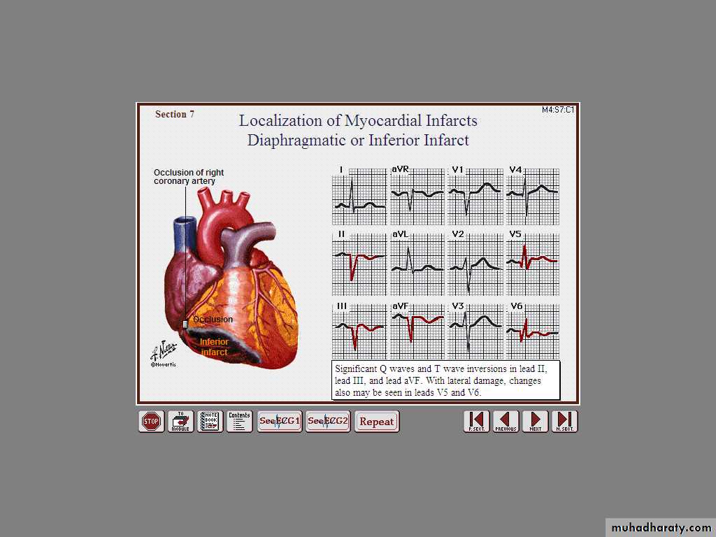
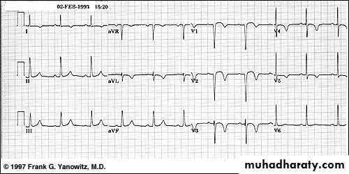
Acute Inferior wall MI
30
What is striking ?
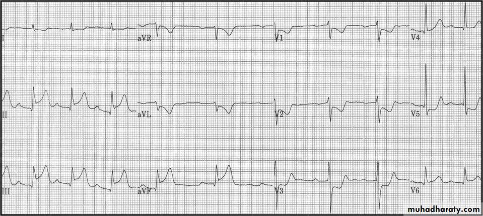
Acute Inf Post
31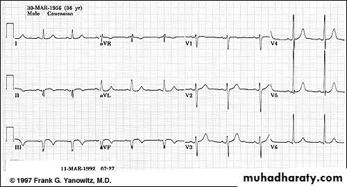
Where are the ST ↑ ?
Inf Lysed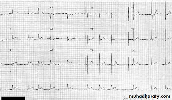
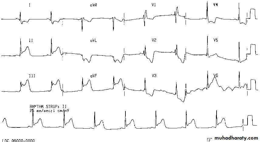
Acute ST segment elevation
Reciprocal ST segment depression33
What changes we see ?
34
Why Acute changes disappeared ?
r TPA
35
Guess How Old is this MI !
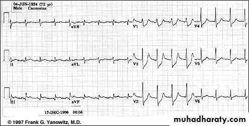
Acute True Posterior MI
36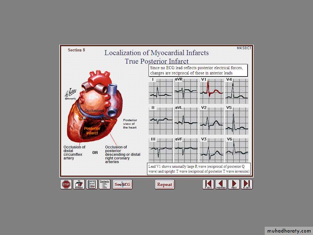
37
Decipher V1, V2, V3
Identify the Double wall MI
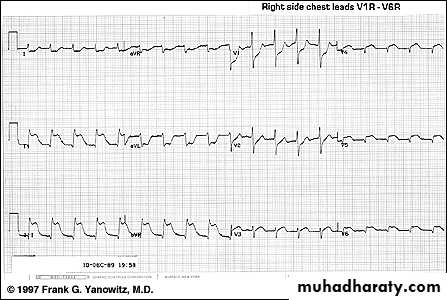
Inferior STEMI + Hypotension = ??
Next ??
Look at the Right Chest Leads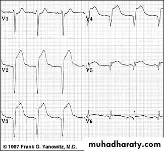
R
R
R
R
R
R
Management
Prehospital care:
Major elements includeRecognition of symptoms by the patient and
prompt medical attention
Rapid deployment of EMS capable of
resuscitation and defibrillation
Goals of Initial management in ED
Control of cardiac painRapid identification of patients suitable for reperfusion
Triage of low risk patients for subsequent careAvoiding inappropriate discharge of patients with MI
Initial management
• Focused history and Focused examination
• Reassurance• Ensure IV access + Basic investigations
• Aspirin: 160-325 mg chewable aspirin + Clopidogril
• Oxygen by nasal cannula if hypoxemia is present
• Sublingual nitroglycerine followed by IV infusion if needed
• Intravenous beta blockers (decrease myocardial oxygen demand, control chest pain and reduce mortality)
• Morphine for pain relief (given IV in small doses)+ Metelopromide
• Monitor
• 12 Leads ECG
• Consider Reperfusion
Reperfusion therapy
Primary percutaneous coronary intervention (PCI).Thrombolysis.
Absolute Contraindications
Any prior intracranial hemorrhageKnown structural cerebral vascular lesion (e.g., AV malformation)
Malignant intracranial neoplasm
Ischemic stroke in last 3 months
Suspected aortic dissection
Active bleeding or bleeding diathesis
Closed head or facial trauma in last 3 months
Relative Contraindications
Recent (3 weeks) major surgery
Recent (3 weeks) trauma
Cardiopulmonary resuscitation of >10min
BP > 180/110
Ischemic stroke more than 3 months old
Internal bleeding in last month
Noncompressible vascular punctures
For streptokinase/Anistreplase: prior exposure or allergy
Pregnancy
Active peptic ulcer
Currently on anticoagulants (sodium warfarin, Coumadin); the higher the INR, the higher the risk
Complications of acute coronary syndrome
Arrhythmias VF,AF, BRADYCARDIAIschemia
Acute circulatory failure
Pericarditis
Mechanical complications
Embolism
Impaired ventricular function HF
Ventricular aneurysm
Maintenance Therapy
Life style changes
Aspirin
Clopidogril
B blocker
ACE inhibitors
Calcium channel blocker
Statins ( Antilipids)
Normal initial ECG exclude STEMI??
23 min. later
1 hr post revascularization