Pathology dr.(rasha3)
1
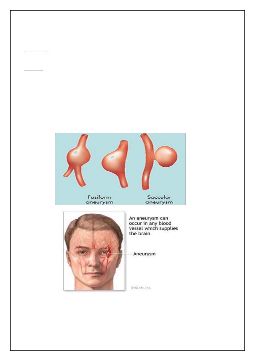
Aneurysm
:-
Abnormal dilation of arteries or veins, they develop wherever there's a
-
:
Definition
marked weakening of the wall of vessel.
Cause of weakness in vessel wall either :
-
:
Etiology
1- Congenital defect e.g. intracranial arteries as saccular Berry aneurysm.
2- Local infection (mycotic aneurysm or due to syphilis).
3- Trauma (traumatic aneurysm).
4- Systemic disease such as occur in aorta due to atherosclerosis and cystic medial
necrosis.
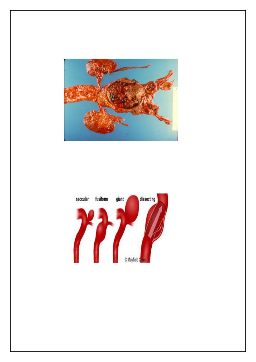
Atherosclerotic aneurysm:
Whcich's usually occur in the abdominal aorta below
renal arteries, its etiology occur due to genetic defects in connective tissue
component for strength of blood vessels in atherosclerotic and hypertensive which
will cause weakness of aortic wall.
Pathology dr.(rasha3)
2
This is take the form of saccular (ballon-like), cylindroid or fusiform swellings
sometimes up to 15 cm in greatest diameter and to 25 cm length. It's complicated by
thrombus formation in the wall and even rupture.
Aortic dissection:-
Definition:- dissection of blood along the laminar planes of the media along with
formation of a blood-filled channel within the aortic wall, such a channel often
ruptures, causing massive hemorrhage, it's unusual to occur in severe
atherosclerosis.
Venous disorders:
(I) Varicose veins:-
Are abnormally dilated tortuous veins, this condition caused by increase in
intraluminal pressure and loss of support of vessel wall.

Pathology dr.(rasha3)
3
Most common veins affected:
1-
Superficial veins of leg:-
which are most frequently involved due to high venous
pressure in the legs with relatively poor tissue support for superficial veins, the
venous pressure occur especially due to:
a- Pregnancy, so female are more often affected than males.
b- Pelvic pressure due to abdominal neoplasm or encircling surgical dressing.
2-
Hemorroids:-
Which's varicose dilation of hemorroidal plexes of the anorectal
junction, this occur due to prolonged pelvic congestion result from repeated
pregnancies or chronic constipation and straining at stools.
3-
Varicosity of esophagus:-
This occur in patients have cirrhosis of the liver because
of portal hypertension, rupture of esophagus varix may be more serious than the
primary liver disease itself.
(II) Phlebothrombosis and thrombophlebitis:
These 2 names for same condition characterized by thrombus formation in deep
veins of lower extremities, this condition is silent clinically but its complication is
more serious by giving rise to emboli that travel to the lung to produce pulmonary
embolism and infarction which will cause death when it's massive.
Lymphatic disorder:-
The lymphatic disorders are divided into 2 categories:-
1- Priamry diseases which are uncommon.
2- Secondary processes result from inflammation or cancer to give rise 2 lymphatic
diseases→lymphangitis and lymphedema.
Lymphangitis:-
It results from drainage of infection by the lymphatic channels, the most common
causative agent is group Aβ-hemolytic streptococci, but any virulent pathogen may
become the cause, this lead to redness and dilation of the channels (red streaks) and
local tenderness, infection may be transformed to drainage LN to cause
lymphadenitis or even to venous drainage to cause septicaemia.
Lymphedema:-
It's divided to:-
Pathology dr.(rasha3)
4
1- Primary
result from congenital defect or it may be familial.
2- Secondary
that result from post-(1)inflammatory scarring of lymphatic channels,
(2) spread of malignant neoplasm with obstruction of either the lymphatic channels
or nodes of drainage, (3) post surgical procedure with excision of drainge LN e.g.
removal of axillary LN in radical mastectomy and (4) filariasis.
Vascular tumors:
It's divided into benign and malignant vascular tumor with intermediate grade
between the two.
Benign tumors:-
The most common benign tumor of blood vessel is hemangioma which's also divided
into types: Cavernous and capillary hemangioma.
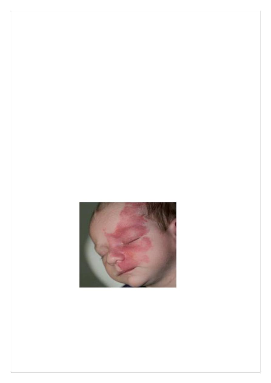
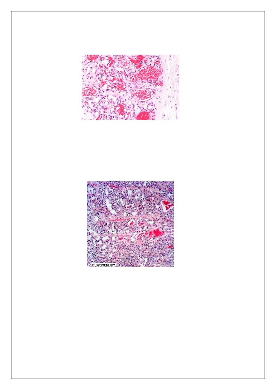
Cavernous hemangioma:-
This occur in the skin, mucous membrane but it may arise in viscera, in infant
cavernous hemangioma sometimes constitute large lesions of the skin of the face or
scalp, the so-called port-wine stains or birth mark, it's usually red-blue,
compressible, spongy lesions, sharply defined at margins
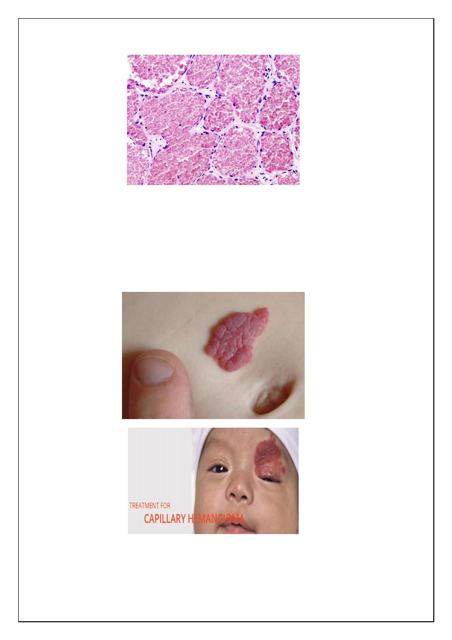
Microscopically
show large cavernous spaces filled by blood or lymphatics.
Cavernous hemangioma in skin of children may be regress, but its importance when
it occurs in brain because it causes pressure symptoms and rupture.
Pathology dr.(rasha3)
5
Capillary hemangioma:
It's also occur in skin, subcutaneous tissue or mucous membrane of oral cavity and
lips, it appear as bright red to blue lesion, it's slightly elevated and even
pedunculated, occasionally the hemangioma takes the form of large, flat, map-like
discoloration that cover large areas of face or upper parts of body producing port
wine stain like cavernous hemangioma.
Microscopically
, it consists of uncapsulated closely packed capillaries separated by a
scant connective tissue stroma, the endothelium is usually plumpy but no atypia
Pathology dr.(rasha3)
6
present.The channels filled by fluid or thrombosed blood . In the skin and mucous
membrane, it may cause traumatic ulceration and bleeding.
Hemangioendothelioma:-
It represents the intermediate grade between benign hemangioma and malignant
anaplastic angiosarcoma.
Microscopically
: consist of vascular channels with masses consist of proliferating
well-differntiated endothelial cells.
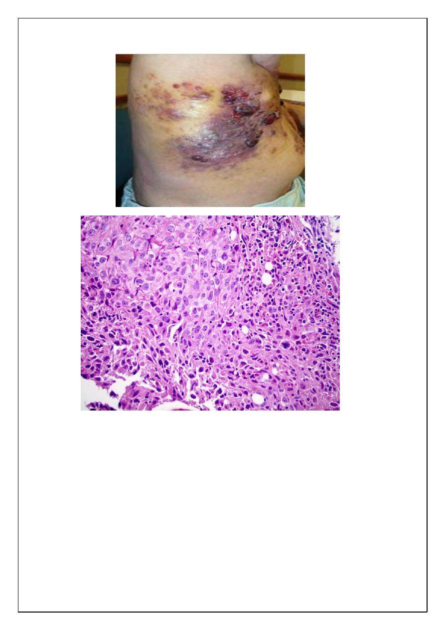
Angiosarcoma:-
Microscopically
appear as masses of anaplastic cells with few poorly formed vascular
channels or may be not seen and in this case, it can not be differentiated from other
malignant soft tissue tumor as fibrosarcoma or leiomyosarcoma only by
immunohistochemical study.
These 2 malignant tumors distruption in the same location of benign tumor but it's
usually larger, more solid, less obviously vascular and more invasive, there is special
type of angiosarcoma called Kaposi's sarcoma, the type of epidemic KS seen in AIDS
patients.
Pathology dr.(rasha3)
7