Fifth stage
MedicineLec-4
د.بشار
4/12/2016
مكتب الجامعه للطباعه والاستنساخعدد الاوراق 5 السعر 250
مكتب الجامعه للطباعه والاستنساخ
عدد الاوراق 5 السعر 250
Brain SOL
Traumatic
Subdural haematomaExtradural haematoma
Vascular
Intracerebral haematoma
Infective
Cerebral abscess ;pyogenic, Toxoplasma
Tuberculoma
Cysticercosis
Hydatid cyst
Schistomiasis
Inflammatory
Sarcoid mass
Neoplastic
Cerebral neoplasms (benign or malignant )
Other
Embryonic dysplastic lesions e.g. craniopharyngioma &hamartomas
Arachnoid cyst
CNS tumours
Primary or Secondary
Account for 2% of all deaths
The majority are metastatic
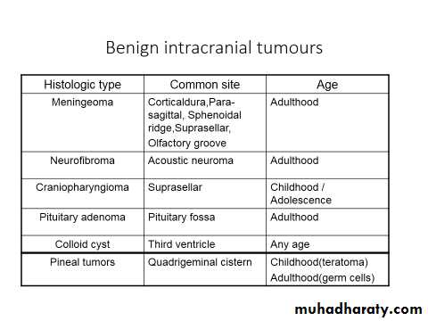
Meningeomas account for 20 %of all intracranial tumours

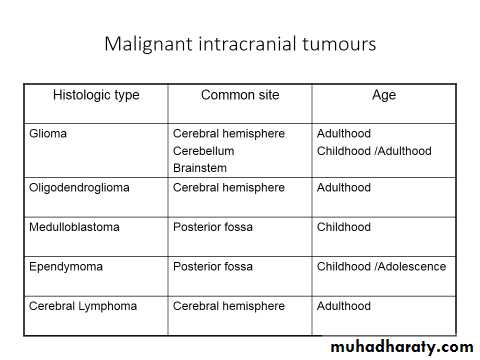
Even malignant tumours don’t metastasize outside the CNS
Secondaries (metastases)Usually located in the white matter of cerebral or cerebellar hemispheres;common sources are bronchus ,breast &gastrointestinal tract.
CLINICAL FEATURES:
Insidious Onset = May be acute with cystic degeneration, hemorrhage or with seizures.
Progressive course.
Space- occupying effect
Site of the tumor
False localizing signs
Space occupying effectRaised intracranial pressure
HEADACHE: Non specific;dull aching, eventually in most patients, more severe in early morning in 10-15 % , aggravated by cough, sneezing, straining &change of posture (bending,lying ).
It is ipsilateral to supratentorial tumors in 80% of cases
Nausea &vomiting
Seizures : more if tumor in ant. Cranium e.g. more than 50% of frontal lobe tumours have seizures ;generalized or partial .
Papilloedema : more in infratentorial tumours, leading to transient visual obscurations.
Altered mentation.
SITE OF TUMOR (FOCAL SIGNS &SYMPTOMS)
Frontal lobe : Altered mood &behavior, contra. Motor deficit, incontinence,primitive reflexes
Parietal lobe : Sensory s.&s. may predominate, contra. Visual field &motor deficit
Non dominant parietal l. :sensory or visual inattention, dressing apraxia
Temporal lobe : Wernicke aphasia, sup. Quadrantanopia, temporal lobe epilepsy
Occipital lobe : visual field abnormality
FALSEAC LOCALIZING SIGNS:
Pupillary dilatation.6th cranial nerve palsy(unilateral or bilateral).
Hemiparesis (ipsilateral to the lesion ).
Bilateral extensor plantar responses.
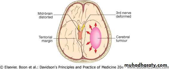
The rise in intracranial pressure from a mass lesion is not usually uniform within the cerebral substance and alterations in pressure relationships within the skull may lead to displacement of parts of the brain between its various compartments. Downward displacement of the temporal lobes through the tentorium due to a large hemisphere mass may cause 'temporal coning
This may stretch the 3rd and/or 6th cranial nerves, or cause pressure on the contralateral cerebral peduncle (causing ipsilateral upper motor neuron signs).
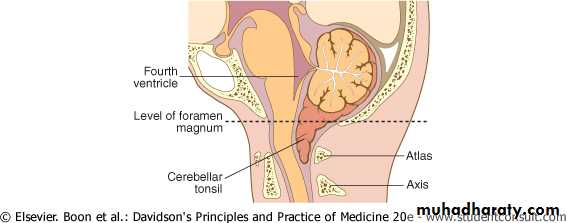
Downward movement of the cerebellar tonsils through the foramen magnum may compress the medulla-(tonsillar coning)
This coning may result in brain-stem haemorrhage and/or acute obstruction of the CSF pathways. As coning progresses, the patient may adopt a decerebrate posture and, unless rapidly treated, death almost invariably ensues. The process may be acutely accelerated if the pressure dynamics are suddenly disturbed by lumbar puncture
INVESTIGATIONS
Plain X Ray ; Signs of raised ICP, Calcification…EEG : Focal slowing
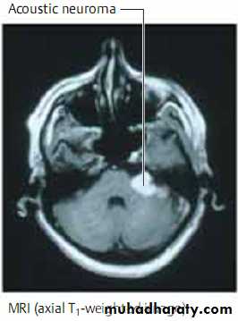
CT Scan ,MRI &MRA
TREATMENT:
Reduce ICP : Osmotic diuretics, SteroidsOften required when surgery is not possible or when life is threatened.
Dexamethasone 8- 12 mg 12-hourly orally or by injection ;a striking improvement in consciousness is often produced &focal deficits may regress.
Mannitol 0.25 – 1 mg /Kg /Dose IVI.
Surgery
Mainstay of treatmentOnly partial excision may be possible if the the tumour is inaccesible or its removal is likely to cause unacceptable damage.
Biopsy should be considered even if the tumour is not removable ?prognosis &management.
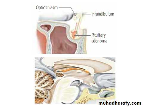
Meningeomas,acoustic neuromas &pituitary adenomas.
Radiotherapy &Chemotherapy:
Marginal effect on survival in metastases &malignant gliomas.
Combined therapy has improved prognosis in medulloblastomas in children.
Radiotherapy reduces the risk of recurrence of pituitary adenoma after surgery.
Ependymomas,some pineal tumours &low gradegliomas in children &young adults are often radiosensitive.
Prognosis:
For benign tumours is good if removed completely.Ependymomas &Medulloblastomas may recur with seeding via the CSF.
Oligodendrogliomas may transform to more malignant form ---- glioma .
Related to histologic grade ;
G1&2 may survive for years
G 4 –only 20 %survive for 1 year.
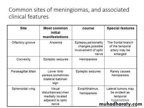
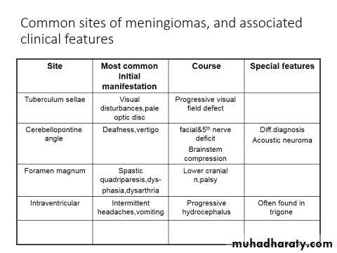
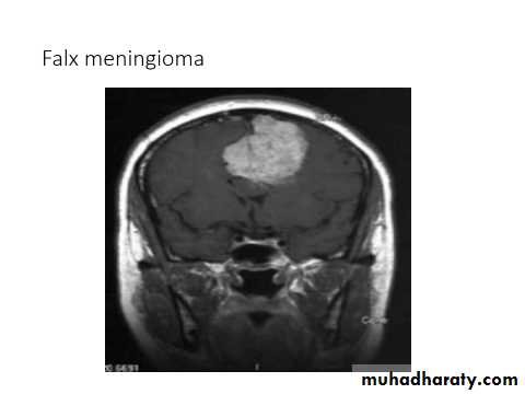
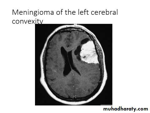
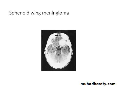
Meningiomas:
Arise from the dura mater and are nearly always benign, well-demarcated lesions that displace rather than invade the adjacent neural tissue as they grow.These mesodermal tumors most often become clinically evident between the ages of 40 and 50.
They are diagnosed by MRI or CT scanning which reveals marked, homogeneous contrast enhancement.
Meningiomas tend to appear in certain classic locations with corresponding typical neurological manifestations.
They often grow very slowly and are not uncommonly discovered as an incidental radiological finding.
The indications for treatment must then be carefully considered: resection may be desirable in younger patients, but unnecessary in older ones.