GENETICS
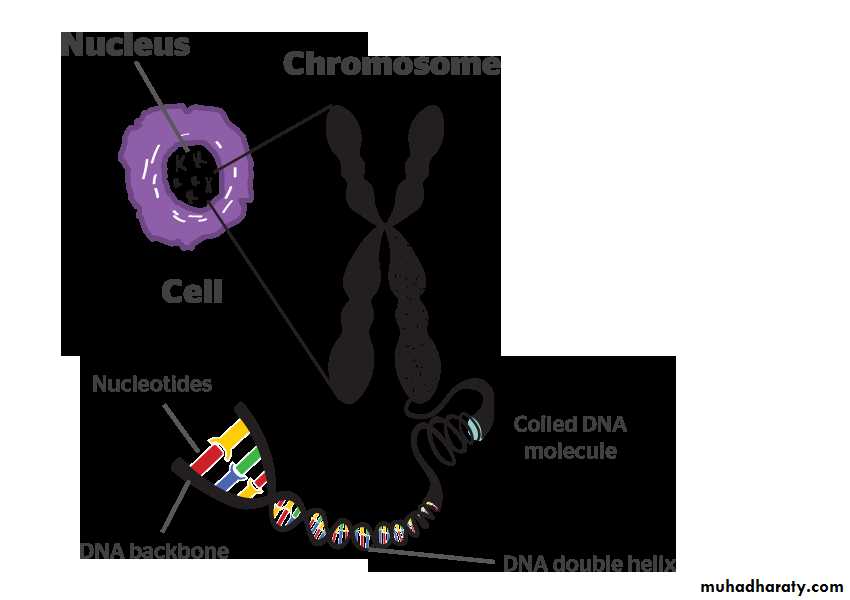
Heredity is the transmission of information required to construct multiple proteins. These proteins have diverse roles and different subsets, they are utilized by different cell types but all are encoded in the cells DNA which is organized into discrete structures called chromosomes.Chromosomes: every cell nucleus contains a set of chromosomes. Each chromosome consists of a single molecule of DNA (deoxyribonucleic acid) together with associated acidic and basic proteins.
Most human cells contain 46 chromosomes (the diploid number) with 22 pairs of autosomes which are a like in males and females and a pair of sex chromosomes: XX in female and XY in a male.
Each chromosome has a narrow waist called the centromere which has a constant position for a given chromosome. The centromere divides each chromosome into short and long arms.
At mitosis each chromosome replicates to form a pair of sister chromatids which are held together at the centromere. Although exchanges of genetic material can occur by crossing over (sister chromatid exchange) during mitosis, as each sister chromatid is identical, clinical consequences do not arise. Thus at the end of cell division each daughter cell has identical set of 46 chromosomes.
In contrast, reduction cell division or meiosis results in cell with a half set (haploid number) of 23 chromosomes. Meiosis, which is confined to gonadal cells involved in gametogenesis, consist of two successive division in which the DNA replicates only once before the first division. Each mature egg thus normally contains one of each pair of autosomes and one X and each mature sperm has one of each pair of autosomes and the X or Y chromosomes. At fertilization the diploid number is restored and in consequence half of each individual's autosomes are derived from each parent and a female has an X from each parent. Whereas a male has a maternal X and a paternal Y sex chromosome.
DNA: each molecule of DNA is composed of two nucleotide chains which are coiled clockwise around one another to form double helix. Each nucleotide consists of a nitrogenous base, a molecule of deoxyribose and a phosphate molecule.
The nitrogenous bases are of two types, purines and pyrimidines. In DNA there are two purine bases, adenine (A) and guanine (G) and two pyrimidine bases, thymine (T) and cytosine (C). The nucleotide chains run in opposite direction and are held together by hydrogen bonds between A and T or between G and C since A: T and G: C pairing is obligatory the parallel strands must be complementary to one another.
Gene: is the unit of the DNA which codes for a protein.
Genetic diseases:-Are large group of diseases can be subdivided into:-
Single gene defect (dominant & recessive).
Chromosomal diseases (numerical & structural)
Multifactorial diseases & non traditional disorders.
Mutation refers to permanent changes in the DNA. Those that affect germ cells are transmitted to the progeny and may give rise to inherited diseases. Mutations in somatic cells are not transmitted to the progeny but are important in the causation of cancers and some congenital malformations
Thus there is certain changes occur in nitrogenous bases may lead to abnormal protein synthesis.
Single gene diseases (Unifactorial diseases):-These are caused by a mutation in a gene. Genes may behave as dominant, i.e. when one of the alleles becomes mutated it results in a genetic disease; or they may behave as recessive, i.e. the diseases does not manifest unless both alleles are affected by the same mutation.
A third category of genes are those which determine an autosomal character but are situated on the sex chromosome (sex-linked)
The question arises why some genes act in a dominant manner while others behave in a recessive fashion, i.e. the problem of dominance and recessiveness. To answer this question, one has to consider and always remember the following principles:
A Single gene is responsible for formation of a single type of protein, but since proteins are made of units of polypeptides that could be the same or different in one molecule of protein, the principle becomes:
A Single gene is responsible for the formation of a single type of polypeptides, and if we know that our body structures and functions from the moment of post-fertilization to the full maturity and later on are determined by proteins one can understand how genes function. These types of proteins are varied; they could be
Structural proteins, like fibrous tissue and elastic tissue proteins
Immunoglobulins
Signal proteins produced by many oncogenes
Receptors
Enzymes
Hormones
Therefore, the action of the gene being dominant or recessive is determined by the type of protein it produces and its function.
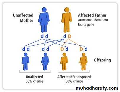
Autosomal dominant disorders:
are manifested in a heterozygous state (when one of the alleles becomes mutated)one of the parents of the affected individual should be affected and the child appears as diseased individual.
both males and females are affected and both can transmitted the condition. When affected person marries unaffected one, every child has one chance in two of having the disease.
Sometimes the parents are normal but the child is diseased that happened because the mutation occur in the cell of that child alone while their parents are completely normal. The siblings of this child are neither affected nor at risk of developing the disease
Some trait is seen in all individuals that carrying the mutant gene but it is expressed differently among different individuals: phenomena called variable expressivity.e.g. polydectaly may be expressed in toes or in fingers as one or more digitis.
Dominant genes usually produce two types of proteins:
Major structural proteins, which form or are present in many parts of the body e.g.is Marfan syndrome in which there is mutation in fibrillin gene leading to a qualitative & quantitative defects in fibrillin which result in skeletal abnormality .
Enzymes, which are key enzymes in metabolic pathways, under feedback mechanism, or receptors regulating metabolic pathways.
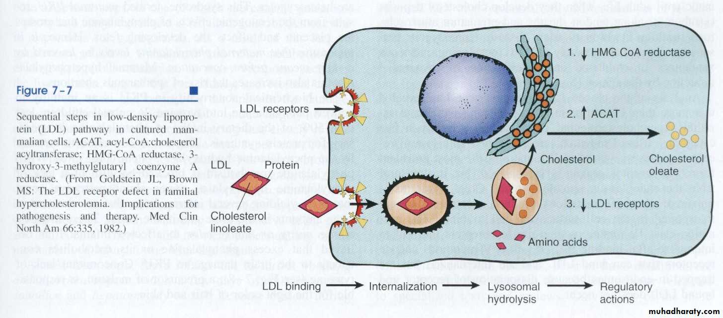
Example of the second is AD familial hypercholesterolemia [] disease where the receptors for LDL are mutated. They are responsible for regulation of LDL in the cells and the circulation. To explain the latter example and how the action of this pathway is executed, let us consider the pathway of circulating LDL: usually it should enter the cells of the body for building cellular membranes and nuclear membranes to replace the old ones that are affected by wear and tear. LDL could not enter the cells unless it is complexed with receptors on the cell membrane. Once it is inside the cell, the complex will be degraded into free cholesterol and amino acid. The latter is the remnant of the proteinaceous coat of the lipoprotein. The free cholesterol in the cell constitutes the cholesterol pool of the cell and its level is regulated by three systems of enzymes, the HMG-CoA reductase, which forms cholesterol from fatty acids, ACAT, which hydrolyzes cholesterol into esters and thus rendering it inactive and the number of the receptors on the surface of the cell. If the pool concentration is low, messages are sent to activate the HMG-CoA, to inactivate ACAT and increase the number of receptors on the cell surface. Therefore, when one of the two alleles responsible for the formation of the receptor protein becomes mutated, half of the number of receptors are formed only, so 50% of LDL which is used to be internalized inside the cell will remain in the circulation unable to enter the cells and a state of hypercholesterolemia results with reading of 400-500 iu/dl of cholesterol in the blood (normal value 180-220 iu/dl)
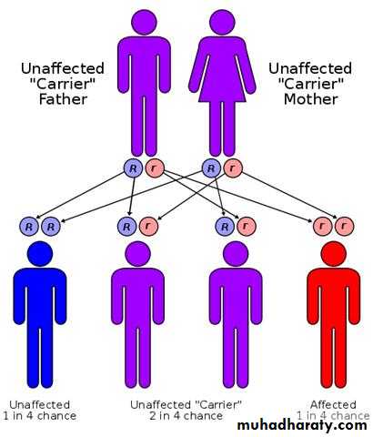
Autosomal recessive disorders:
are manifested in a homozygous state (they occur when both of the alleles at a given gene locus are mutants).usually the parents are unaffected clinically because each has only one mutant gene and so they are a carrier or heterozygote.
For two carrier parents the chance for getting an affected child is 1 to 4.
As for recessive genes, they are protein enzymes, which usually share in catabolic pathways and when both alleles are ?defective, there is no protein, i.e. no enzyme and therefore the catabolic pathway is obstructed with the accumulation of the biochemical substrate. Examples of these are mucopolysaccharidosis & phenylketonuria (PKU) and most of inborn errors of metabolism.
Athird category of single gene disorders are Sex-linked diseases in which genes that determine an autosomal character are situated on the sex chromosome (sex-linked) and because most of the genes are carried on the “X” and very few are present on the “Y”, usually sex-linked is used for “X”-linked both dominant and recessive. Most of the X-linked disorders are X-linked recessive and are characterized by the following features:
They are transmited by heterozygous female carriers only to sons.
An affected male does not transmitted the disorder to sons but all daughters are carriers.
Sons of heterozygous women have one chance in two of receiving the mutant gene.
Sex linked dominant disease e.g. is the vit D resistant rickets
Y inheritance disease is the hairy ears in males
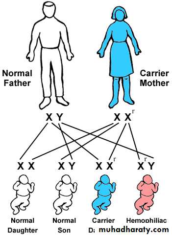
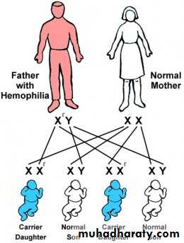
Sex-linked diseases, because most of the genes are carried on the “X” and very few are present on the “Y”, usually sex-linked is used for “X”-linked both dominant and recessive. In this type of inheritance, there is a lot of deviation from the expected and their explanation is forwarded by a hypothesis known as Lyon’s hypothesis, e.g. in clinical practice, both haemophilia and G6PD-deficiencies are diseases caused by sex linked genes recessive in nature, i.e. only males who carry the mutated gene on their “X” are affected clinically while carrier females are usually silent clinically but transfer the disease to their sons. But it happens that some cases of both diseases present in female by Lyon’s hypothesis, which states that in a female’s autosomal cells, all the “X” chromosomes will be inactivated except one which remains active during inetrphase.
This process of inactivation takes place early in the post-fertilization period, 19-20 days P.F.
The process of inactivation is random concerning the origin of the “X” inactivated, i.e. paternal “X”, which comes from the father or maternal; “X” that comes from the mother.
In a cell, all the daughter cells that descend from it, the same “X” will remain inactive.
This means that 50% of the “X” chromosomes are inactivated but this does not necessarily involve all the paternal “X” or all the maternal “X”. in some areas the paternal X is being inactivated while in other areas, it is the maternal “X” that are being inactivated, therefore, the body of the female is a mosaic concerning the function of the active “X”. So, a heterozygote female for type G6PD enzyme A & B; if we examine different parts of her body for the type of the enzyme, we either find type A or type B and never both in one part of the body. In contrast males could either be A or B.
A female who carries the mutated gene for “X” linked and presents clinically the disease; it happens by chance that in most parts of her body the “X” that carries the mutated gene remains active, which results in deficiency of the product of the gene disease. This is because of the randomness of the inactivation. Those females are known as manifesting carriers in clinical practice.