5. Examine and sample the organs
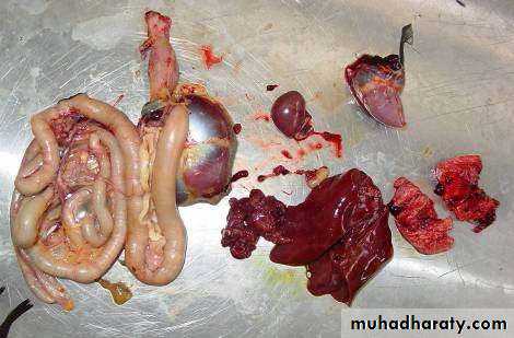
lymphoid tissue, brain, lungs, heart, kidneys, reproductive tract, liver, intestinal tract.
Note any abnormalities for each (color, consistency, distribution, and size). Be sure to examine both capsular and cut surface. Make several cuts in each organ. Collect specimens for further diagnostic work.
Brain
Often the brain is sliced down the middle to create symmetrical halves for frozen and formalin specimens.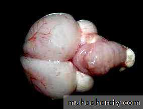
Lymphoid system
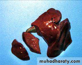
ThymusThe principal gross lesion in this organ is a notable decrease in size known as thymic atrophy (fig. 1). This is a significant lesion in broilers and may have multiple causes, but the most common cause is an infection by avian infectious anaemia virus.
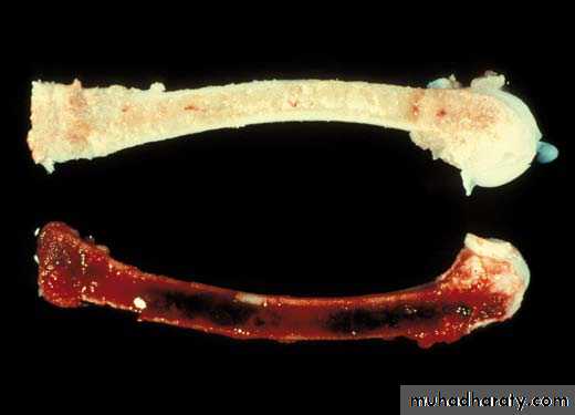
Usually this disease causes pale pink or yellowish lesions in the affected animal's bone marrow (fig. 2). Hemorrhaging, normally observed due to the presence of petechiae, is often found in the thymus although it is nonspecific.
Fig. 1 Marked thymic atrophy in a bird affected by avian infectious anaemia.
Fig. 2 The femur bone marrow is significantly pale in a bird affected by avian infectious anaemia.
The spleen in birds is a small round organ that should be a uniform mahogany color on capsular and cut surfaces.
Spleen
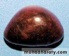
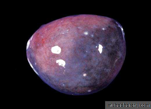
• Like the thymus, a change in size is the most common gross alteration to be observed. An increase in the relative size of the spleen, known as splenomegaly, is normal as a primary response to circulating antigen. This size increase is usually accompanied by miliary white spots seen both on the organ surface as well as at the section (fig. 3). This lesion is often observed in animals suffering from septicaemia, mainly due to Escherichia coli. Inflammation of the spleen, known as splenitis, is quite rare.Fig. 3 Splenomegaly in a bird affected by colisepticaemia.
It is seen in cases of tuberculosis in which the spleen has multiple white nodules that correspond to granulomas.
Finally, the spleen may be enlarged due to the presence of a lymphoma in animals affected by Marek's disease or avian leukosis.
The bursa changes dramatically with age. Birds older than10 weeks will have a bursa that may even be difficult to locate. A normal bursa in a young bird has an accordion-like structure and is a homogeneous tan color.
Cardiovascular system
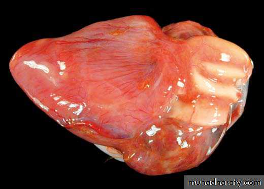
Heart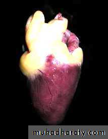
The heart can now be examined. Make one incision into each ventricle, and examine muscles and valves.
Pericarditis:
it is an inflammation of the serous membranes surrounding the heart and forming the pericardium sac. This lesion is characterised by the presence of exudate, usually fibrinous or fibrino-purulent, in the pericardial cavity or on the surface of the visceral pericardium (fig. 4). This lesion is frequent in animals suffering from septicaemia, usually due to Escherichia coli. It is normal to observe perihepatitis and airsacculitis (polyserositis) in addition to pericarditis (fig. 5).Fig. 4 Fibrinous pericarditis showing the presence of fibrinous exudate on the
pericardial surface.Fig. 5 Fibrinous polyserositis with fibrin exudate on the serous membranes
of the coelomic cavity.
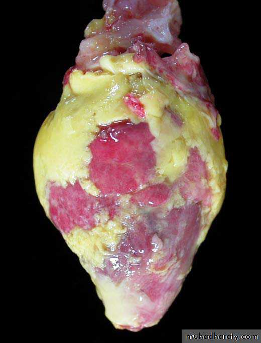
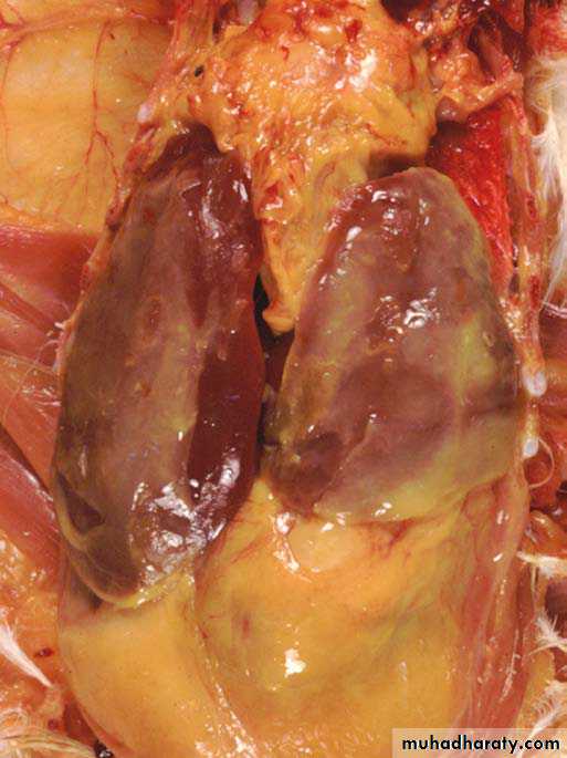
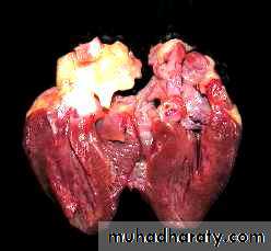
Fig. 6 Myocardial lymphoma in a bird affected by an acute or visceral form (arrows) of Marek's disease.
Neoplasm of the myocardium:
it is relatively rare, but the most common tumour is lymphoma, seen as white areas or nodules in the myocardium (fig. 6) associated to Marek's disease virus.
Aortic rupture in turkeys:
it is an uncommon lesion but can lead to elevated mortality losses in male turkeys. The rupture of the aorta and subsequent massive haemorrhage in the coelomic cavity often originates from an aneurysm which forms in the abdominal aorta and produces the bird's death.Respiratory system
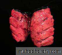
Begin by cutting through the larynx, trachea, and syrinx making note of any mucus, froth, or petechiae. Lungs should be pink, “spongy”, and free of any fluid.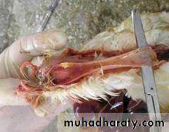
Urinary system
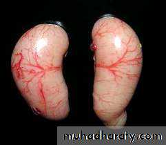
KidneysThese should be
smooth and homogeneous. A reticular pattern is an indication of dehydration.
Reproductive system
Reproductive tract in maleTestes are homogeneous on capsular and cut surfaces. Ovaries should be free of inflammation. Sterile egg yolk peritonitis is a common finding in “spent” layers.
The main changes to be observed in the female reproductive system are:
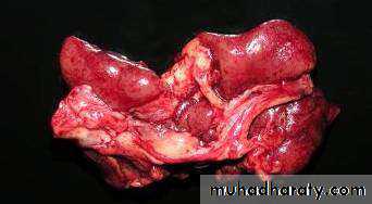
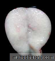
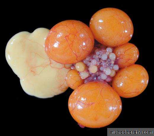
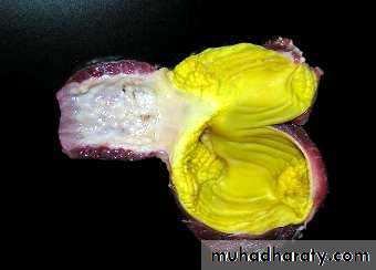
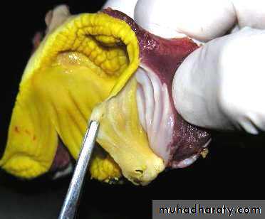
Ovarian regression: it is associated with the hen reaching the end of the laying cycle or phase, or with various factors such as nutritional changes, infectious diseases, toxicity, hormonal manipulations or environmental factors which lead to cessation of ovulation. This process is called ovarian regression or atrophy (fig. 7). In the affected ovary new follicles will not develop and those present will suffer follicular atresia. Ovarian follicular atresia is the process whereby an ovule which has failed to ovulate, disappears and is reabsorbed.These follicles lose the characteristic shape and stiffness of developing follicles. The yolk they contain becomes more watery, less dense, and eventually are reabsorbed into the bloodstream (fig. 8). This may be due to a physiological process since not all developing ova arrive to ovulation, or, due to a pathological process that induces ovarian regression.
Fig. 7 Ovarian regression. The ovary shown has virtually no large yellow follicle close to ovulation, but conversely it does have some atretic follicles.
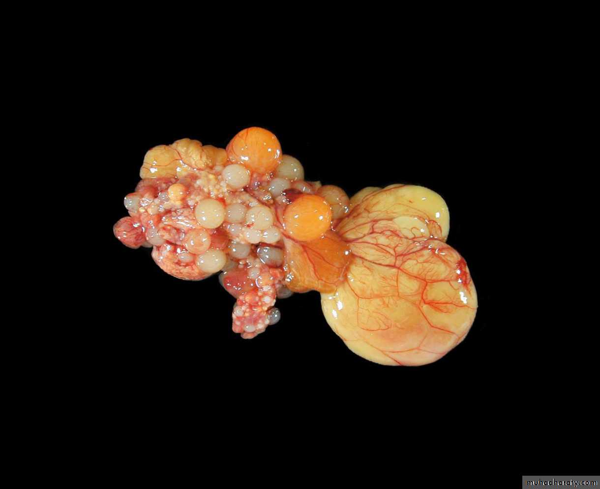
Fig. 8 Functional ovary of a 35 week old hen. Six large yellow follicles are seen alongside one atretic follicle. This has lost its turgor and displays a paler colour.
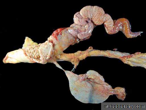
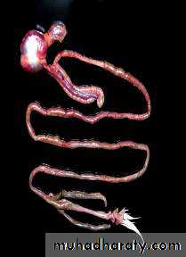
Persistent right oviduct: in some cases the right-side ovary and oviduct may not disappear but accumulate clear liquid which forms a cyst on the right side of the cloaca.
This persistent or cystic right oviduct is observed in birds that have not yet reached sexual maturity (fig. 9), as well as in adult birds (figs. 10 and 11).
Fig. 9 A persistent right oviduct in a bird (arrow). A cyst with clear fluid can be seen in the region to the right in the cloaca.
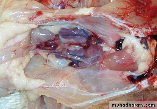
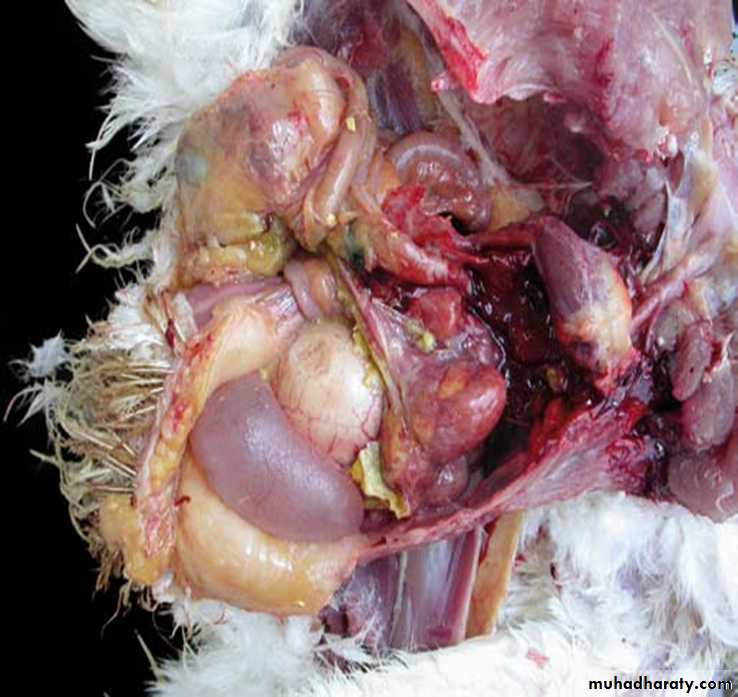
Fig. 10 Coelomic cavity of a 40 week old chicken with a persistent right oviduct (arrow). A fluid-filled diverticulum in the caudal region can also be seen.
Fig. 11 Persistent right oviduct and a functional left oviduct.
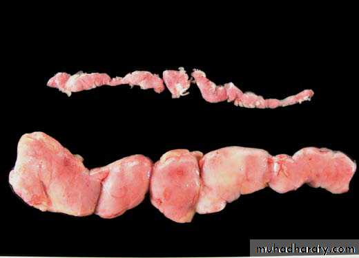
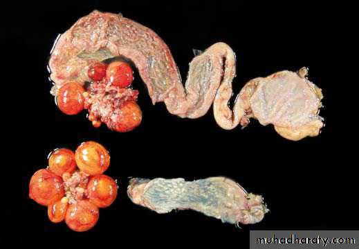
Oviduct hypoplasia: is an abnormality characterised by an incomplete development of the oviduct. Macroscopically, a shorter oviduct is seen, and also in some cases, the end of the lumen becomes occluded (figs. 12 and 13). This may be due to genetic causes or early infections before the bird reaches sexual maturity (e.g., infectious bronchitis virus).
Fig. 12 Hypoplastic oviduct occluded in the caudal region.
Fig. 13 Oviduct hypoplasia. The top part of the image shows a normal oviduct, while underneath is a shorter oviduct. Here it is impossible to distinguish the different regions.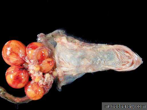
Digestive system
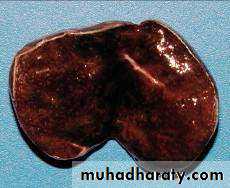
LiverThe liver’s surface should be examined for any abnormalities. It should be palpated for any nodules, friable areas, or other abnormal changes. Several slices are made into the liver in order to examine the deeper structure of the liver.
Intestinal tract
Look in the mouth for any abnormalities. Cut down the esophagus and see how much food is in the crop.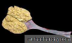
Open the proventriculus and make note of the lining which is normally bumpy due to the presence of digestive glands. Note any abnormalities.
Proventriculus-ventriculus The junction is an area with abundant lymphoid tissue and should be examined carefully for lesions.
The ventriculus, or gizzard, should be examined next. Because the
gizzard is responsible for grinding ingested material, it has a thick external muscularis layer and contains small stones or grit. The ventricular glands secrete a thick protective gel, known as koilin, which has a yellowish color. The gizzard thickness should be examined and the surface examined for erosions, ulcerations, discoloration, or other abnormalities.
The small intestine of birds is typically arranged into several loops before entering the colon. The first loop is the duodenum. It is easily identified by the location of the pancreas within the duodenal loop mesentery.
The remaining loops make up the jejunum and ileum. The colon is relatively short with two long ceca and connects to the cloaca via the colorectum.
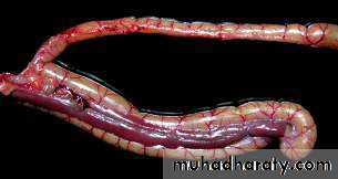
In this picture you see the two ceca and the large colon in between.
There are large lymphoid patches in the proximal portions of the ceca.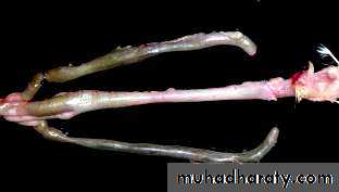
6. Write the report
No necropsy is complete until all findings have been recorded in written form. The report should include at least the following information:Species, breed, age, sex
HistoryDied or euthanized?
Nutritional, hydration status
Findings from external examination
Findings by organ system: Lymphoid (spleen, bursa), Respiratory, Digestive, Urogenital, Musculoskeletal, Nervous