Muscle Tissue; Learning Objectives
• 1. Be able to identify the three types of muscle at the light microscope level, including distinctive features of each, such as the intercalated disk of cardiac muscle.• 2. Be able to describe the structural basis of muscle striation.
• 3. Know the mechanism by which muscle cells contract.
• Understand the function and organization of the connective tissue in skeletal muscle (endo-, peri-, and epimysium).
• Be familiar with the regenerative potential of each muscle type.
.
I. Striated Muscle - regularly arranged contractile units
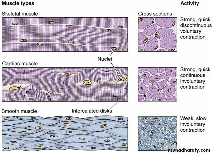
A. Skeletal Muscle - long, cylindrical multinucleated cells with peripherally placed nuclei. Contraction is typically quick and vigorous and under voluntary control. Used for locomotion and mastication.
B. Cardiac Muscle - elongated, branched cells with a single centrally placed nucleus and intercalated discs at the ends. Contraction is involuntary, vigorous, and rhythmic.
II. Smooth Muscle - possesses contractile machinery, but it is irregularly arranged (thus, non-striated). Cells are fusiform with a central nucleus. Contraction is involuntary, slow, and long lasting.
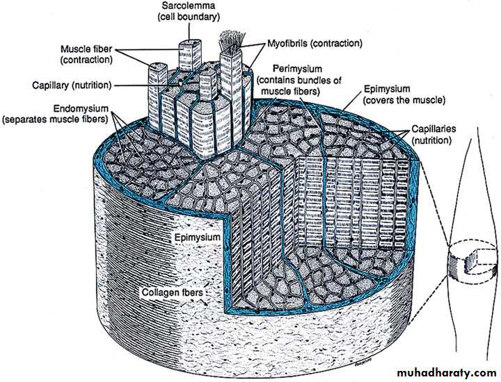
Epimysium - dense irr. co.t.
Perimysium -
less dense irr. co.t.Endomysium - basal lamina and reticular fibers
Skeletal Muscle InvestmentsALL MUSCLE CELLS HAVE BASAL LAMINAE!
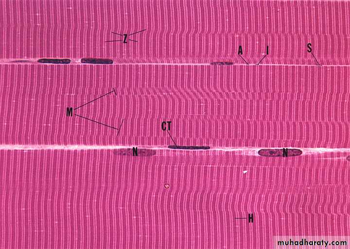
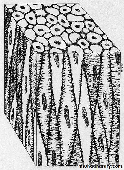
Skeletal Muscle as seen in longitudinal section in the light microscope...
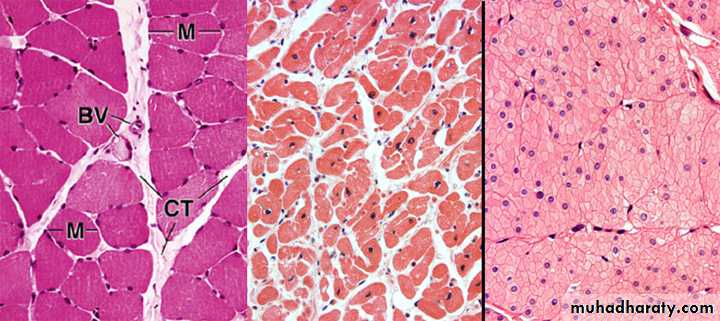
Skeletal Muscle as seen in transverse section (TS) in the LM.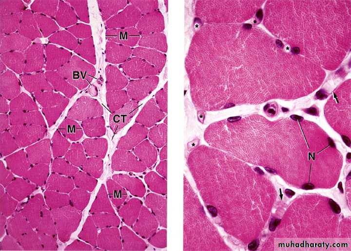
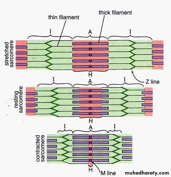
Sliding Filament Theory
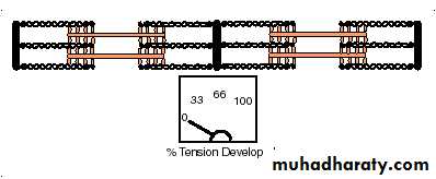
Sarcomere
Muscle fibers are composed of many contractile units (sarcomeres)Changes in the amount of overlap between thick and thin filaments allows for contraction and relaxation of muscle fibers
Many fibers contracting together result in gross movement
Note: Z lines move closer together; I band and H band become smaller during contraction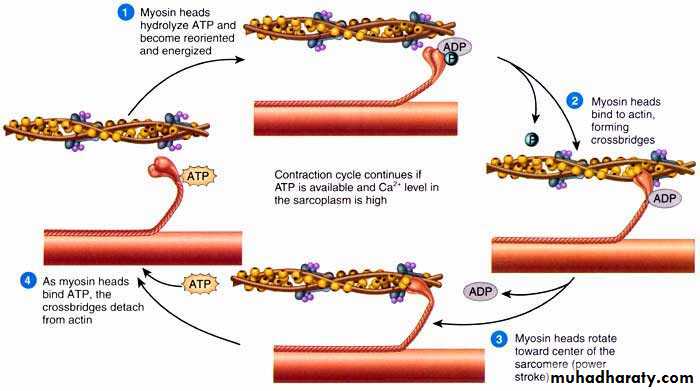
Contraction is Ca+ dependent
• In resting state, free ATP is bound to myosin• ATP hydrolysis induces conformational change – myosin head cocks forward 5nm (ADP+Pi remain bound to myosin).
• Stimulation by nerves cause release of calcium (green) into cytoplasm; calcium binds troponin (purple) and reveals myosin binding site (black) on actin (yellow)
• Myosin binds weakly to actin, causing release of Pi
• Release of Pi from myosin induces strong binding to actin, power stroke, and release of ADP
• Cycle continues if ATP is available and cytoplasmic Ca+ level is high
1
2
4
5
3
&
Cardiac Muscle
Tissue Features:Striated (same contractile machinery)
Self-excitatory and electrically coupled
Rate of contractions modulated by autonomic nervous system
innervation is neuroendocrine in nature (i.e. no “motor end plates”)
Cell Features:
1 or 2 centrally placed nuclei
Branched fibers with intercalated discs
Numerous mitochondria (up to 40% of cell volume)
Sarcoplasmic reticulum & T-tubules appear as diads at Z lines
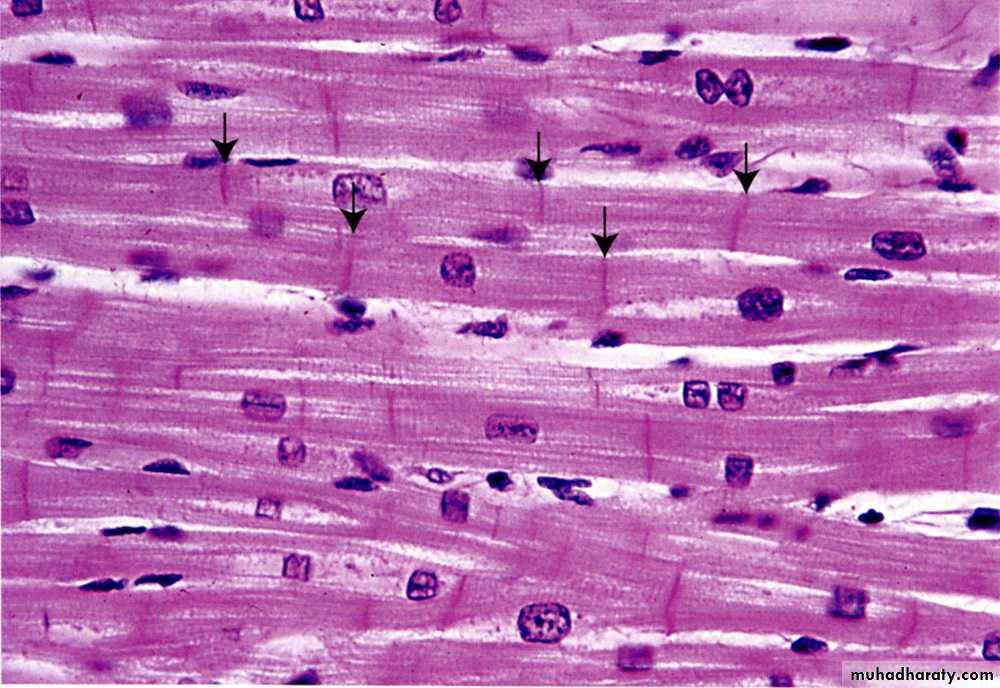
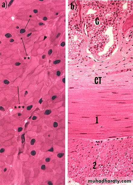
Cardiac Muscle (LS)
Cardiac Muscle TS)Cardiac Muscle (LS)
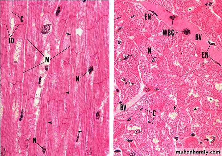
TS of Cardiac Muscle versus Skeletal Muscle
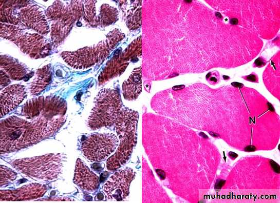
As with skeletal muscle, delicate, highly vascularized connective tissue (endomysium) surrounds each cardiac muscle cell. Fibers are bundled into fascicles, so there is also perimysium. However, there really isn’t an epimysium; instead, Co.T. ensheathing the muscle of the heart is called the epicardium.
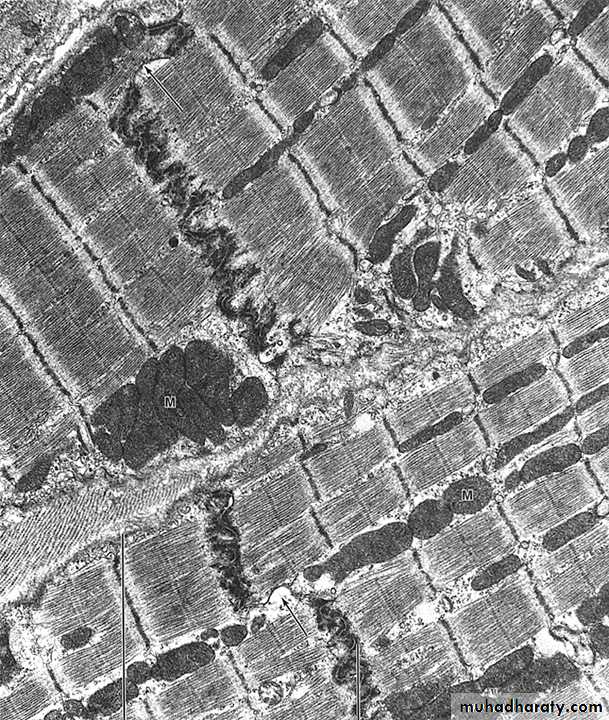
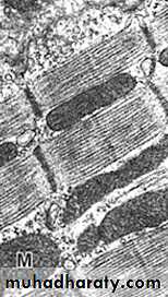
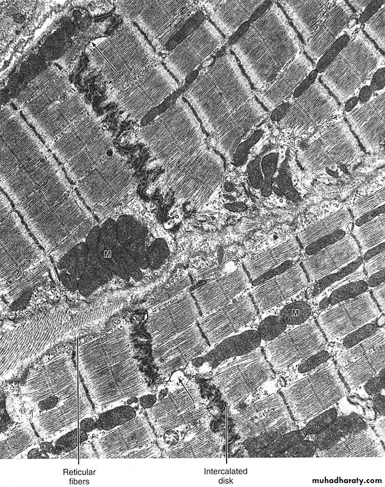
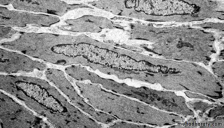
Cardiac Muscle (TEM)
Intercalated Discs Couple Heart Muscle Mechanically and Electrically
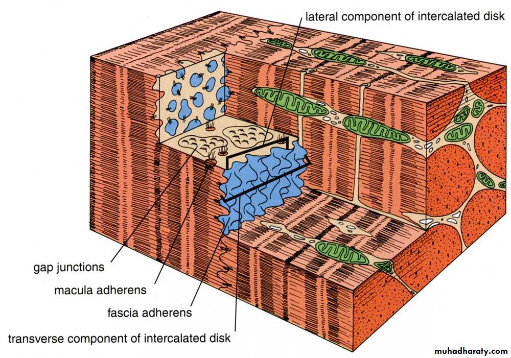
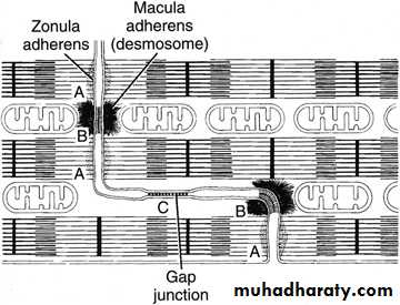
Transverse portion:
forms mechanical couplingLateral Portion:
forms electrical couplingaka “Fascia adherens”
Smooth Muscle
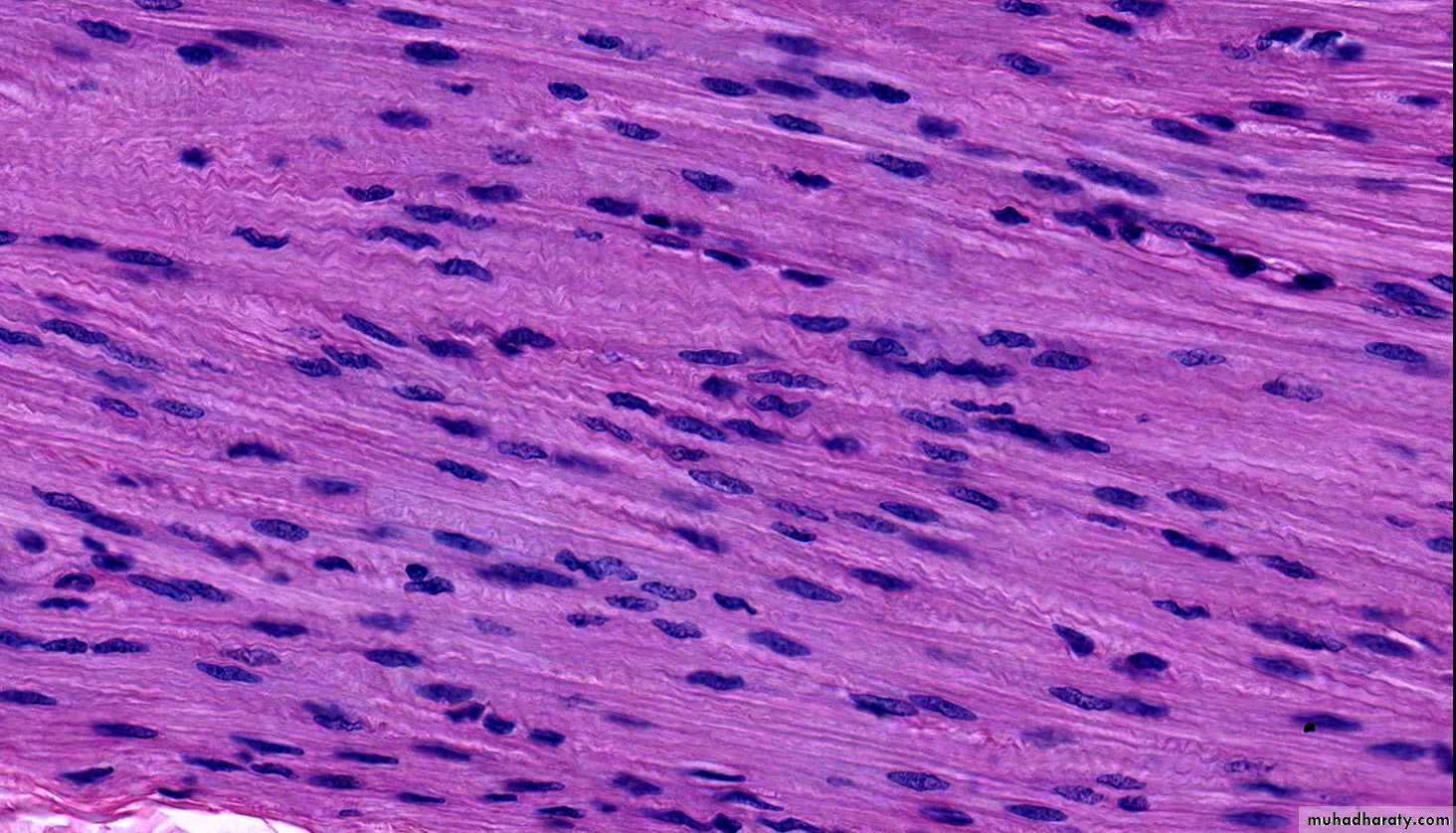
Fusiform, non-striated cellsSingle, centrally-placed nucleus
Contraction is non-voluntary
Contraction is modulated in a neuroendocrine manner
Found in blood vessels, GI and urogenital organ walls, dermis of skin
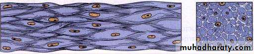
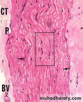
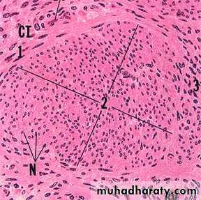
Smooth Muscle (longitudinal section)
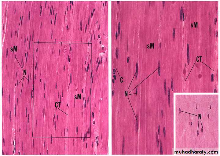
Smooth Muscle Viewed in Transverse and Longitudinal Section
actin and myosin filaments
intermediate filaments of desmin (also vimentin in vascular smooth muscle)membrane associated and cytoplasmic dense bodies containing actinin (similar to Z lines)
relatively active nucleus (smooth muscle cells make collagen, elastin, and proteoglycans)
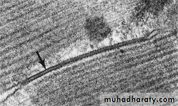
Ultrastructure of Smooth Muscle:
*
Smooth Muscle CS (TEM)
What is the structure marked by * ?
Also, note collagen –SMC secrete ECM:
collagen (I,III, IV), elastin, and proteoglycans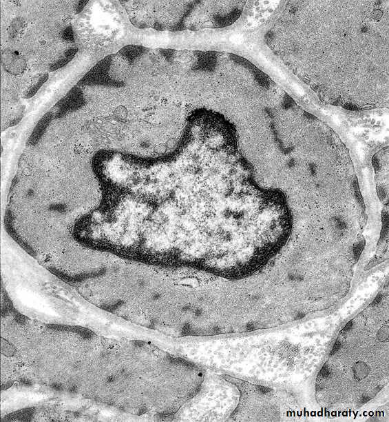
*
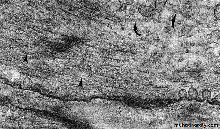
microtubules (curved arrows)
actin filament (arrowheads)intermediate filaments
dense bodies (desmin/vimentin plaques)
caveoli (membrane invaginations & vesicular system contiguous with SER –functionally analogous to sarcoplasmic reticulum)
More Ultrastructure of Smooth Muscle Cells:
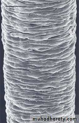
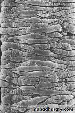
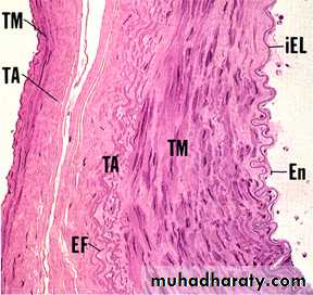
Smooth Muscle (vascular)
RelaxedContracted
10-100mm in diameter
Up to 30cm in length10-15mm in diameter
80-100mm in length
0.2-2mm in diameter
20-200mm in length
Skeletal Muscle
Cardiac MuscleSmooth Muscle
Skeletal Muscle
Cardiac MuscleSmooth Muscle
Smooth Muscle
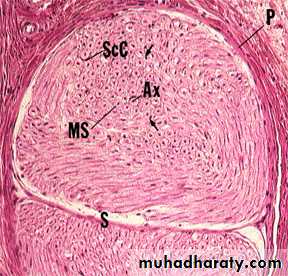
VERSUSNerve
VERSUS
Connective Tissue
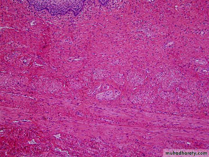
How these tissues actually appear…
Nerve
SMSM
SM
CT
CT
CT
Epithelium
Epithelium
B.V.
B.V.