
Contacts • Phone/E-Mail
Name:
Ph:
e-mail:
Name:
Ph:
e-mail:
Name:
Ph:
e-mail:
Name:
Ph:
e-mail:
Name:
Ph:
e-mail:
Name:
Ph:
e-mail:
Name:
Ph:
e-mail:
Name:
Ph:
e-mail:
Name:
Ph:
e-mail:
Name:
Ph:
e-mail:
Name:
Ph:
e-mail:
Name:
Ph:
e-mail:
00ECG-FM 2/10/05 7:45 PM Page 2
Copyright © 2005 F. A. Davis.

F. A. Davis Company • Philadelphia
ECG
N
otes
Purchase additional copies of this book
at your health science bookstore or
directly from F. A. Davis by shopping
online at www.fadavis.com or by calling
800-323-3555 (US) or 800-665-1148 (CAN)
A Davis’s Notes Book
Shirley A. Jones, MS Ed, MHA, EMT-P
Interpretation and Management Guide
ECG
N
otes
Interpretation and Management Guide
00ECG-FM 2/10/05 7:45 PM Page i
Copyright © 2005 F. A. Davis.

F. A. Davis Company
1915 Arch Street
Philadelphia, PA 19103
www.fadavis.com
Copyright © 2005 by F. A. Davis Company
All rights reserved. This book is protected by copyright. No part of it may be
reproduced, stored in a retrieval system, or transmitted in any form or by any
means, electronic, mechanical, photocopying, recording, or otherwise, with-
out written permission from the publisher.
Printed in China by Imago
Last digit indicates print number: 10 9 8 7 6 5 4 3 2 1
Publisher, Nursing: Lisa Deitch
Project Editor: Ilysa H. Richman
Developmental Editor: Anne-Adele Wight
Design Manager: Joan Wendt
Cover Design: Paul Fry
Consultant: Dawn McKay, RN, MSN, CCRN
As new scientific information becomes available through basic and clinical
research, recommended treatments and drug therapies undergo changes. The
author(s) and publisher have done everything possible to make this book
accurate, up to date, and in accord with accepted standards at the time of
publication. The author(s), editors, and publisher are not responsible for
errors or omissions or for consequences from application of the book, and
make no warranty, expressed or implied, in regard to the contents of the book.
Any practice described in this book should be applied by the reader in accor-
dance with professional standards of care used in regard to the unique
circumstances that may apply in each situation. The reader is advised always
to check product information (package inserts) for changes and new informa-
tion regarding dose and contraindications before administering any drug.
Caution is especially urged when using new or infrequently ordered drugs.
Authorization to photocopy items for internal or personal use, or the internal
or personal use of specific clients, is granted by F. A. Davis Company for users
registered with the Copyright Clearance Center (CCC) Transactional Reporting
Service, provided that the fee of $.10 per copy is paid directly to CCC, 222
Rosewood Drive, Danvers, MA 01923. For those organizations that have been
granted a photocopy license by CCC, a separate system of payment has been
arranged. The fee code for users of the Transactional Reporting Service is:
8036-1347-4/05 0 + $.10.
00ECG-FM 2/10/05 7:45 PM Page ii
Copyright © 2005 F. A. Davis.

BASICS
ECGS
12-LEAD
MEDS/
SKILLS
CPR
ACLS
TEST
STRIPS
TOOLS
Waterproof and Reusable
Wipe-Free Pages
Write directly onto any page of ECG Notes with
a ballpoint pen. Wipe old entries off with an
alcohol pad and reuse.
Place 2
7
/
8
2
7
/
8
Sticky Notes
here
for a convenient and refillable note pad
HIPAA Compliant
OSHA Compliant
✓
✓
00ECG-FM 2/10/05 7:45 PM Page iii
Copyright © 2005 F. A. Davis.

Look for our other
Davis’s Notes titles
Available Now!
RNotes
®
:
Nurse’s Clinical Pocket Guide
ISBN: 0-8036-1060-2
LPN Notes:
Nurse’s Clinical Pocket Guide
ISBN: 0-8036-1132-3
MedNotes:
Nurse’s Pharmacology Pocket Guide
ISBN: 0-8036-1109-9
MedSurg Notes:
Nurse’s Clinical Pocket Guide
ISBN: 0-8036-1115-3
NutriNotes:
Nutrition & Diet Therapy Pocket Guide
ISBN: 0-8036-1114-5
IV Therapy Notes:
Nurse’s Clinical Pocket Guide
ISBN: 0-8036-1288-5
PsychNotes:
Clinical Pocket Guide
ISBN: 0-8036-1286-9
LabNotes:
Pocket Guide to Lab & Diagnostic Tests
ISBN: 0-8036-1265-6
OrthoNotes:
A Clinical Examination Pocket Guide
ISBN: 0-8036-1350-4
MA Notes:
Medical Assistant’s Pocket Guide
ISBN: 0-8036-1281-8
00ECG-FM 2/10/05 7:45 PM Page iv
Copyright © 2005 F. A. Davis.
1
BASICS
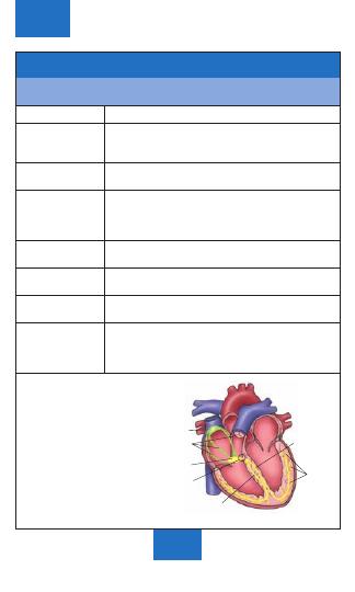
Anatomy of the Heart
The heart, located in the mediastinum, is the central structure of
the cardiovascular system. It is protected by the bony structures
of the sternum anteriorly, the spinal column posteriorly, and the
rib cage.
♥
Clinical Tip: The cone-shaped heart has its tip (apex) just
above the diaphragm to the left of the midline. This is why we
may think of the heart as being on the left side, since the
strongest beat can be heard or felt here.
01ECG-Tab 01 2/4/05 3:57 PM Page 1
Copyright © 2005 F. A. Davis.

2
BASICS
Endocardium
Parietal
pericardium
Myocardium
(heart muscle)
Epicardium
(visceral pericardium)
Fibrous pericardium
(pericardial sac)
Pericardial cavity
Layers of the Heart
The pericardial cavity contains a small amount of lubricating fluid to
prevent friction during heart contraction.
01ECG-Tab 01 2/4/05 3:57 PM Page 2
Copyright © 2005 F. A. Davis.
3
BASICS
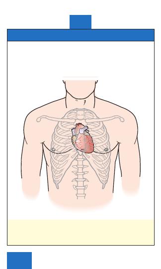
Pulmonary semilunar
valve
Aortic semilunar
valve
Tricuspid
valve
Fibrous
skeleton
Mitral valve
Posterior
Coronary artery
Heart Valves
Properties of Heart Valves
■
Fibrous connective tissue prevents enlargement of valve
openings and anchors valve flaps.
■
Valve closure prevents backflow of blood during and after
contraction.
The atria have been removed in this superior view.
01ECG-Tab 01 2/4/05 3:57 PM Page 3
Copyright © 2005 F. A. Davis.
4
BASICS
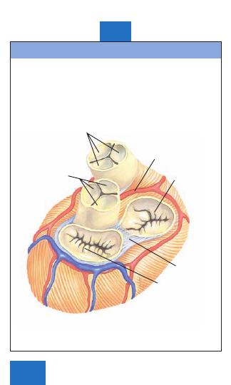
Brachiocephalic
artery
Superior vena cava
Left common carotid artery
Left subclavian artery
Aortic arch
Right
pulmonary artery
Right
pulmonary veins
Right atrium
Inferior vena cava
Tricuspid
valve
Pulmonary
semilunar valve
Left pulmonary artery
Left atrium
Left pulmonary veins
Mitral valve
Left ventricle
Aortic semilunar
valve
Interventricular
septum
Apex
Chordae
tendineae
Right
ventricle
Papillary
muscles
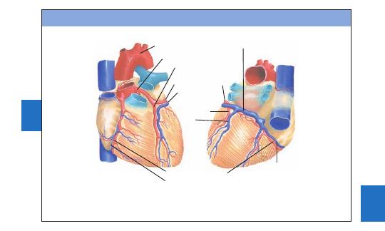
Heart Chambers and Great Vessels
01ECG-Tab 01 2/4/05 3:57 PM Page 4
Copyright
©
2005
F.
A.
Davis.
5
BASICS
Aorta
Left coronary artery
Anterior
descending branch
Coronary sinus
Posterior
artery and
vein
Small
cardiac vein
Right coronary artery
A
B
Circumflex branch
Great cardiac
vein
Right coronary vein
Coronary Arterial Circulation
(A) Anterior view
(B) Posterior view
01ECG-Tab 01 2/4/05 3:57 PM Page 5
Copyright
©
2005
F.
A.
Davis.
6
Anatomy of the Cardiovascular System
The cardiovascular system is a closed system consisting of
blood vessels and the heart. Arteries and veins are connected
by smaller structures in which electrolytes are exchanged
across cell membranes.
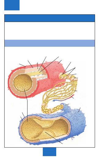
Blood Vessel Structures
BASICS
Tunica
externa
External elastic
lamina
Tunica
media
Internal elastic
lamina Endothelium (lining)
Artery
Arteriole
Endothelial
cells
Smooth
muscle
Precapillary
sphincter
Capillary
Blood flow
Venule
Vein
Valve
Tunica
intima
Tunica
externa
Tunica
media
01ECG-Tab 01 2/4/05 3:57 PM Page 6
Copyright © 2005 F. A. Davis.
7
Arterial Circulation
Arteries (excluding the pulmonary artery) transport oxygenated blood.
BASICS
Occipital
Internal carotid
Vertebral
Brachiocephalic
Aortic arch
Maxillary
Facial
External carotid
Common carotid
Subclavian
Axillary
Pulmonary
Celiac
Left gastric
Hepatic
Splenic
Superior
mesenteric
Abdominal aorta
Right common
iliac
Internal iliac
External iliac
Femoral
Popliteal
Anterior tibial
Posterior tibial
Intercostal
Brachial
Renal
Gonadal
Inferior mesenteric
Radial
Ulnar
Deep palmar arch
Superficial
palmar arch
Deep femoral
01ECG-Tab 01 2/4/05 3:58 PM Page 7
Copyright © 2005 F. A. Davis.
8
BASICS
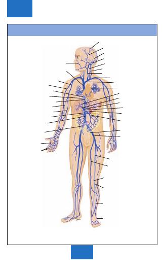
Superior sagittal sinus
Inferior sagittal sinus
Straight sinus
Transverse sinus
Vertebral
External jugular
Internal jugular
Subclavian
Brachiocephalic
Pulmonary
Hepatic
Hepatic portal
Left gastric
Renal
Splenic
Inferior
mesenteric
Internal iliac
Femoral
External iliac
Great saphenous
Popliteal
Small saphenous
Anterior tibial
Anterior facial
Superior vena cava
Axillary
Cephalic
Hemiazygos
Intercostal
Inferior vena cava
Brachial
Basilic
Gonadal
Superior
mesenteric
Dorsal arch
Volar digital
Dorsal arch
Common iliac
Venous Circulation
Veins (excluding the pulmonary vein) carry blood low in oxygen and high
in carbon dioxide.
01ECG-Tab 01 2/4/05 3:58 PM Page 8
Copyright © 2005 F. A. Davis.
Physiology of the Heart
Mechanics of Heart Function
Process
Action
Cardiac cycle
Systole
Diastole
Stroke
volume (SV)
Cardiac
output (CO)
Properties of Cardiac Cells
Property
Ability
Automaticity
Excitability
Conductivity
Contractility
9
BASICS
Sequence of events in 1 heartbeat. Blood is
pumped through the entire cardiovascular
system.
Contraction phase—usually refers to
ventricular contraction.
Relaxation phase—the atria and ventricles are
filling. Lasts longer than systole.
Amount of blood ejected from either ventricle
in a single contraction. Starling’s Law of the
Heart states that degree of cardiac muscle
stretch can increase force of ejected blood.
More blood filling the ventricles
↑ SV.
Amount of blood pumped through the
cardiovascular system per min.
CO
SV Heart rate (HR)
Generates electrical impulse independently,
without involving the nervous system.
Responds to electrical stimulation.
Passes or propagates electrical impulses
from cell to cell.
Shortens in response to electrical
stimulation.
01ECG-Tab 01 2/4/05 3:58 PM Page 9
Copyright © 2005 F. A. Davis.
10
BASICS
Dominant pacemaker of the heart, located in
upper portion of right atrium. Intrinsic rate
60–100 bpm.
Direct electrical impulses between SA and AV
nodes.
Part of AV junctional tissue. Slows
conduction, creating a slight delay before
impulses reach ventricles. Intrinsic rate
40–60 bpm.
Transmits impulses to bundle branches.
Located below AV node.
Conducts impulses that lead to left ventricle.
Conducts impulses that lead to right ventricle.
Network of fibers that spreads impulses
rapidly throughout ventricular walls.
Located at terminals of bundle branches.
Intrinsic rate 20–40 bpm.
Bundle of His
Left bundle
branch
Purkinje
fibers
Right bundle
branch
AV Node
SA node
Internodal
pathways
Electrical Conduction System of the Heart
Conduction System Structures and Functions
Structure
Function and Location
Sinoatrial (SA)
node
Internodal
pathways
Atrioventricular
(AV) node
Bundle of His
Left bundle
branch
Right bundle
branch
Purkinje system
Conduction system of the heart.
01ECG-Tab 01 2/4/05 3:58 PM Page 10
Copyright © 2005 F. A. Davis.
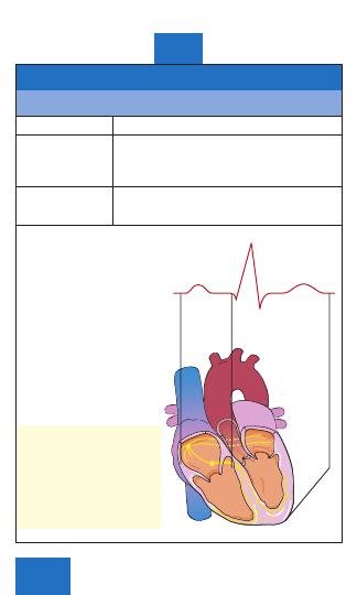
Electrical Conduction System of the Heart
Electrophysiology
Action
Effect
Depolarization
The electrical charge of a cell is altered
by a shift of electrolytes on either side
of the cell membrane. This change
stimulates muscle fiber to contract.
Repolarization
Chemical pumps re-establish an internal
negative charge as the cells return to
their resting state.
Depolarization and
repolarization of the heart.
♥
Clinical Tip: Mechanical
and electrical functions of
the heart are influenced by
proper electrolyte balance.
Important components of
this balance are sodium,
calcium, potassium, and
magnesium.
11
BASICS
P
R
T
Q
S
Ventricular
depolarization
Ventricular
repolarization
Atrial
depolarization
01ECG-Tab 01 2/4/05 3:58 PM Page 11
Copyright © 2005 F. A. Davis.

12
BASICS
The Electrocardiogram (ECG)
■
An ECG is a series of waves and deflections recording the
heart’s electrical activity from a certain “view.”
■
Many views, each called a lead, monitor voltage changes
between electrodes placed in different positions on the body.
■
Leads I, II, and III are bipolar leads, which consist of two
electrodes of opposite polarity (positive and negative). The
third (ground) electrode minimizes electrical activity from
other sources.
■
Leads aVR, aVL, and aVF are unipolar leads and consist of a
single positive electrode and a reference point (with zero
electrical potential) that lies in the center of the heart’s
electrical field.
■
Leads V
1
–V
6
are unipolar leads and consist of a single positive
electrode with a negative reference point found at the
electrical center of the heart.
■
Voltage changes are amplified and visually displayed on an
oscilloscope and graph paper.
■
An ECG tracing looks different in each lead because the
recorded angle of electrical activity changes with each lead.
■
Several different angles allow a more accurate perspective
than a single one would.
■
The ECG machine can be adjusted to make any skin electrode
positive or negative. The polarity depends on which lead the
machine is recording.
■
A cable attached to the patient is divided into several
different-colored wires: three, four, or five for monitoring
purposes, or ten for a 12-lead ECG.
■
Incorrect placement of electrodes may turn a normal ECG
tracing into an abnormal one.
♥
Clinical Tip: Patients should be treated according to their
symptoms, not merely their ECG.
♥
Clinical Tip: To obtain a 12-lead ECG, four wires are attached
to each limb and six wires are attached at different locations on
the chest. The total of ten wires provides twelve views (12
leads).
01ECG-Tab 01 2/4/05 3:58 PM Page 12
Copyright © 2005 F. A. Davis.
13
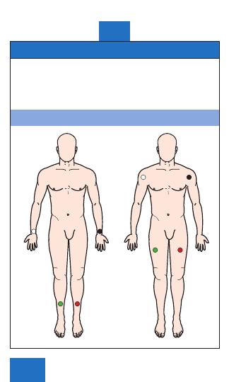
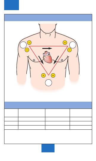
Limb Leads
Electrodes are placed on the right arm (RA), left arm (LA), right
leg (RL), and left leg (LL). With only four electrodes, six leads
are viewed.
■
Standard leads: I, II, III
■
Augmented leads: aVR, aVL, aVF
Standard Limb Lead Electrode Placement
BASICS
LA
RA
or
RL
LL
RA
LA
LL
RL
01ECG-Tab 01 2/4/05 3:58 PM Page 13
Copyright © 2005 F. A. Davis.
14
BASICS
I
III
II
LL
RA
LA
Standard Limb Leads
Elements of Standard Limb Leads
Positive
Negative
View of
Lead
Electrode
Electrode
Heart
I
LA
RA
Lateral
II
LL
RA
Inferior
III
LL
LA
Inferior
01ECG-Tab 01 2/4/05 3:58 PM Page 14
Copyright © 2005 F. A. Davis.
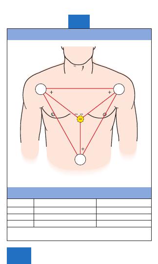
Augmented Limb Leads
Elements of Augmented Limb Leads
Lead
Positive Electrode
View of Heart
aVR
RA
None
aVL
LA
Lateral
aVF
LL
Inferior
15
BASICS
LL
RA
LA
aVR
aVL
aVF
01ECG-Tab 01 2/4/05 3:58 PM Page 15
Copyright © 2005 F. A. Davis.
16
BASICS
Midclavicular
line
V
1
V
2
V
3
V
4
V
5
V
6
Anterior
axillary line
Midaxillary
line
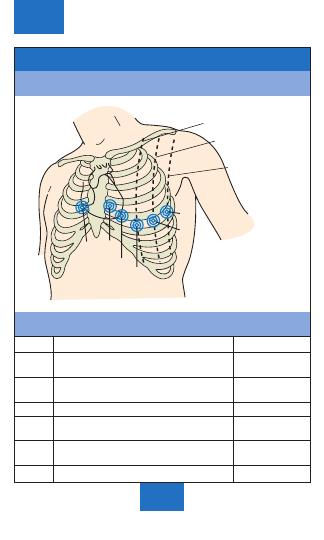
Chest Leads
Standard Chest Lead Electrode Placement
Elements of Chest Leads
Lead
Positive Electrode Placement
View of Heart
V
1
4th Intercostal space to
Septum
right of sternum
V
2
4th Intercostal space to
Septum
left of sternum
V
3
Directly between V
2
and V
4
Anterior
V
4
5th Intercostal space at
Anterior
left midclavicular line
V
5
Level with V
4
at left anterior
Lateral
axillary line
V
6
Level with V
5
at left midaxillary line
Lateral
01ECG-Tab 01 2/4/05 3:58 PM Page 16
Copyright © 2005 F. A. Davis.
17
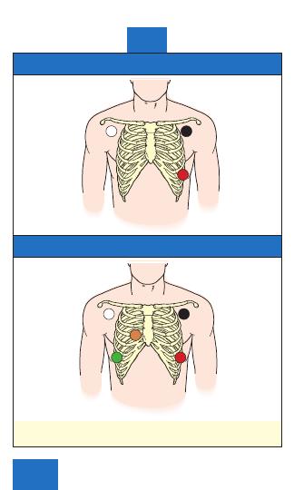
Electrode Placement Using a 3-Wire Cable
Electrode Placement Using a 5-Wire Cable
♥
Clinical Tip: Five-wire telemetry units are commonly used to monitor
leads I, II, III, aVR, aVL, aVF, and V
1
in critical care settings.
BASICS
RA
LA
LL
RA
LA
LL
RL
V
1
01ECG-Tab 01 2/4/05 3:58 PM Page 17
Copyright © 2005 F. A. Davis.
18
BASICS
G
G
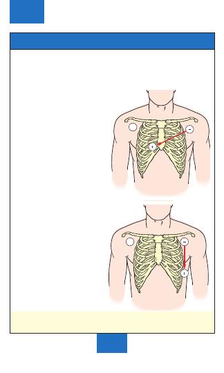
Modified Chest Leads
■
Modified chest leads (MCL) are useful in detecting bundle branch
blocks and premature beats.
■
Lead MCL
1
simulates chest lead V
1
and views the ventricular septum.
■
Lead MCL
6
simulates chest lead V
6
and views the lateral wall of the left
ventricle.
Lead MCL
1
electrode placement.
Lead MCL
6
electrode placement.
♥
Clinical Tip: Write on the rhythm strip which simulated lead
was used.
01ECG-Tab 01 2/4/05 3:58 PM Page 18
Copyright © 2005 F. A. Davis.
19
The Right-Sided 12-Lead ECG
■
The limb leads are placed as usual but the chest leads are a
mirror image of the standard 12-lead chest placement.
■
The ECG machine cannot recognize that the leads have been
reversed. It will still print “V
1
–V
6
” next to the tracing. Be sure
to cross this out, and write the new lead positions on the ECG
paper.
The Right-Sided 12-Lead ECG
Chest Leads
Position
V
1R
4th Intercostal space to left of sternum
V
2R
4th Intercostal space to right of sternum
V
3R
Directly between V
2R
and V
4R
V
4R
5th Intercostal space at right midclavicular line
V
5R
Level with V
4R
at right anterior axillary line
V
6R
Level with V
5R
at right midaxillary line
♥
Clinical Tip: Patients with an acute inferior MI should have
right-sided ECGs to assess for possible right ventricular
infarction.
BASICS
Midclavicular
line
Anterior
axillary line
Midaxillary
line
V
6R
V
4R
V
5R
V
1R
V
2R
V
3R
01ECG-Tab 01 2/4/05 3:58 PM Page 19
Copyright © 2005 F. A. Davis.
20
BASICS
V
9
Spinal
column
Left
shoulder
V
8
V
6
V
6
V
8
V
9
V
4R
The 15-Lead ECG
Areas of the heart that are not well visualized by the six chest
leads include the wall of the right ventricle and the posterior
wall of the left ventricle. A 15-lead ECG, which includes the
standard 12 leads plus leads V
4R
, V
8
, and V
9
, increases the
chance of detecting an MI in these areas.
The 15-Lead ECG
Chest
Electrode
View
Leads
Placement
of Heart
V
4R
5th Intercostal space in right
Right ventricle
anterior midclavicular line
V
8
Posterior 5th intercostal space
Posterior wall
in left midscapular line
of left ventricle
V
9
Directly between V
8
and spinal
Posterior wall
column at posterior 5th
of left ventricle
intercostal space
♥
Clinical Tip: Use a 15-lead ECG when the 12-lead is normal
but the history is still suggestive of an acute infarction.
01ECG-Tab 01 2/4/05 3:58 PM Page 20
Copyright © 2005 F. A. Davis.
21
BASICS
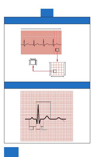
1 mm
0.1 mv
0.04 sec
Constant speed of 25 mm/sec
0.20 sec
5 mm
0.5 mv
Small
box
Large
box
R
Q S
QT Interval
P
U
T
PR
Interval
ST
Segment
QRS
Interval
Isoelectric
line
Recording of the ECG
Components of an ECG Tracing
01ECG-Tab 01 2/4/05 3:58 PM Page 21
Copyright © 2005 F. A. Davis.
22
BASICS
First wave seen
Small rounded, upright (positive) wave
indicating atrial depolarization (and
contraction)
Distance between beginning of P wave and
beginning of QRS complex
Measures time during which a depolariza-
tion wave travels from the atria to the
ventricles
Three deflections following P wave
Indicates ventricular depolarization (and
contraction)
Q Wave: First negative deflection
R Wave: First positive deflection
S Wave: First negative deflection after R wave
Distance between S wave and beginning of
T wave
Measures time between ventricular
depolarization and beginning of
repolarization
Rounded upright (positive) wave following
QRS
Represents ventricular repolarization
Measured from beginning of QRS to end of
T wave.
Represents total ventricular activity.
Small rounded, upright wave following
T wave
Most easily seen with a slow HR.
Represents repolarization of Purkinje fibers.
Electrical Components
Deflection
Description
P Wave
PR Interval
QRS Interval
ST Segment
T Wave
QT Interval
U Wave
01ECG-Tab 01 2/4/05 3:58 PM Page 22
Copyright © 2005 F. A. Davis.
23
Methods for Calculating Heart Rate
Heart rate is calculated as the number of times the heart beats
per minute. It usually measures ventricular rate (the number of
QRS complexes) but can refer to atrial rate (the number of P
waves). The method chosen to calculate HR varies according to
rate and regularity on the ECG tracing.
Method 1: Count Large Boxes
Regular rhythms can be quickly determined by counting the
number of large graph boxes between two R waves. That
number is divided into 300 to calculate bpm. The rates for the
first one to six large boxes can be easily memorized.
Remember: 60 sec/min divided by 0.20 sec/large box
300
large boxes/min.
Counting large boxes for heart rate. The rate is 60 bpm.
BASICS
50
60
75
100
300 150
01ECG-Tab 01 2/4/05 3:58 PM Page 23
Copyright © 2005 F. A. Davis.
24
BASICS
Method 2: Count Small Boxes
Sometimes it is necessary to count the number of small boxes
between two R waves for fast heart rates. That number is
divided into 1500 to calculate bpm. Remember: 60 sec/min
divided by 0.04 sec/small box
1500 small boxes/min.
Examples: If there are six small boxes between two R waves:
1500/6
250 bpm.
If there are ten small boxes between two R waves:
1500/10
150 bpm.
Methods 1 and 2 for Calculating Heart Rate
Number of
Number of
Large Boxes
Rate/Min
Small Boxes
Rate/Min
1
300
2
750
2
150
3
500
3
100
4
375
4
75
5
300
5
60
6
250
6
50
7
214
7
43
8
186
8
38
9
167
9
33
10
150
10
30
11
136
11
27
12
125
12
25
13
115
13
23
14
107
14
21
15
100
15
20
16
94
♥
Clinical Tip: Approximate rate/min is rounded to the next-
highest number.
01ECG-Tab 01 2/4/05 3:58 PM Page 24
Copyright © 2005 F. A. Davis.
25
BASICS
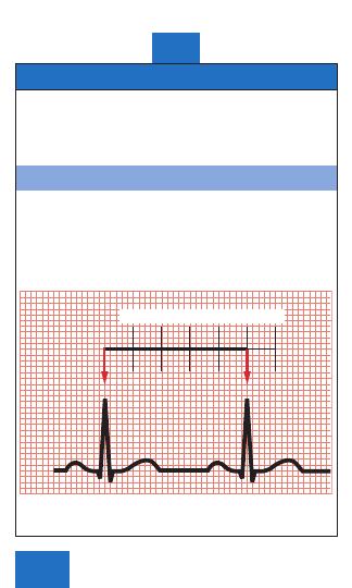
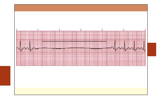
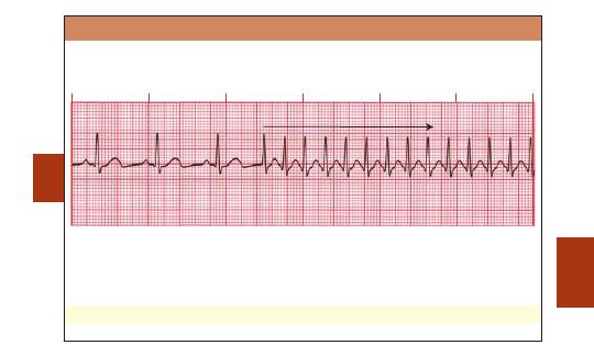
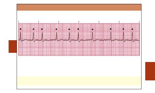
Method 3: Six-Second ECG Rhythm Strip
The best method for measuring irregular rates with varying R-R intervals is to count the
number of R waves in a 6-sec strip and multiply by 10. This gives the average number of bpm.
Using 6-sec ECG rhythm strip to calculate heart rate. Formula: 7
10 70 bpm
♥
Clinical Tip:
If a rhythm is extremely irregular, it is best to count the number of R-R intervals
per 60 sec (1 min).
01ECG-Tab 01 2/4/05 3:58 PM Page 25
Copyright
©
2005
F.
A.
Davis.
26
BASICS
ECG Interpretation
Analyzing a Rhythm
Component
Characteristic
Rate
Regularity
P Waves
PR Interval
QRS Interval
QT Interval
Dropped beats
The bpm is commonly the ventricular rate.
If atrial and ventricular rates differ, as in a
3
rd
-degree block, measure both rates.
Normal: 60–100 bpm
Slow (bradycardia):
60 bpm
Fast (tachycardia):
100 bpm
Measure R-R intervals and P-P intervals.
Regular: Intervals consistent
Regularly irregular: Repeating pattern
Irregular: No pattern
If present: Same in size, shape, position?
Does each QRS have a P wave?
Normal: Upright (positive) and uniform
Inverted: Negative
Notched: P
′
None: Rhythm is junctional or ventricular.
Constant: Intervals are the same.
Variable: Intervals differ.
Normal: 0.12–0.20 sec and constant
Normal: 0.06–0.10 sec
Wide:
0.10 sec
None: Absent
Beginning of R wave to end of T wave
Varies with HR.
Normal: Less than half the R-R interval
Occur in AV blocks.
Occur in sinus arrest.
01ECG-Tab 01 2/4/05 3:58 PM Page 26
Copyright © 2005 F. A. Davis.

27
Component
Characteristic
Pause
QRS Complex
grouping
Notes:
BASICS
Compensatory: Complete pause following a
premature atrial contraction (PAC),
premature junctional contraction (PJC), or
premature ventricular contraction (PVC)
Noncompensatory: Incomplete pause
following a PAC, PJC, or PVC
Bigeminy: Repeating pattern of normal
complex followed by a premature
complex
Trigeminy: Repeating pattern of 2 normal
complexes followed by a premature
complex
Quadrigeminy: Repeating pattern of 3
normal complexes followed by a
premature complex
Couplets: 2 Consecutive premature
complexes
Triplets: 3 Consecutive premature
complexes
01ECG-Tab 01 2/4/05 3:58 PM Page 27
Copyright © 2005 F. A. Davis.
28
ECGs
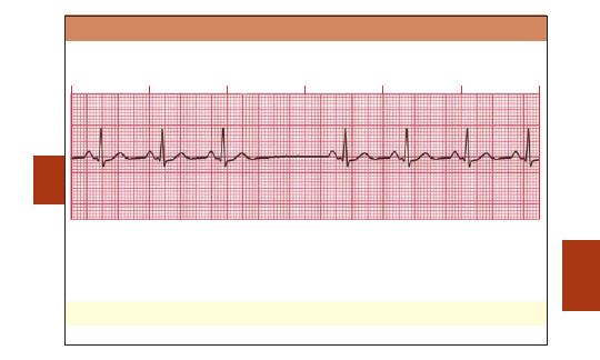
Sinoatrial (SA) Node Arrhythmias
■
Upright P waves all look similar.
Note: All ECG strips in this tab were recorded in lead II.
■
PR intervals and QRS complexes are of normal duration.
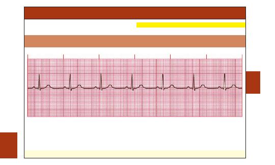
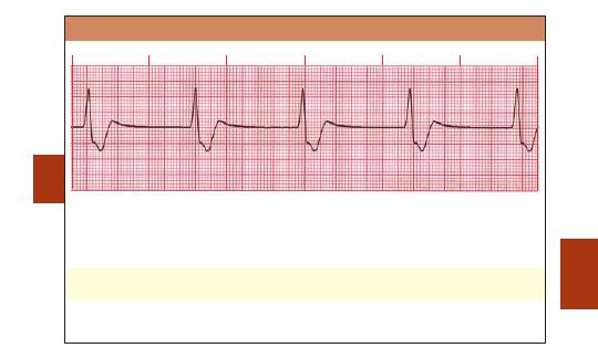
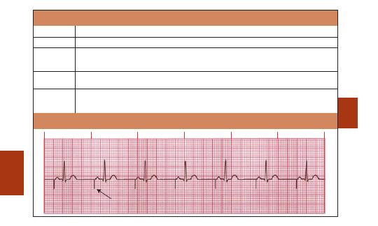
Normal Sinus Rhythm (NSR)
Rate: Normal (60–100 bpm)
Rhythm: Regular
P Waves: Normal (upright and uniform)
PR Interval: Normal (0.12–0.20 sec)
QRS: Normal (0.06–0.10 sec)
♥
Clinical Tip:
A normal ECG does not exclude heart disease.
02ECG-Tab 02 2/4/05 3:58 PM Page 28
Copyright
©
2005
F.
A.
Davis.

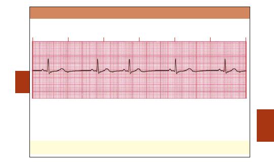
Sinus Bradycardia
■
Results from slowing of the SA node.
Rate: Slow (
60 bpm)
Rhythm: Regular
P Waves: Normal (upright and uniform)
PR Interval: Normal (0.12–0.20 sec)
QRS: Normal (0.06–0.10 sec)
♥
Clinical Tip:
Sinus bradycardia is normal in athletes and during sleep. In acute MI, it may be
protective and beneficial or the slow rate may compromise cardiac output. Certain
medications, such as beta blockers, may also cause sinus bradycardia.
29
ECGs
02ECG-Tab 02 2/4/05 3:58 PM Page 29
Copyright
©
2005
F.
A.
Davis.
30
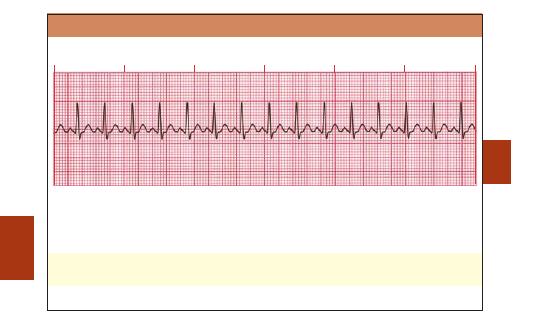
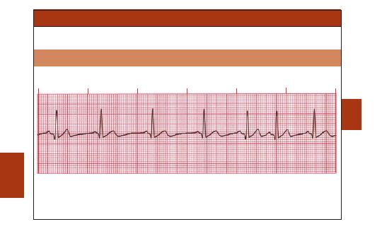
Sinus Tachycardia
■
Results from increased SA node discharge.
Rate: Fast (
100 bpm)
Rhythm: Regular
P Waves: Normal (upright and uniform)
PR Interval: Normal (0.12–0.20 sec)
QRS: Normal (0.06–0.10 sec)
♥
Clinical Tip:
Sinus tachycardia may be caused by exercise, anxiety, fever, hypoxemia,
hypovolemia, or cardiac failure.
ECGs
02ECG-Tab 02 2/4/05 3:58 PM Page 30
Copyright
©
2005
F.
A.
Davis.
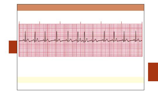
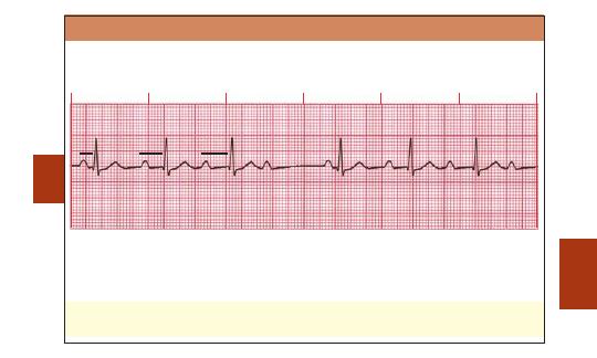
Sinus Arrhythmia
■
The SA node discharges irregularly.
■
The R-R interval is irregular.
Rate: Usually normal (60–100 bpm); frequently increases with inspiration and decreases with
expiration
Rhythm: Irregular; varies with respiration
P Waves: Normal (upright and uniform)
PR Interval: Normal (0.12–0.20 sec)
QRS: Normal (0.06–0.10 sec)
♥
Clinical Tip:
The pacing rate of the SA node varies with respiration, especially in children
and elderly people.
31
ECGs
02ECG-Tab 02 2/4/05 3:58 PM Page 31
Copyright
©
2005
F.
A.
Davis.
32
ECGs
3 - Sec pause/ arrest
Sinus Pause (Sinus Arrest)
■
The SA node fails to discharge and then resumes.
■
Electrical activity resumes either when the SA node resets itself or when a lower latent
pacemaker begins to discharge.
■
The pause (arrest) time interval is not a multiple of the normal P-P interval.
Rate: Normal to slow; determined by duration and frequency of sinus pause (arrest)
Rhythm: Irregular whenever a pause (arrest) occurs
P Waves: Normal (upright and uniform) except in areas of pause (arrest)
PR Interval: Normal (0.12–0.20 sec)
QRS: Normal (0.06–0.10 sec)
♥
Clinical Tip:
Cardiac output may decrease, causing syncope or dizziness.
02ECG-Tab 02 2/4/05 3:58 PM Page 32
Copyright
©
2005
F.
A.
Davis.
33
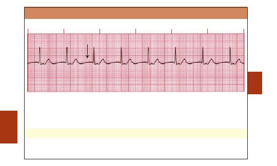
Sinoatrial (SA) Block
■
The block occurs in some multiple of the P-P interval.
■
After the dropped beat, cycles continue on time.
Rate: Normal to slow; determined by duration and frequency of SA block
Rhythm: Irregular whenever an SA block occurs
P Waves: Normal (upright and uniform) except in areas of dropped beats
PR Interval: Normal (0.12–0.20 sec)
QRS: Normal (0.06–0.10 sec)
♥
Clinical Tip:
Cardiac output may decrease, causing syncope or dizziness.
ECGs
Dropped beat
X
02ECG-Tab 02 2/4/05 3:58 PM Page 33
Copyright
©
2005
F.
A.
Davis.
Atrial Arrhythmias
■
P Waves differ in appearance from sinus P waves.
■
QRS Complexes are of normal duration.
Wandering Atrial Pacemaker (WAP)
■
Pacemaker site transfers from the SA node to other latent pacemaker sites in the atria and
the AV junction and then moves back to the SA node.
Rate: Normal (60–100 bpm)
Rhythm: Irregular
P Waves: At least three different forms, determined by the focus in the atria
PR Interval: Variable; determined by focus
QRS: Normal (0.06–0.10 sec)
34
ECGs
02ECG-Tab 02 2/4/05 3:58 PM Page 34
Copyright
©
2005
F.
A.
Davis.
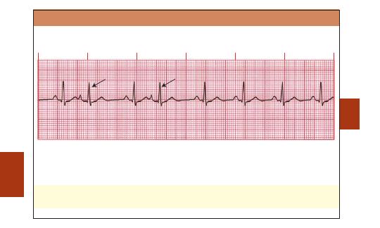
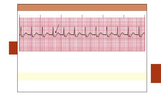
Multifocal Atrial Tachycardia (MAT)
■
This form of WAP is associated with a ventricular response of
100 bpm.
■
MAT may be confused with atrial fibrillation (A-fib); however, MAT has a visible P wave.
Rate: Fast (
100 bpm)
Rhythm: Irregular
P Wave: At least three different forms, determined by the focus in the atria
PR Interval: Variable; depends on focus
QRS: Normal (0.06–0.10 sec)
♥
Clinical Tip:
MAT is commonly seen in patients with COPD but may also occur in acute MI.
35
ECGs
02ECG-Tab 02 2/4/05 3:58 PM Page 35
Copyright
©
2005
F.
A.
Davis.
36
Premature Atrial Contraction (PAC)
■
A single complex occurs earlier than the next expected sinus complex.
■
After the PAC, sinus rhythm usually resumes.
Rate: Depends on rate of underlying rhythm
Rhythm: Irregular whenever a PAC occurs
P Waves: Present; in the PAC, may have a different shape
PR Interval: Varies in the PAC; otherwise normal (0.12–0.20 sec)
QRS: Normal (0.06–0.10 sec)
♥
Clinical Tip:
In patients with heart disease, frequent PACs may precede paroxysmal
supraventricular tachycardia (PSVT), A-fib, or A-flutter.
ECGs
PAC
PAC
02ECG-Tab 02 2/4/05 3:58 PM Page 36
Copyright
©
2005
F.
A.
Davis.
37
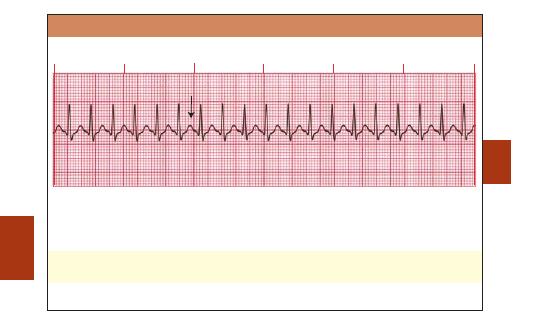
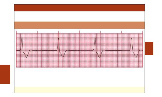
Atrial Tachycardia
■
A rapid atrial rate overrides the SA node and becomes the dominant pacemaker.
■
Some ST wave and T wave abnormalities may be present.
Rate: 150–250 bpm
Rhythm: Regular
P Waves: Normal (upright and uniform) but differ in shape from sinus P waves
PR Interval: May be short (
0.12 sec) in rapid rates
QRS: Normal (0.06–0.10 sec) but can be aberrant at times
ECGs
02ECG-Tab 02 2/4/05 3:58 PM Page 37
Copyright
©
2005
F.
A.
Davis.
38
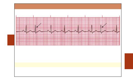
Supraventricular Tachycardia (SVT)
■
This arrhythmia has such a fast rate that the P waves may not be seen.
Rate: 150–250 bpm
Rhythm: Regular
P Waves: Frequently buried in preceding T waves and difficult to see
PR Interval: Usually not possible to measure
QRS: Normal (0.06–0.10 sec) but may be wide if abnormally conducted through ventricles
♥
Clinical Tip:
SVT may be related to caffeine intake, nicotine, stress, or anxiety in healthy
adults.
ECGs
P wave buried in T wave
02ECG-Tab 02 2/4/05 3:58 PM Page 38
Copyright
©
2005
F.
A.
Davis.
39
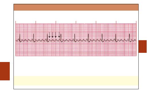
Paroxysmal Supraventricular Tachycardia (PSVT)
■
PSVT is a rapid rhythm that starts and stops suddenly.
■
For accurate interpretation, the beginning or end of the PSVT must be seen.
■
PSVT is sometimes called paroxysmal atrial tachycardia (PAT).
Rate: 150–250 bpm
Rhythm: Regular
P Waves: Frequently buried in preceding T waves and difficult to see
PR Interval: Usually not possible to measure
QRS: Normal (0.06–0.10 sec) but may be wide if abnormally conducted through ventricles
♥
Clinical Tip:
The patient may feel palpitations, dizziness, lightheadedness, or anxiety.
ECGs
Sudden onset of SVT
02ECG-Tab 02 2/4/05 3:58 PM Page 39
Copyright
©
2005
F.
A.
Davis.
40
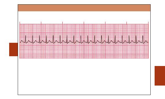
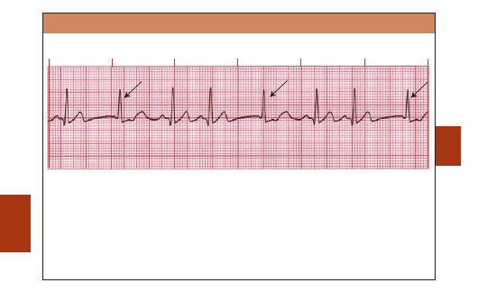
Atrial Flutter (A-flutter)
■
AV node conducts impulses to the ventricles at a 2:1, 3:1, 4:1, or greater ratio (rarely 1:1).
■
Degree of AV block may be consistent or variable.
Rate: Atrial: 250–350 bpm; ventricular: slow or fast
Rhythm: Usually regular but may be variable
P Waves: Flutter waves have a saw-toothed appearance
PR Interval: Variable
QRS: Usually normal (0.06–0.10 sec), but may appear widened if flutter waves are buried in QRS
♥
Clinical Tip:
The presence of A-flutter may be the first indication of cardiac disease.
♥
Clinical Tip:
Signs and symptoms depend on ventricular response rate.
ECGs
Flutter waves
02ECG-Tab 02 2/4/05 3:58 PM Page 40
Copyright
©
2005
F.
A.
Davis.
41
Atrial Fibrillation (A-fib)
■
Rapid, erratic electrical discharge comes from multiple atrial ectopic foci.
■
No organized atrial contractions are detectable.
Rate: Atrial: 350 bpm or greater; ventricular: slow or fast
Rhythm: Irregular
P Waves: No true P waves; chaotic atrial activity
PR Interval: None
QRS: Normal (0.06–0.10 sec)
♥
Clinical Tip:
A-fib is usually a chronic arrhythmia associated with underlying heart disease.
♥
Clinical Tip:
Signs and symptoms depend on ventricular response rate.
ECGs
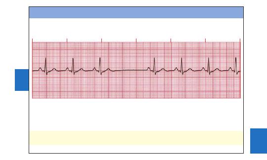
Irregular R-R intervals
02ECG-Tab 02 2/4/05 3:58 PM Page 41
Copyright
©
2005
F.
A.
Davis.

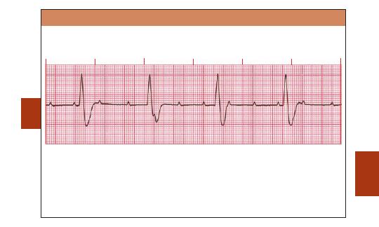
42
ECGs
Delta
wave
Wolff-Parkinson-White (WPW) Syndrome
■
In WPW an accessory conduction pathway is present between the atria and the ventricles.
Electrical impulses are rapidly conducted to the ventricles.
■
These rapid impulses create a slurring of the initial portion of the QRS called the delta wave.
Rate: Depends on rate of underlying rhythm
Rhythm: Regular unless associated with A-fib
P Waves: Normal (upright and uniform) unless A-fib is present
PR Interval: Short (
0.12 sec) if P wave is present
QRS: Wide (
0.10 sec); delta wave present
♥
Clinical Tip:
WPW is associated with narrow-complex tachycardias, including A-flutter and
A-fib.
02ECG-Tab 02 2/4/05 3:58 PM Page 42
Copyright
©
2005
F.
A.
Davis.
43
ECGs
Inverted P wave
Absent P wave
Junctional Arrhythmias
■
The atria and SA node do not perform their normal pacemaking functions.
■
A junctional escape rhythm begins.
Junctional Rhythm
Rate: 40–60 bpm
Rhythm: Regular
P Waves: Absent, inverted, buried, or retrograde
PR Interval: None, short, or retrograde
QRS: Normal (0.06–0.10 sec)
02ECG-Tab 02 2/4/05 3:58 PM Page 43
Copyright
©
2005
F.
A.
Davis.
44
Accelerated Junctional Rhythm
Rate: 61–100 bpm
Rhythm: Regular
P Waves: Absent, inverted, buried, or retrograde
PR Interval: None, short, or retrograde
QRS: Normal (0.06–0.10 sec)
♥
Clinical Tip:
Monitor the patient, not just the ECG, for clinical improvement.
ECGs
Absent P wave
02ECG-Tab 02 2/4/05 3:58 PM Page 44
Copyright
©
2005
F.
A.
Davis.
45
Junctional Tachycardia
Rate: 101–180 bpm
Rhythm: Regular
P Waves: Absent, inverted, buried, or retrograde
PR Interval: None, short, or retrograde
QRS: Normal (0.06–0.10 sec)
♥
Clinical Tip:
Signs and symptoms of decreased cardiac output may be seen in response to
the rapid rate.
ECGs
Retrograde P wave
02ECG-Tab 02 2/4/05 3:58 PM Page 45
Copyright
©
2005
F.
A.
Davis.
46
Junctional Escape Beat
■
An escape complex comes later than the next expected sinus complex.
Rate: Depends on rate of underlying rhythm
Rhythm: Irregular whenever an escape beat occurs
P Waves: None, inverted, buried, or retrograde in the escape beat
PR Interval: None, short, or retrograde
QRS: Normal (0.06–0.10 sec)
ECGs
Junctional escape beats
02ECG-Tab 02 2/4/05 3:58 PM Page 46
Copyright
©
2005
F.
A.
Davis.
47
Premature Junctional Contraction (PJC)
■
Enhanced automaticity in the AV junction produces PJCs.
Rate: Depends on rate of underlying rhythm
Rhythm: Irregular whenever a PJC occurs
P Waves: Absent, inverted, buried, or retrograde in the PJC
PR Interval: None, short, or retrograde
QRS: Normal (0.06–0.10 sec)
♥
Clinical Tip:
Before deciding that isolated PJCs may be insignificant, consider the cause.
ECGs
PJC
PJC
02ECG-Tab 02 2/4/05 3:58 PM Page 47
Copyright
©
2005
F.
A.
Davis.
48
ECGs
Ventricular Arrhythmias
■
QRS complex is
0.10 sec. P Waves are absent or, if visible, have no consistent relationship
to the QRS complex.
Idioventricular Rhythm
Rate: 20–40 bpm
Rhythm: Regular
P Waves: None
PR Interval: None
QRS: Wide (
0.10 sec), bizarre appearance
♥
Clinical Tip:
Idioventricular rhythm may also be called agonal rhythm.
02ECG-Tab 02 2/4/05 3:58 PM Page 48
Copyright
©
2005
F.
A.
Davis.
49
Accelerated Idioventricular Rhythm
Rate: 41–100 bpm
Rhythm: Regular
P Waves: None
PR Interval: None
QRS: Wide (
0.10 sec), bizarre appearance
♥
Clinical Tip:
Idioventricular rhythms appear when supraventricular pacing sites are
depressed or absent. Diminished cardiac output is expected if the heart rate is slow.
ECGs
02ECG-Tab 02 2/4/05 3:58 PM Page 49
Copyright
©
2005
F.
A.
Davis.
50
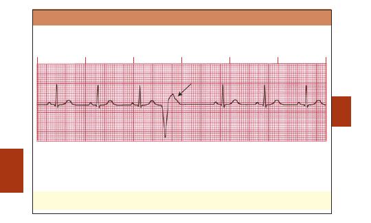
Premature Ventricular Contraction (PVC)
■
Usually PVCs result from an irritable ventricular focus.
■
PVCs may be uniform (same form) or multiform (different forms).
■
The pause following a PVC may be compensatory or noncompensatory.
Rate: Depends on rate of underlying rhythm
Rhythm: Irregular whenever a PVC occurs
P Waves: None associated with the PVC
PR Interval: None associated with the PVC
QRS: Wide (
0.10 sec), bizarre appearance
♥
Clinical Tip:
Patients may sense the occurrence of PVCs as skipped beats. Because the
ventricles are only partially filled, the PVC frequently does not generate a pulse.
ECGs
PVC
02ECG-Tab 02 2/4/05 3:58 PM Page 50
Copyright
©
2005
F.
A.
Davis.
51
ECGs
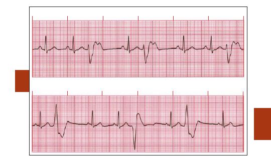
Premature Ventricular Contraction: Uniform (same form)
Premature Ventricular Contraction: Multiform (different forms)
02ECG-Tab 02 2/4/05 3:58 PM Page 51
Copyright
©
2005
F.
A.
Davis.
52
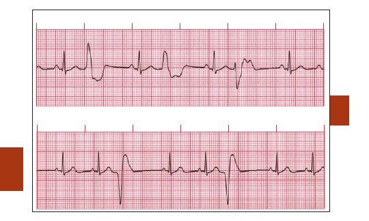
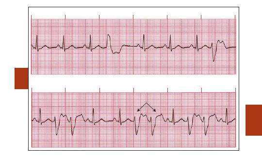
Premature Ventricular Contraction: Ventricular Bigeminy (PVC every other beat)
Premature Ventricular Contraction: Ventricular Trigeminy (PVC every 3rd beat)
ECGs
02ECG-Tab 02 2/4/05 3:58 PM Page 52
Copyright
©
2005
F.
A.
Davis.
53
Premature Ventricular Contraction: Ventricular Quadrigeminy (PVC every 4th beat)
Premature Ventricular Contraction: Couplets (paired PVCs)
ECGs
Couplets
02ECG-Tab 02 2/4/05 3:58 PM Page 53
Copyright
©
2005
F.
A.
Davis.
54
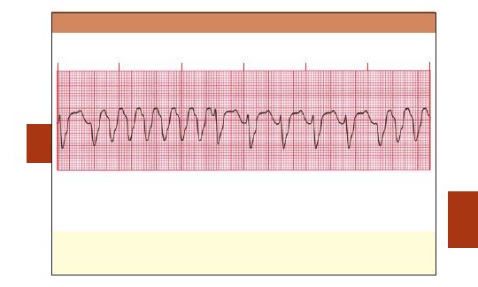
Ventricular Tachycardia (VT): Monomorphic
■
QRS complexes in monomorphic VT have the same shape and amplitude.
Rate: 100–250 bpm
Rhythm: Regular
P Waves: None or not associated with the QRS
PR Interval: None
QRS: Wide (
0.10 sec), bizarre appearance
♥
Clinical Tip:
It is important to confirm the presence or absence of pulses because
monomorphic VT may be perfusing or nonperfusing.
♥
Clinical Tip:
Monomorphic VT will probably deteriorate into VF or unstable VT if sustained
and not treated.
ECGs
02ECG-Tab 02 2/4/05 3:58 PM Page 54
Copyright
©
2005
F.
A.
Davis.
55
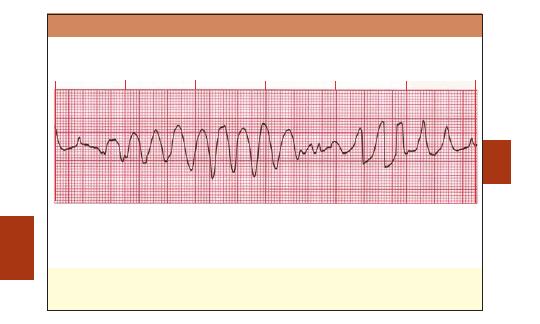
Ventricular Tachycardia (VT): Polymorphic
■
QRS complexes in polymorphic VT vary in shape and amplitude.
■
The QT interval is normal or long.
Rate: 100–250 bpm
Rhythm: Regular or irregular
P Waves: None or not associated with the QRS
PR Interval: None
QRS: Wide (
0.10 sec), bizarre appearance
♥
Clinical Tip:
It is important to confirm the presence or absence of pulses because
polymorphic VT may be perfusing or nonperfusing.
♥
Clinical Tip:
Consider electrolyte abnormalities as a possible etiology.
ECGs
02ECG-Tab 02 2/4/05 3:58 PM Page 55
Copyright
©
2005
F.
A.
Davis.
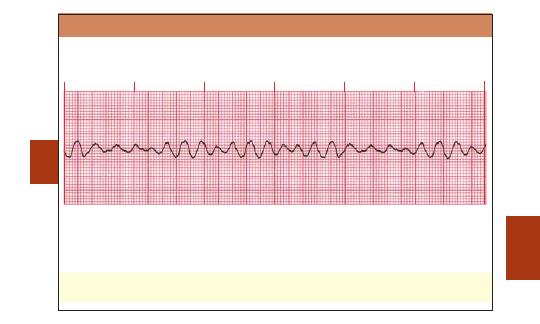
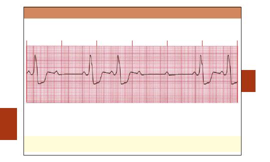
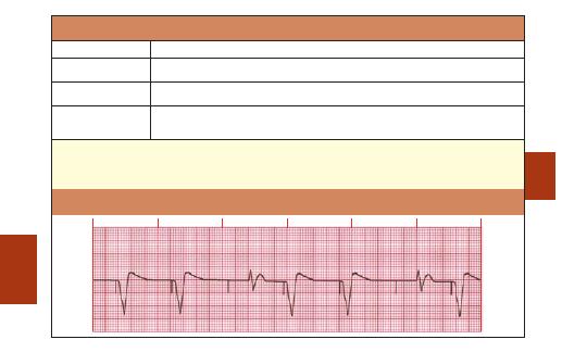
56
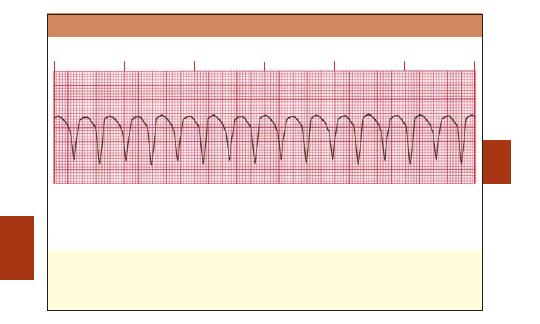
Torsade de Pointes
■
The QRS reverses polarity and the strip shows a spindle effect.
■
This rhythm is an unusual variant of polymorphic VT with normal or long QT intervals.
■
In French the term means “twisting of the points.”
Rate: 200–250 bpm
Rhythm: Irregular
P Waves: None
PR Interval: None
QRS: Wide (
0.10 sec), bizarre appearance
♥
Clinical Tip:
Torsade de pointes may deteriorate to VF or asystole.
♥
Clinical Tip:
Frequent causes are drugs that prolong QT interval and electrolyte
abnormalities such as hypomagnesemia.
ECGs
02ECG-Tab 02 2/4/05 3:58 PM Page 56
Copyright
©
2005
F.
A.
Davis.
57
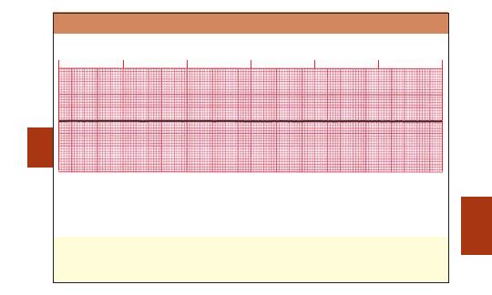
Ventricular Fibrillation (VF)
■
Chaotic electrical activity occurs with no ventricular depolarization or contraction.
■
The amplitude and frequency of the fibrillatory activity can be used to define the type of
fibrillation as coarse, medium, or fine.
Rate: Indeterminate
Rhythm: Chaotic
P Waves: None
PR Interval: None
QRS: None
♥
Clinical Tip:
There is no pulse or cardiac output. Rapid intervention is critical. The longer the
delay, the less the chance of conversion.
ECGs
02ECG-Tab 02 2/4/05 3:58 PM Page 57
Copyright
©
2005
F.
A.
Davis.
58
Pulseless Electrical Activity (PEA)
■
Monitor shows an identifiable electrical rhythm, but no pulse is detected.
■
Rhythm may be sinus, atrial, junctional, or ventricular in origin.
■
PEA is also called electromechanical dissociation (EMD).
Rate, rhythm, P waves, P-R interval, and QRS: Reflect underlying rhythm.
♥
Clinical Tip:
Potential causes of PEA are pulmonary embolism, MI, acidosis, tension
pneumothorax, hyper- and hypokalemia, cardiac tamponade, hypovolemia, hypoxia,
hypothermia, and drug overdose (i.e., cyclic antidepressants, beta blockers, calcium channel
blockers, digoxin).
ECGs
02ECG-Tab 02 2/4/05 3:58 PM Page 58
Copyright
©
2005
F.
A.
Davis.
59
Asystole
■
Electrical activity in the ventricles is completely absent.
Rate: None
Rhythm: None
P Waves: None
PR Interval: None
QRS: None
♥
Clinical Tip:
Always confirm asystole by checking the ECG in two different leads. Also,
search to identify underlying ventricular fibrillation.
♥
Clinical Tip:
Seek to identify the underlying cause as in PEA.
ECGs
02ECG-Tab 02 2/4/05 3:58 PM Page 59
Copyright
©
2005
F.
A.
Davis.
60
Atrioventricular (AV) Blocks
■
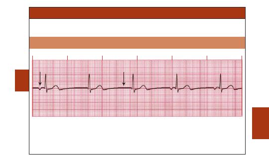
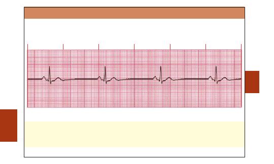
AV blocks are divided into three categories: first-, second-, and third-degree.
First-Degree AV Block
Rate: Depends on rate of underlying rhythm
Rhythm: Regular
P Waves: Normal (upright and uniform)
PR Interval: Prolonged (
0.20 sec)
QRS: Normal (0.06–0.10 sec)
♥
Clinical Tip:
Usually AV block is benign, but if associated with an acute MI, it may lead to
further AV defects.
ECGs
02ECG-Tab 02 2/4/05 3:58 PM Page 60
Copyright
©
2005
F.
A.
Davis.
61
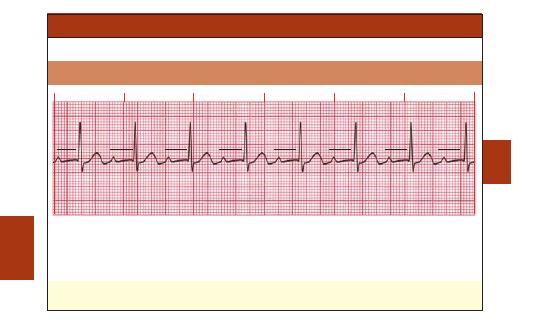
Second-Degree AV Block
Type I (Mobitz I or Wenckebach)
■
P-R intervals become progressively longer until one P wave is totally blocked and produces
no QRS. After a pause, during which the AV node recovers, this cycle is repeated.
Rate: Depends on rate of underlying rhythm
Rhythm: Irregular
P Waves: Normal (upright and uniform)
PR Interval: Progressively longer until one P wave is blocked and a QRS is dropped
QRS: Normal (0.06–0.10 sec)
♥
Clinical Tip:
This rhythm may be caused by medication such as beta blockers, digoxin, and
calcium channel blockers. Ischemia involving the right coronary artery is another cause.
ECGs
Blocked beat
X
02ECG-Tab 02 2/4/05 3:59 PM Page 61
Copyright
©
2005
F.
A.
Davis.
62
Second-Degree AV Block
Type II (Mobitz II)
■
Conduction ratio (P waves to QRS complexes) is commonly 2:1, 3:1, or 4:1.
■
QRS complexes are usually wide because this block usually involves both bundle branches.
Rate: Atrial rate (usually 60–100 bpm); faster than ventricular rate
Rhythm: Atrial regular and ventricular irregular
P Waves: Normal (upright and uniform); more P waves than QRS complexes
PR Interval: Normal or prolonged but constant
QRS: Usually wide (
0.10 sec)
♥
Clinical Tip:
Resulting bradycardia can compromise cardiac output and lead to complete AV
block. This rhythm often occurs with cardiac ischemia or an MI.
ECGs
02ECG-Tab 02 2/4/05 3:59 PM Page 62
Copyright
©
2005
F.
A.
Davis.
63
ECGs
Third-Degree AV Block
■
Conduction between atria and ventricles is absent because of electrical block at or below the
AV node.
■
“Complete heart block” is another name for this rhythm.
Rate: Atrial: 60–100 bpm; ventricular: 40–60 bpm if escape focus is junctional,
40 bpm if
escape focus is ventricular
Rhythm: Usually regular, but atria and ventricles act independently
P Waves: Normal (upright and uniform); may be superimposed on QRS complexes or T waves
PR Interval: Varies greatly
QRS: Normal if ventricles are activated by junctional escape focus; wide if escape focus is
ventricular
02ECG-Tab 02 2/4/05 3:59 PM Page 63
Copyright
©
2005
F.
A.
Davis.
64
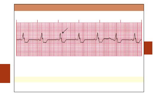
Bundle Branch Block (BBB)
■
Either the left or the right ventricle may depolarize late, creating a “notched” QRS complex.
Rate: Depends on rate of underlying rhythm
Rhythm: Regular
P Waves: Normal (upright and uniform)
PR Interval: Normal (0.12–0.20 sec)
QRS: Usually wide (
0.10 sec) with a notched appearance
♥
Clinical Tip:
Commonly, BBB occurs in coronary artery disease.
ECGs
Notched QRS
02ECG-Tab 02 2/4/05 3:59 PM Page 64
Copyright
©
2005
F.
A.
Davis.
65
Artificial Cardiac Pacemakers
■
Electronically stimulate the heart in place of the heart’s own
pacemaker.
■
May be preset to stimulate the heart’s activity continuously or
intermittently.
Temporary Pacemaker
■
Paces the heart through epicardial, transvenous, or
transcutaneous routes. The pulse generator is located externally.
Permanent Pacemaker
■
Its circuitry sealed in an airtight case, the pacemaker is
implanted in the body. Uses sensing and pacing device leads.
Single-Chamber Pacemaker
■
One lead is placed in the heart and paces a single heart chamber
(either atrium or ventricle).
Dual-Chamber Pacemaker
■
One lead is placed in the right atrium and the other in the right
ventricle. The atrial electrode generates a spike that should be
followed by a P wave, and the ventricular electrode generates a
spike followed by a wide QRS complex.
Pacemaker Modes
■
Fixed rate (asynchronous): Discharges at a preset rate (usually
70—80 bpm) regardless of the patient’s own electrical activity.
■
Demand (synchronous): Discharges only when the patient’s heart
rate drops below the pacemaker’s preset (base) rate.
♥
Clinical Tip: Pacemaker patients may receive defibrillation, but
avoid placing the defibrillator paddles or pads closer than 5 inches
from the pacemaker battery pack.
ECGs
02ECG-Tab 02 2/4/05 3:59 PM Page 65
Copyright © 2005 F. A. Davis.
66
ECGs
Artificial Pacemaker Rhythm
Rate:
Varies according to preset pacemaker rate
Rhythm:
Regular for asynchronous pacemaker; irregular for demand pacemaker
P waves:
None produced by ventricular pacemaker. Sinus P waves may be seen but are unrelated
to QRS. Atrial or dual-chamber pacemaker should have P waves following each atrial
spike.
P-R interval:
None for ventricular pacer. Atrial or dual-chamber pacemaker produces ventricular spike
at constant interval from P wave.
QRS:
Wide (
0.10 sec) following each ventricular spike in paced rhythm. Patient’s own
electrical activity may generate QRS that looks different from paced QRS complexes. If
atrially paced only, may be within normal limits.
Single-Chamber Pacemaker Rhythm—Atrial
Pacemaker spike
02ECG-Tab 02 2/4/05 3:59 PM Page 66
Copyright
©
2005
F.
A.
Davis.
67
ECGs
Pacemaker spike
Atrial pacemaker spike
Ventricular pacemaker spike
Single-Chamber Pacemaker Rhythm—Ventricular
Dual-Chamber Pacemaker Rhythm—Atrial and Ventricular
02ECG-Tab 02 2/4/05 3:59 PM Page 67
Copyright
©
2005
F.
A.
Davis.
68
Pacemaker Malfunctions
Malfunction
Reason
Failure to fire
Failure to capture
Failure to sense
♥
Clinical Tip:
A pacemaker spike—a mark on the ECG projecting upward or downward from the baseline—
indicates that the pacemaker has fired.
♥
Clinical Tip:
A pacemaker is said to be in capture when a spike produces an ECG wave or complex.
Pacemaker Failure to Sense
ECGs
Pacemaker spikes are absent. The cause may be a dead battery or a disruption in the
connecting wires.
Pacemaker spikes are present, but no P wave or QRS complex follows the spike. Turning
up the pacemaker’s voltage often corrects this problem.
The pacemaker fires because it fails to detect the heart’s intrinsic beats, resulting in
abnormal complexes. The cause may be a dead battery, decrease of P wave or QRS
voltage, or damage to a pacing lead wire.
02ECG-Tab 02 2/4/05 3:59 PM Page 68
Copyright
©
2005
F.
A.
Davis.
69
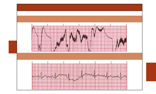
Artifact
■
Artifacts are ECG deflections caused by influences other than the heart’s electrical activity.
Loose Electrodes
Baseline Varies with Respiration
ECGs
02ECG-Tab 02 2/4/05 3:59 PM Page 69
Copyright
©
2005
F.
A.
Davis.
70
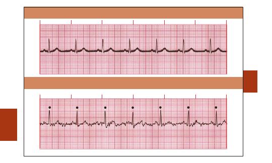
ECGs
Regular R-R intervals
60-Cycle Interference
Muscle Artifact
02ECG-Tab 02 2/4/05 3:59 PM Page 70
Copyright
©
2005
F.
A.
Davis.
71
ECGs
♥
Clinical Tip:
Never confuse muscle ar
tifact with
A-fib if the rh
ythm is regular
.
Notes:
02ECG-Tab 02 2/4/05 3:59 PM Page 71
Copyright © 2005 F. A. Davis.

72
The 12-Lead ECG
■
The most commonly used clinical ECG system is the 12-lead
ECG. It consists of the following leads: I, II, III, aVR, aVL, aVF,
V
1
, V
2
, V
3
, V
4
, V
5
, and V
6
. Both limb and chest electrodes are
used to record 12-lead ECGs.
■
Measurements are central to 12-lead ECG analysis. The height
and depth of waves can be important in the diagnosis of
certain conditions, including MI or ventricular hypertrophy.
■
The direction of ventricular depolarization is an important
factor in determining the axis of the heart.
■
In the case of MI, multiple leads are necessary to recognize its
presence and determine its location. If large areas of the heart
are affected, the patient can develop cardiogenic shock.
■
ECG signs of an MI are best seen in the reflecting leads—
those facing the affected surface of the heart. Reciprocal leads
are in the same plane but opposite the area of the MI; they
show a “mirror image” of the electrical complex.
■
Prehospital EMS systems may use 12-lead ECGs to discover
signs of acute myocardial infarction, such as ST segment
elevation, in preparation for in-hospital administration of
thrombolytic drugs.
■
Once a 12-lead ECG is performed, a 15-lead, or right-sided,
ECG may be used for an even more comprehensive view if it
appears that the right ventricle or posterior portion of the
heart has been affected.
♥
Clinical Tip: Always compare the patient’s current 12-lead
ECG with the previous one.
12-LEAD
03ECG-Tab 03 2/4/05 3:59 PM Page 72
Copyright © 2005 F. A. Davis.
73
12-LEAD
V
1
V
2
V
3
V
4
V
5
V
6
Left
lung
Right
lung
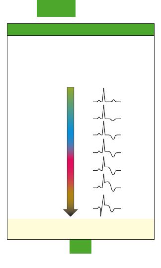
R Wave Progression
■
Normal ventricular depolarization in the heart progresses
from right to left and from front to back.
■
In a normal heart the R wave becomes taller and the S wave
becomes smaller as electrical activity moves across the heart
from right to left. This phenomenon is called R wave
progression.
■
Alteration in the normal progression of the R wave may be
seen in left ventricular hypertrophy, COPD, left bundle branch
block, or anteroseptal MI.
Normal R wave progression in chest leads V
1
–V
6
.
03ECG-Tab 03 2/4/05 3:59 PM Page 73
Copyright © 2005 F. A. Davis.
74
12-LEAD
30
°
0
°
180
°
90
°
–90
°
–30
°
60
°
120
°
150
°
–150
°
III
II
I
aVF
aVR
aVL
I
I
aVF
I
aVF
aVF
I
aVF
No
rm
al
A
xi
s
R
ig
h
t
A
xis
D
evia
tion
A
xi
s
D
ev
iat
ion
E
xt
re
m
e
Ri
gh
t
Left
Ax
is
Devia
tio
n
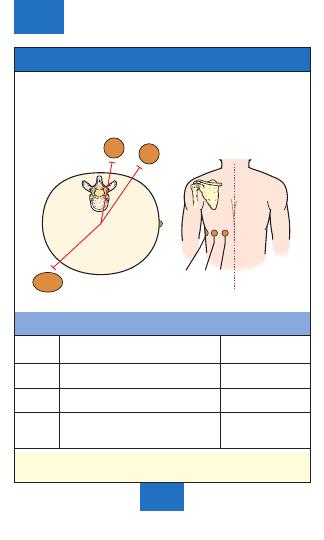
Electrical Axis of the Heart
The electrical axis is the sum total of all electrical currents
generated by the ventricular myocardium during depolarization.
Analysis of the axis may help to determine the location and
extent of cardiac injury, such as ventricular hypertrophy, bundle
branch block, or changes in the position of the heart in the chest
(from, e.g., pregnancy or ascites).
The direction of the QRS complex in leads I and aVF determines
the axis quadrant in relation to the heart.
♥
Clinical Tip: Extreme right axis deviation is also called
indeterminate, “no man’s land,” and “northwest.”
03ECG-Tab 03 2/4/05 3:59 PM Page 74
Copyright © 2005 F. A. Davis.
75
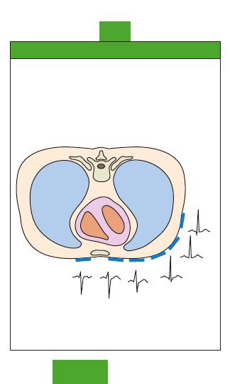
12-LEAD
Anterior wall
Anterior view
Lateral
wall
Septal wall
Inferior wall
Anterior view
Posterior view
I lateral
aVR
V1 septal
V
4
anterior
II inferior
aVL lateral
V2 septal
V
5
lateral
III inferior
aVF inferior
V3 anterior
V
6
lateral
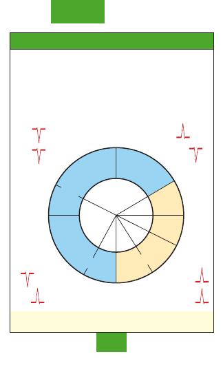
Ischemia, Injury, or Infarction in
Relation to the Heart
Location of MI by ECG Leads
♥
Clinical Tip: Lead aVR is a nondiagnostic lead and does not
show any change in an MI.
♥
Clinical Tip: An MI may not be limited to just one region of
the heart. For example, if there are changes in leads V
3
and V
4
(anterior) and in I, aVL, V
5
, and V
6
(lateral), the MI is called an
anterolateral infarction.
03ECG-Tab 03 2/4/05 3:59 PM Page 75
Copyright © 2005 F. A. Davis.
76
Progression of an Acute Myocardial Infarction
An acute MI is a continuum that extends from the normal state
to a full infarction:
■
Ischemia—Lack of oxygen to the cardiac tissue, represented
by ST segment depression, T wave inversion, or both
■
Injury—An arterial occlusion with ischemia, represented by ST
segment elevation
■
Infarction—Death of tissue, represented by a pathological Q
wave
♥
Clinical Tip: Once the acute MI has ended, the ST segment
returns to baseline and the T wave becomes upright, but the Q
wave remains abnormal because of scar formation.
12-LEAD
Infarction
Injury
Ischemia
Normal
03ECG-Tab 03 2/4/05 3:59 PM Page 76
Copyright © 2005 F. A. Davis.
77
ST Segment Elevation and Depression
■
A normal ST segment represents early ventricular repolarization.
■
Displacement of the ST segment can be caused by various conditions
listed below.
ST segment is at baseline.
ST segment is elevated.
ST segment is depressed.
Primary Causes of ST Segment Elevation
■
ST segment elevation
1 mm in the limb leads and 2 mm in the
chest leads indicates an evolving acute MI until there is proof to the
contrary. Other primary causes:
◆ Early repolarization (normal variant in young adults)
◆ Pericarditis
◆ Ventricular aneurysm
◆ Pulmonary embolism
◆ Intracranial hemorrhage
Primary Causes of ST Segment Depression
■
Myocardial ischemia
■
Left ventricular hypertrophy
■
Intraventricular conduction defects
■
Medication (e.g., digitalis)
■
Reciprocal changes in leads opposite the area of acute injury
12-LEAD
03ECG-Tab 03 2/4/05 3:59 PM Page 77
Copyright © 2005 F. A. Davis.
78
12-LEAD
I
aVR
V
1
V
4
II
aVL
V
2
V
5
III
aVF
V
3
V
6
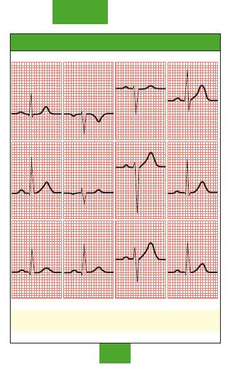
Normal 12-Lead ECG
♥
Clinical Tip: A normal ECG does not rule out any acute
coronary syndrome.
03ECG-Tab 03 2/4/05 3:59 PM Page 78
Copyright © 2005 F. A. Davis.
79
12-LEAD
I
aVR
V
1
V
4
II
aVL
V
2
V
5
III
aVF
V
3
V
6
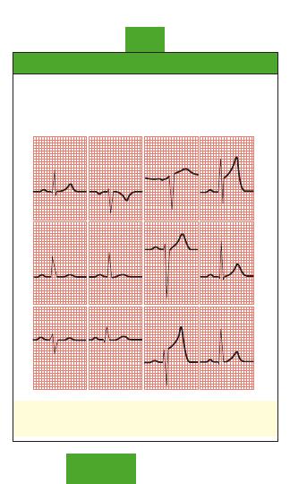
Anterior Myocardial Infarction
■
Occlusion of the left coronary artery—left anterior descending
branch
■
ECG changes: ST segment elevation with tall T waves and
taller-than-normal R waves in leads V
3
and V
4
♥
Clinical Tip: Anterior MI frequently involves a large area of
the myocardium and can present with cardiogenic shock,
second-degree AV block type II, or third-degree AV block.
03ECG-Tab 03 2/4/05 3:59 PM Page 79
Copyright © 2005 F. A. Davis.
80
12-LEAD
I
aVR
V
1
V
4
II
aVL
V
2
V
5
III
aVF
V
3
V
6
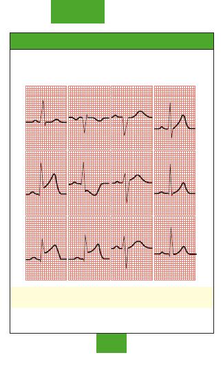
Inferior Myocardial Infarction
■
Occlusion of the right coronary artery—posterior descending
branch
■
ECG changes: ST segment elevation in leads II, III, and aVF
♥
Clinical Tip: Be alert for symptomatic sinus bradycardia, AV
blocks, hypotension, and hypoperfusion.
03ECG-Tab 03 2/4/05 3:59 PM Page 80
Copyright © 2005 F. A. Davis.
81
12-LEAD
I
aVR
V
1
V
4
II
aVL
V
2
V
5
III
aVF
V
3
V
6
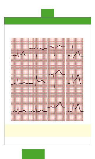
Lateral Myocardial Infarction
■
Occlusion of the left coronary artery—circumflex branch
■
ECG changes: ST segment elevation in leads I, aVL, V
5
, and V
6
♥
Clinical Tip: Lateral MI is often associated with anterior or
inferior wall MI. Be alert for changes that may indicate
cardiogenic shock or congestive heart failure.
03ECG-Tab 03 2/4/05 3:59 PM Page 81
Copyright © 2005 F. A. Davis.
82
12-LEAD
I
aVR
V
1
V
4
II
aVL
V
2
V
5
III
aVF
V
3
V
6
Septal Myocardial Infarction
■
Occlusion of the left coronary artery—left anterior descending
branch
■
ECG changes: pathological Q waves; absence of normal R
waves in leads V
1
and V
2
♥
Clinical Tip: Septal MI is often associated with an anterior
wall MI.
03ECG-Tab 03 2/4/05 3:59 PM Page 82
Copyright © 2005 F. A. Davis.
83
12-LEAD
I
aVR
V
1
V
4
II
aVL
V
2
V
5
III
aVF
V
3
V
6
Posterior Myocardial Infarction
■
Occlusion of the right coronary artery (posterior descending
branch) or the left circumflex artery
■
Tall R waves and ST segment depression possible in leads V
1
,
V
2
, V
3
, and V
4
■
ST segment elevation in true posterior leads, V
8
and V
9
♥
Clinical Tip: Diagnosis may require a 15-lead ECG because a
standard 12-lead does not look directly at the posterior wall.
03ECG-Tab 03 2/4/05 3:59 PM Page 83
Copyright © 2005 F. A. Davis.
84
12-LEAD
I
aVR
V
1
V
4
II
aVL
V
2
V
5
III
aVF
V
3
V
6
Left Bundle Branch Block
■
QRS
0.10 sec
■
QRS predominantly negative in leads V
1
and V
2
■
QRS predominantly positive in V
5
and V
6
and often notched
■
Absence of small, normal Q waves in I, aVL, V
5
, and V
6
■
Wide monophasic R waves in I, aVL, V
1
, V
5
, and V
6
♥
Clinical Tip: Patients may have underlying heart disease,
including coronary artery disease, hypertension,
cardiomyopathy, and ischemia.
03ECG-Tab 03 2/4/05 3:59 PM Page 84
Copyright © 2005 F. A. Davis.
85
12-LEAD
I
aVR
V
1
V
4
II
aVL
V
2
V
5
III
aVF
V
3
V
6
Right Bundle Branch Block
■
QRS
0.10 sec
■
QRS normal or deviated to the right
■
Slurred S wave in leads I and V
6
■
RSR’ pattern in lead V
1
with R’ taller than R
♥
Clinical Tip: Patients may have underlying right ventricular
hypertrophy, pulmonary edema, cardiomyopathy, congenital
heart disease, or rheumatic heart disease.
03ECG-Tab 03 2/4/05 3:59 PM Page 85
Copyright © 2005 F. A. Davis.
86
MEDS/
SKILLS
Emergency Medications
This list is a reference list only. It is not meant to be exhaustive
in clinical content.
♥
Clinical Tip: Always consult an authoritative, current
reference about dose, dilution, route and rate of administration,
and interactions before administering medications, especially
IV medications. Have a second licensed person independently
check dose calculations, preparation, original orders, and
infusion pump programming.
ACE INHIBITORS
(Angiotensin-converting Enzyme Inhibitors)
(Antihypertensive)
Common Agents: Captopril, enalapril, lisinopril, ramipril.
Indications: MI, hypertension (HTN), congestive heart failure
(CHF), heart failure without hypotension, ST segment
elevation, left ventricular dysfunction after MI.
Dose: See individual order and drug for route and dosage.
Usually not started in emergency department, but within 24
hr after fibrinolytic therapy has been completed and blood
pressure (BP) has stabilized.
Contraindications: Lactation, pregnancy, angioedema,
hypersensitivity to ACE inhibitors, serum potassium
5
mEq/L.
Side Effects: Tachycardia, dizziness, headache, fatigue,
hypotension, hyperkalemia.
Precautions: Reduce dose in renal failure.
ADENOSINE
(Adenocard, Adenoscan) (Antiarrhythmic)
Indications: Narrow-complex tachycardias and PSVT.
Dose: 6 mg rapid intravenous push (IVP) over 1–3 sec followed
by a 20-mL bolus of normal saline. Give 12 mg by IVP in 1–2
min if needed. A third dose of 12 mg IVP may be given in 1–2
min, max. 30 mg.
04ECG-Tab 04 2/4/05 4:00 PM Page 86
Copyright © 2005 F. A. Davis.

87
MEDS/
SKILLS
Contraindications: Hypersensitivity, sick sinus syndrome, 2nd-
or 3rd-degree AV block (unless a functional artificial
pacemaker is present), drug- or poison-induced tachycardia.
Side Effects: Flushing, dizziness, bronchospasm, chest pain or
tightness, bradycardia, AV block, asystole, ventricular ectopic
beats, VF.
Precautions: Ineffective in treating A-fib, A-flutter, or VT. Avoid
in patients receiving dipyridamole and in patients with
asthma or unstable angina.
AMIODARONE
(Cordarone, Pacerone) (Antiarrhythmic)
Indications: Wide- and narrow-complex tachycardia,
polymorphic VT, shock-refractory VF or pulseless VT, SVT,
PSVT.
Dose: Cardiac arrest 300 mg (diluted in 20–30 mL D5W) IVP;
consider additional 150 mg IVP in 3–5 min. Wide- and narrow-
complex tachycardia (stable) 150 mg IVP over first 10 min (15
mg/min)—may repeat infusion of 150 mg IVP every 10 min as
needed; slow infusion of 360 mg IV over next 6 hr (1 mg/min);
maintenance infusion of 540 mg over next 18 hr (0.5 mg/min).
Max. cumulative dose: 2.2 g IV in 24 hr.
Contraindications: Bradycardia, hypersensitivity, cardiogenic
shock, 2nd- or 3rd-degree AV block.
Side Effects: Vasodilation, hypotension, visual impairment,
hepatotoxicity, pulmonary toxicity, CHF; may prolong QT
interval, producing torsade de pointes.
Precautions: Avoid concurrent use with procainamide. Correct
hypokalemia and hypomagnesemia if possible before use.
Draw up amiodarone through a large-gauge needle to reduce
foaming. For slow or maintenance IV infusion, mix
medication only in glass bottle containing D5W and
administer through an in-line filter.
04ECG-Tab 04 2/4/05 4:00 PM Page 87
Copyright © 2005 F. A. Davis.
88
MEDS/
SKILLS
ASPIRIN
(Acetylsalicylic Acid) (Antiplatelet)
Indications: Acute coronary syndrome, symptoms suggestive
of cardiac ischemia.
Dose: 162–325 mg PO non-enteric coated for antiplatelet effect.
Give within minutes of onset.
Contraindications: Known allergy to aspirin, pregnancy.
Side Effects: Anorexia, nausea, epigastric pain, anaphylaxis.
Precautions: Active ulcers and asthma, bleeding disorders, or
thrombocytopenia.
ATROPINE
(Antiarrhythmic, Anticholinergic)
Indications: Symptomatic sinus bradycardia, asystole, PEA with
rate
60 bpm, cholinergic drug toxicity and mushroom
poisoning (antidote).
Dose: Cardiac arrest 1 mg IVP every 3–5 min (may give through
endotracheal (ET) tube at 2.0–3.0 mg diluted in 10 mL normal
saline, max. 0.03–0.04 mg/kg. Bradycardia 0.5–1.0 mg IVP
every 3–5 min, max. 0.03–0.04 mg/kg.
Contraindications: A-fib, A-flutter, glaucoma, asthma.
Side Effects: Tachycardia, headache, dry mouth, dilated pupils,
VF or VT.
Precautions: Use caution in myocardial ischemia and hypoxia.
Avoid in hypothermic bradycardia and in 2nd-degree (Mobitz
type II) and 3rd-degree AV block.
BETA BLOCKERS
(Antihypertensive)
Common Agents: Atenolol, esmolol, labetalol, metoprolol,
propranolol.
Indications: MI, unstable angina, PSVT, A-fib, A-flutter, HTN.
Dose: See individual order and drug for route and dosage.
Contraindications: HR
60 bpm, systolic BP 100 mm Hg,
2nd- or 3rd-degree AV block, left ventricular failure.
Side Effects: Hypotension, dizziness, bradycardia, headache,
nausea and vomiting.
04ECG-Tab 04 2/4/05 4:00 PM Page 88
Copyright © 2005 F. A. Davis.
89
MEDS/
SKILLS
Precautions: Concurrent use with calcium channel blockers,
such as verapamil or diltiazem, can cause hypotension. Use
caution in patients with a history of bronchospasm or cardiac
failure.
CALCIUM CHLORIDE
(Minerals/Electrolytes/Calcium Salt)
Indications: Hyperkalemia, hypocalcemia, hypermagnesemia;
antidote to calcium channel blockers and beta blockers; given
prophylactically with calcium channel blockers to prevent
hypotension.
Dose: Hyperkalemia and antidote to calcium channel blocker
8–16 mg/kg (usually 5–10 mL) slow IVP, may be repeated as
needed. Given prophylactically prior to IV calcium channel
blockers 2–4 mg/kg (usually 2 mL) slow IVP.
Contraindications: Hypercalcemia, VF, digoxin toxicity, renal
calculi.
Side effects: Bradycardia, asystole, hypotension, VF, nausea
and vomiting.
Precautions: Incompatible with sodium bicarbonate.
DIGOXIN IMMUNE FAB
(Fragment Antigen Binding) (Digibind)
(Antidote to Digoxin, Digitoxin)
Indications: Symptomatic digoxin toxicity or acute ingestion of
unknown amount of digoxin.
Dose: Dependent on serum digoxin levels. One 40-mg vial binds
to approximately 0.6 mg of digoxin. Dose typically
administered over 30 min.
Contraindications: Allergy only, otherwise none known.
Side Effects: Worsening of CHF, A-fib, hypokalemia; increased
serum digoxin levels.
Precautions: Allergies to sheep proteins or other sheep
products.
04ECG-Tab 04 2/4/05 4:00 PM Page 89
Copyright © 2005 F. A. Davis.
90
MEDS/
SKILLS
DIGOXIN
(Lanoxin) (Inotropic, Antiarrhythmic)
Indications: To slow ventricular response in A-fib or A-flutter, as
a positive inotrope in CHF, pulmonary edema. May be used as
an alternative drug for PSVT.
Dose: Loading dose of 10–15
g/kg, administered over 5 min.
Maintenance dose determined by body size and renal
function.
Contraindications: Hypersensitivity, uncontrolled ventricular
arrhythmias, AV block, idiopathic hypertrophic subaortic
stenosis (IHSS), constrictive pericarditis.
Side Effects: Arrhythmias, particularly VF and AV block;
bradycardia; fatigue; nausea and vomiting; blurred or yellow
vision; headache; hypersensitivity; hypokalemia.
Precautions: Avoid electrical cardioversion of stable patients. If
the patient’s condition is unstable, use lower current settings
such as 10–20 J. Use cautiously in elderly patients. Correct
electrolyte abnormalities, monitor digoxin levels, monitor for
clinical signs of toxicity.
DILTIAZEM
(Cardizem) (Calcium Channel Blocker)
Indications: A-fib, A-flutter, PSVT refractory to adenosine with
narrow QRS complex and adequate BP.
Dose: 15–20 mg (0.25 mg/kg) IVP over 2 min. May repeat in 15
min at 20–25 mg (0.35 mg/kg) IVP over 2 min. Start
maintenance drip at 5–15 mg/hr and titrate to HR.
Contraindications: Drug- or poison-induced tachycardia, wide-
complex tachycardia of uncertain origin, rapid A-fib and A-
flutter with Wolff-Parkinson-White syndrome, sick sinus
syndrome, 2nd- and 3rd-degree AV block (unless a functional
artificial pacemaker is present).
Side Effects: Hypotension, bradycardia (including AV block),
chest pain, ventricular arrhythmias.
Precautions: Severe hypotension in patients receiving beta
blockers, hepatic injury, renal disease.
04ECG-Tab 04 2/4/05 4:00 PM Page 90
Copyright © 2005 F. A. Davis.
91
MEDS/
SKILLS
DOPAMINE (INTROPIN)
(Vasopressor, Inotropic)
Indications: Symptomatic bradycardia and hypotension,
cardiogenic shock.
Dose: Continuous infusions (titrate to patient response): Low
dose 1–5
g/kg/min; moderate dose 5–10 g/kg/min (cardiac
doses); high dose 10–20
g/kg/min (vasopressor doses). Mix
400 mg/250 mL in normal saline, lactated Ringer’s solution, or
D5W (1600
g/mL).
Contraindications: Pheochromocytoma, uncorrected
tachycardia, cardiogenic shock with CHF.
Side Effects: Tachyarrhythmias, angina, hypotension,
palpitations, vasoconstriction, dyspnea, nausea and vomiting.
Precautions: Hypovolemia, MI. Adjust dosage in elderly
patients and in those with occlusive vascular disease. Ensure
adequate hydration prior to infusion. Taper slowly. Do not mix
with sodium bicarbonate. Use care with peripheral
administration; infiltration can cause tissue necrosis. Central
line is preferred.
EPINEPHRINE
(Adrenalin) (Adrenergic Agonist)
Indications: Cardiac arrest: PEA, asystole, pulseless VT, VF;
severe hypotension; symptomatic bradycardia; anaphylaxis;
severe allergic reactions.
Dose: Cardiac arrest 1 mg IVP (10 mL of 1:10,000 solution) every
3–5 min; follow each dose with 20 mL IV flush; higher doses
(up to 0.2 mg/kg) may be used if 1-mg dose fails. Give 2.0–2.5
mg diluted in 10 mL normal saline if administering by ET
tube. For continuous infusion add 30 mg (30 mL of 1:1000
solution) to 250 mL normal saline or D5W, run at 100 mL/hr,
and titrate to response. Profound bradycardia or hypotension
2–10
g/min IV (add 1 mg of 1:1000 solution to 500 mL
normal saline or D5W and infuse at 1–5 mL/min).
Anaphylaxis/asthma 0.1–0.5 mg SC or IM of 1:1000 solution
every 5–15 min, may be followed by 1–4
g/min continuous
infusion.
Contraindications: Hypersensitivity to adrenergic amines,
hypovolemic shock, coronary insufficiency.
04ECG-Tab 04 2/4/05 4:00 PM Page 91
Copyright © 2005 F. A. Davis.
92
MEDS/
SKILLS
EPINEPHRINE
(Continued)
Side Effects: Angina, HTN, tachycardia, VT, VF, nervousness,
restlessness, tremors, weakness, headache, nausea.
Precautions: Use caution in HTN and increasing heart rate
(may cause increased myocardial oxygen demand). Higher
doses can contribute to postarrest cardiac impairment, but
they may be required to treat poison- or drug-induced shock.
FIBRINOLYTIC AGENTS
(Thrombolytic, Fibrinolytic)
Common Agents: Alteplase (Activase, t-PA), anistreplase
(Eminase), reteplase (Retavase), streptokinase (Streptase),
tenecteplase (TNKase).
Indications: Within
12 hr from onset of symptoms of acute
MI. Alteplase is the only fibrinolytic agent approved for acute
ischemic stroke and must be started
3 hr from onset of
symptoms.
Dose: See individual order and drug for route and dosage.
Contraindications: Active internal bleeding within 21 days
(except menses), neurovascular event within 3 months, major
surgery or trauma within 2 weeks, aortic dissection, severe
(uncontrolled) HTN, bleeding disorders, prolonged
cardiopulmonary resuscitation (CPR), lumbar puncture within
1 week.
Side Effects: Hypotension, reperfusion, arrhythmias, heart
failure, headache, increased bleeding time, deep or superficial
hemorrhage, flushing, urticaria, anaphylaxis.
Precautions: Use cautiously in patients with severe renal or
hepatic disease.
FUROSEMIDE
(Lasix) (Diuretic, Loop Diuretics)
Indications: CHF with acute pulmonary edema, hypertensive
crisis, postarrest cerebral edema, hepatic or renal disease.
Dose: 0.5–1.0 mg/kg slow IVP over 1–2 min, may repeat at 2
mg/kg slow IVP over 1–2 min.
04ECG-Tab 04 2/4/05 4:00 PM Page 92
Copyright © 2005 F. A. Davis.
93
MEDS/
SKILLS
Contraindications: Hypersensitivity (cross-sensitivity with
thiazides and sulfonamides may occur), uncontrolled
electrolyte imbalance, hepatic coma, anuria, hypovolemia.
Side Effects: Severe dehydration, hypovolemia, hypotension,
hypokalemia, hyponatremia, hypochloremia, hyperglycemia,
dizziness, ototoxicity.
Precautions: Use cautiously in severe liver disease
accompanied by cirrhosis or ascites, electrolyte depletion,
diabetes mellitus, pregnancy, lactation, risk for ototoxicity
with increased dose or rapid injection. Monitor electrolytes
closely.
IBUTILIDE
(Corvert) (Antiarrhythmic)
Indications: SVT, including A-fib and A-flutter; most effective for
conversion of A-fib or A-flutter of short duration.
Dose: Patients
≥60 kg 1 mg IVP over 10 min, may repeat same
dose in 10 min. Patients
60 kg 0.01 mg/kg IVP over 10 min,
may repeat same dose in 10 min.
Contraindications: Known hypersensitivity, history of
ventricular arrhythmias including torsade de pointes.
Side Effects: Headache, nausea and vomiting.
Precautions: Monitor ECG for 4–6 hr after administration, with
defibrillator nearby. Correct electrolyte abnormalities prior to
use. If A-fib
48 hr, anticoagulation is required before
cardioversion with ibutilide.
ISOPROTERENOL
(Isuprel) (Sympathomimetic, Beta-
Adrenergic Agonist)
Indications: Symptomatic bradycardia, refractory torsade de
pointes unresponsive to magnesium, bradycardia in heart
transplant patients, beta blocker poisoning.
Dose: IV infusion: mix 1 mg/250 mL in normal saline, lactated
Ringer’s solution, or D5W, run at 2–10
g/min, and titrate to
patient response. In torsade de pointes titrate to increase
heart rate until VT is suppressed.
04ECG-Tab 04 2/4/05 4:00 PM Page 93
Copyright © 2005 F. A. Davis.

94
MEDS/
SKILLS
ISOPROTERENOL
(Continued)
Contraindications: Cardiac arrest, concurrent use with
epinephrine (can cause VF or VT), poison- or drug-induced
shock (exception: beta blocker poisoning).
Side Effects: Anxiety, tachycardia, palpitations, skin flushing.
Precautions: May increase myocardial ischemia, tachycardia,
restlessness. High doses are harmful except in beta blocker
overdose.
LIDOCAINE
(Xylocaine) (Antiarrhythmic, Anesthetic)
Indications: VF or pulseless VT, stable VT, wide-complex
tachycardia of uncertain origin, wide-complex PSVT.
Dose: Cardiac arrest from VF or VT 1.0–1.5 mg/kg IVP (or 2–4
mg/kg via ET tube), may repeat 0.5–0.75 mg/kg IVP every 5–10
min, max. 3 mg/kg. Stable VT, wide-complex tachycardia of
uncertain origin use 0.5–0.75 mg/kg and up to 1.0–1.5 mg/kg,
may repeat 0.5–0.75 mg/kg every 5–10 min; max. total dose
3.0 mg/kg. If conversion is successful, start an IV infusion of
1–4 mg/min (30–50
g/kg/min) in normal saline or D5W.
Contraindications: Prophylactic use in acute MI, advanced AV
block, hypotension, Wolff-Parkinson-White syndrome,
hypersensitivity to amide-type local anesthetics.
Side Effects: Confusion, seizures, hypotension, bradycardia,
cardiovascular collapse, respiratory arrest.
Precautions: CHF, respiratory depression, shock. Reduce
maintenance dose (not loading dose) in presence of impaired
liver function or left ventricular dysfunction or in the elderly.
Stop infusion if signs of toxicity (prolonged PR interval, QRS
widening, or CNS changes) develop.
04ECG-Tab 04 2/4/05 4:00 PM Page 94
Copyright © 2005 F. A. Davis.
95
MEDS/
SKILLS
MAGNESIUM SULFATE
(Electrolyte, Antiarrhythmic)
Indications: Torsade de pointes, VF refractory to lidocaine,
digoxin-induced VT/VF.
Dose: Cardiac arrest (in hypomagnesemia or torsade de
pointes) 1–2 g (2–4 mL of a 50% solution) diluted in 10 mL of
D5W IVP. Digoxin-induced VT or VF 1–2 g IVP. Torsade de
pointes (non-cardiac arrest) load with 1–2 g mixed in 50–100
mL of D5W infused over 5–60 min IV, then infuse 0.5–1.0 g/hr
IV (titrate to control torsade). Acute MI load with 1–2 g mixed
in 50–100 mL of D5W over 5–60 min IV, then infuse 0.5–1.0
g/hr IV for up to 24 hr.
Contraindications: Hypermagnesemia, hypocalcemia, renal
disease, AV block, toxemia of pregnancy 2 hr prior to delivery.
Side Effects: Hypotension, bradycardia, cardiac arrest,
respiratory depression, altered level of consciousness (LOC),
flushed skin, diaphoresis.
Precautions: Renal insufficiency, occasional fall in BP with
rapid administration. Monitor serum magnesium levels.
MORPHINE
(Opioid Agonist Analgesic)
Indications: Chest pain unrelieved by nitroglycerin, CHF and
dyspnea associated with pulmonary edema.
Dose: 2–4 mg IVP (over 1–5 min) every 5–30 min.
Contraindications: Hypersensitivity, heart failure due to
chronic lung disease, respiratory depression, hypotension.
Side Effects: Respiratory depression, hypotension, nausea and
vomiting, bradycardia, altered LOC, seizures.
Precautions: Administer slowly and titrate to effect. Reverse
with naloxone (0.4–2.0 mg IVP). Use caution in cerebral
edema and pulmonary edema with compromised respiration.
04ECG-Tab 04 2/4/05 4:00 PM Page 95
Copyright © 2005 F. A. Davis.
96
MEDS/
SKILLS
NITROGLYCERIN
(Nitrostat, Nitrolingual Pumpspray)
(Antianginal, Nitrate)
Indications: Angina, CHF associated with acute MI,
hypertensive crisis.
Dose: Sublingual route, 0.3–0.4 mg (1 tablet), repeat every 5
min, max. 3 doses/15 min. Aerosol, spray for 0.5–1.0 sec at 5-
min intervals (provides 0.4 mg/dose), max. 3 sprays/15 min.
IVP at 12.5–25.0
g (if no sublingual or spray used). IV
infusion: mix 25 mg/250 mL (100
g/mL) in D5W, run at 5–20
g/min, and titrate to desired response.
Contraindications: Hypersensitivity, systolic BP
90 mm Hg;
severe bradycardia or severe tachycardia; sildenafil (Viagra),
tadalafil (Cialis), vardenafil (Levitra) within 24 hr; right
ventricular infarction.
Side Effects: Hypotension with secondary tachycardia,
syncope, headache, flushed skin.
Precautions: Do not mix with other medications; titrate IV to
maintain systolic BP
90 mm Hg. Mix only in glass IV bottles
and infuse only through tubing provided by manufacturer;
standard polyvinyl chloride tubing can bind up to 80% of the
medication, making it necessary to infuse higher doses.
OXYGEN
(Gas)
Indications: Cardiopulmonary emergencies with shortness of
breath and chest pain, cardiac or respiratory arrest.
Dose: Nasal cannula 1–6 L/min (24%–44% oxygen), Venturi mask
4–8 L/min (24%–40% oxygen), simple mask 5–8 L/min
(40%–60% oxygen), partial rebreathing mask 6–15 L/min
(35%–60% oxygen), nonrebreathing mask 6–15 L/min
(60%–90% oxygen), bag-valve-mask 15 L/min (up to 100%
oxygen).
Contraindications: Emphysema (deliver
35% oxygen unless
severely hypoxic), hyperventilation.
04ECG-Tab 04 2/4/05 4:00 PM Page 96
Copyright © 2005 F. A. Davis.

97
MEDS/
SKILLS
Side Effects: Drying of respiratory mucosa, possible
bronchospasm if oxygen is extremely cold and dry. Oxygen
supports combustion and can fuel a fire.
Precautions: Respiratory arrest in patients with hypoxic drive.
Patient needs an airway and adequate ventilation before
oxygen is effective.
PROCAINAMIDE
(Pronestyl) (Antiarrhythmic)
Indications: Recurrent VT or VF, PSVT refractory to adenosine
and vagal stimulation, rapid A-fib with Wolff-Parkinson-White
syndrome, stable wide-complex tachycardia of uncertain
origin, maintenance after conversion.
Dose: 20 mg/min IV infusion or up to 50 mg/min under urgent
conditions, max. 17 mg/kg loading dose. Maintenance IV
infusion: mix 1 g/250 mL (4 mg/mL) in normal saline or D5W,
run at 1–4 mg/min.
Contraindications: 2nd- and 3rd-degree AV block (unless a
functioning artificial pacemaker is in place), torsade de
pointes, hypersensitivity.
Side Effects: Hypotension, widening QRS, headache, nausea
and vomiting, flushed skin, seizures, ventricular arrhythmias,
AV block, cardiovascular collapse, arrest.
Precautions: Monitor BP every 2–3 min while administering
procainamide. If QRS width increases by 50% or more, or if
BP decreases to
90 systolic, stop drug. Reduce total dose to
12 mg/kg and maintenance infusion to 1–2 mg/min if cardiac
or renal dysfunction is present. Use cautiously in myasthenia
gravis and in hepatic or renal disease and with drugs that
prolong QT interval (e.g., amiodarone, sotalol).
04ECG-Tab 04 2/4/05 4:00 PM Page 97
Copyright © 2005 F. A. Davis.
98
MEDS/
SKILLS
SODIUM BICARBONATE
(Alkalizing Agent, Buffer)
Indications: Prolonged resuscitation with effective ventilation;
hyperkalemia; diabetic ketoacidosis; cocaine toxicity; tricyclic
antidepressant, diphenhydramine, or acetylsalicylic acid
overdose; metabolic acidosis; shock associated with severe
diarrhea.
Dose: 1 mEq/kg IVP, may repeat 0.5 mEq/kg every 10 min.
Contraindications: Metabolic and respiratory alkalosis,
hypocalcemia, renal failure, peptic ulcer, hypertension,
convulsions, hypercarbic acidosis.
Side Effects: Hypokalemia, metabolic alkalosis, seizures,
tetany.
Precautions: CHF, renal disease, cirrhosis, toxemia, concurrent
corticosteroid therapy. Not recommended for routine use in
cardiac arrest patients because adequate ventilation and CPR
are the major “buffer agents” in cardiac arrest. Incompatible
with many drugs; flush line before and after administration.
VASOPRESSIN
(Pitressin Synthetic) (Vasopressor, Hormone)
Indications: Vasodilatory (septic) shock, an alternative to
epinephrine in shock-refractory VF and pulseless VT.
Dose: Cardiac arrest 40 units IVP single dose.
Contraindications: Seizures, heart failure, asthma, coronary
artery disease (CAD), migraine, allergy to beef or pork
protein, chronic renal failure with increased blood urea
nitrogen (BUN).
Side Effects: Dizziness, headache, nausea and vomiting, MI,
chest pain, abdominal cramps, diaphoresis,
bronchoconstriction, anaphylaxis, coma, convulsions.
Precautions: Coronary artery disease (may precipitate angina
or MI), renal impairment; potent peripheral vasoconstrictor.
VERAPAMIL
(Calan, Isoptin) (Calcium Channel Blocker,
Antiarrhythmic, Antihypertensive)
Indications: PSVT (with narrow QRS and adequate BP)
refractory to adenosine, rapid ventricular rates in A-fib,
A-flutter, or MAT.
04ECG-Tab 04 2/4/05 4:00 PM Page 98
Copyright © 2005 F. A. Davis.
99
MEDS/
SKILLS
Dose: 2.5–5.0 mg slow IVP over 2 min; may give second dose, if
needed, of 5–10 mg IVP in 15–30 min, max. dose 20 mg. An
alternative second dose is 5 mg IVP every 15 min, max. dose
30 mg.
Contraindications: A-fib with Wolff-Parkinson-White syndrome,
wide-complex tachycardia of uncertain origin, 2nd- or 3rd-
degree AV block (unless a functioning artificial pacemaker is
in place), sick sinus syndrome, hypotension, severe CHF,
cardiogenic shock.
Side Effects: Hypotension, exacerbation of CHF with left
ventricular dysfunction, bradycardia, AV block.
Precautions: Concurrent oral beta blockers, CHF, impaired
hepatic or renal function; may decrease myocardial
contractility. In geriatric patients administer dose slowly over
3 min.
Emergency Medical Skills
DEFIBRILLATION
Indications: VF or pulseless VT.
Energy Levels: Adult monophasic energy levels first shock 200
J, second shock 200–300 J, third shock 360 J; continue at 360
J for further shocks. Biphasic energy level shocks use lower
energy levels, approximately 150 J.
Application: Use handheld paddles or remote adhesive pads.
Always use a conducting gel with paddles and apply firm
pressure to chest to ensure good skin contact. Dry skin if wet,
shave excessive hair.
Methods: Manual or automated.
Precautions: Place paddles and pads several inches away from
an implanted pacemaker.
♥
Clinical Tip: May be used on children aged 1–8 years. But
always use pediatric paddles or pads and follow pediatric
protocols.
04ECG-Tab 04 2/4/05 4:00 PM Page 99
Copyright © 2005 F. A. Davis.
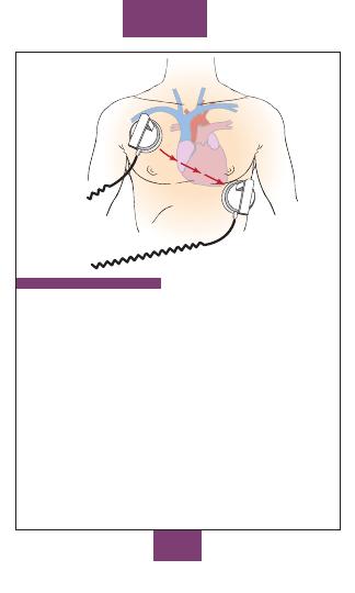
100
MEDS/
SKILLS
+
_
MANUAL DEFIBRILLATION
A manual defibrillator is used to restore a normal heart rhythm.
For a patient experiencing sudden cardiac arrest, first use the
ECG tracing to verify that the rhythm is either VF or pulseless
VT, and then manually deliver an electric shock to the heart.
Procedure
1. Verify patient is in cardiac arrest, with no pulse or
respiration. Have someone provide CPR, if possible, while
the defibrillator is obtained and placed next to the patient.
2. Turn on defibrillator; verify all cables are connected.
3. Turn “lead select” to “paddles” or “defibrillator.”
4. Select initial energy level for an adult to 200 J.
5. Paddles: Use conducting gel and place on apex (lower left
chest, midaxillary) and sternum (right of sternum,
midclavicular).
Pads: Place in same locations as you would put paddles.
6. Verify rhythm as VF or pulseless VT.
7. Say, “Charging defibrillator, stand clear!”
8. Charge defibrillator.
04ECG-Tab 04 2/4/05 4:00 PM Page 100
Copyright © 2005 F. A. Davis.

101
9. Say, “I’m going to shock on three. One, I’m clear; two,
you’re clear; three, everybody’s clear.” Perform visual sweep
to assure all rescue personnel are clear of patient, bed, and
equipment.
10. Discharge defibrillator, reassess rhythm, and refer to
appropriate treatment algorithm for resulting rhythm.
AUTOMATIC EXTERNAL DEFIBRILLATOR (AED)
An AED is a small, lightweight device used by both professionals
and laypersons to assess heart rhythm by computer analysis. If
necessary, it administers an electric shock to restore a normal
rhythm in patients with sudden cardiac arrest. A shock is
administered only if the rhythm detected is VF or VT.
Procedure
1. Verify patient is in cardiac arrest, with no pulse or
respiration. Have someone provide CPR, if possible, while the
AED is obtained and placed next to the patient.
2. Turn on AED. Follow voice prompts or visual messages.
3. Open adhesive pads and attach pads to cables.
4. Attach pads to right sternal border and apex or as
pictured on each of the AED electrodes (see preceding manual
defibrillation figure).
5. Clear patient and stop CPR.
6. Press analyze button, if present.
7. If shock is advised, say, “I’m going to shock on three. One,
I’m clear; two, you’re clear; three, everybody’s clear.” Perform
visual sweep to ensure rescue personnel are not touching
patient or equipment. Press shock button. Reanalyze after
shock and continue as prompted by the AED.
8. If no shock is advised, check for a pulse. If no pulse,
start CPR.
♥
Clinical Tip: Fully automatic AED analyzes the rhythm and
delivers shock if indicated.
♥
Clinical Tip: Semiautomatic AED analyzes the rhythm and
tells operator that shock is indicated. If it is indicated, operator
initiates shock.
MEDS/
SKILLS
04ECG-Tab 04 2/4/05 4:00 PM Page 101
Copyright © 2005 F. A. Davis.
102
CARDIOVERSION
(Synchronized)
Indications: Unstable tachycardia (altered LOC, dizziness, chest
pain, hypotension).
Energy Levels: 100 J, 200 J, 300 J, 360 J.
Application: Use handheld paddles or remote adhesive pads.
Always use a conducting gel with paddles. For conscious
patients explain the procedure and use a medication for
sedation. Consider 2.5–5.0 mg of midazolam (Versed) or 5
mg diazepam (Valium).
Methods: Place defibrillator in synchronized (sync) mode.
Charge to appropriate level. Say, “I’m going to shock on
three. One, I’m clear; two, you’re clear; three, everybody’s
clear.” Perform visual sweep and press shock button. Reassess
and treat according to appropriate algorithm.
Precautions: Reactivation of sync mode is required after each
attempted cardioversion. Defibrillators default to
unsynchronized mode. Place paddles and pads several inches
away from an implanted pacemaker.
♥
Clinical Tip: Sync mode delivers energy just after the R wave
to avoid stimulation during the refractory, or vulnerable, period.
TRANSCUTANEOUS PACING
Indications: Symptomatic bradycardia unresponsive to
atropine, bradycardia with ventricular escape rhythms,
symptomatic 2nd-degree AV block type II, or 3rd-degree AV
block.
Pacing Modes: Demand mode (synchronous) pacer senses the
patient’s heart rate and paces only when the heart rate falls
below the level set by the clinician. Fixed mode
(asynchronous) pacer cannot sense the heart rate, and the
pacer operates at the rate set by the clinician. Rate selection
is 30–180 bpm. Output is adjustable 0–200 mA. Pulse duration
varies from 20 to 40 ms.
Contraindications: Not effective in VF or pulseless VT.
Side Effects: Chest muscle contraction, burns, chest
discomfort.
Precautions: Make sure pads have good skin contact to achieve
capture and avoid burns.
MEDS/
SKILLS
04ECG-Tab 04 2/4/05 4:00 PM Page 102
Copyright © 2005 F. A. Davis.
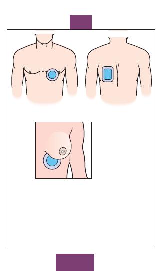
103
MEDS/
SKILLS
Female patients:
Position electrode
under breast
Anterior
Posterior
Placement of anterior-posterior pacemaker pads.
04ECG-Tab 04 2/4/05 4:00 PM Page 103
Copyright © 2005 F. A. Davis.

104
MEDS/
SKILLS
PRECORDIAL THUMP
Indications: VF or pulseless VT; can cause depolarization and
return the heart to an organized rhythm. Cardiac arrest must
be witnessed.
Method: Sharply strike the midsternal area of the chest from a
height of 10–12 inches with a fist. Recheck pulse and rhythm.
Contraindications: Should not be used in infants or children.
Side Effects: Fractured ribs or sternum.
Precautions: Position the fist (thumb side up) and arm parallel
to the long axis of the sternum to avoid injuring adjacent ribs.
CAROTID SINUS MASSAGE
(Vagal Maneuver)
Indications: Can increase vagal nerve stimulation and slow SVT,
or even convert SVT to NSR, without severe hemodynamic
compromise.
Method: Supine position, head tilted to either side with neck
hyperextended. Place index and middle fingers over the
carotid artery below the angle of the jaw, as high on the neck
as possible. Massage the artery for 5–10 sec by firmly
pressing on it and rubbing.
Contraindications: Unequal carotid pulses, carotid bruits,
cervical spine injury, or history of cerebrovascular accident
(CVA) or carotid atherosclerosis.
Side Effects: Slow HR or AV block, PVCs, VT, VF, syncope,
seizure, hypotension, nausea or vomiting, stroke.
Precautions: Be sure patient is receiving oxygen and an IV is in
place. Never massage both arteries simultaneously.
04ECG-Tab 04 2/4/05 4:00 PM Page 104
Copyright © 2005 F. A. Davis.
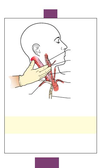
Sternocleido-
mastoid
muscle
Carotid
sinus nerve
Carotid
body
Vagus nerve
Right common
carotid artery
Cardiac
plexus
105
MEDS/
SKILLS
Carotid sinus massage.
♥
Clinical Tip: Each carotid pulse should be palpated and
auscultated before the procedure to maintain safety measures.
♥
Clinical Tip: Alternate vagal maneuvers include coughing,
bearing down, holding breath.
04ECG-Tab 04 2/4/05 4:00 PM Page 105
Copyright © 2005 F. A. Davis.

CPR Skill Performance
Compression/
Rate of
Depth of
Pulse
Hand
CPR
Ventilation
Compressions
Compressions
Check
Position for
Method
Ratio
(min)
(in.)
(artery)
Compressions
Adult, 1
rescuer
Adult, 2
rescuers
Child, 1
rescuer
Child, 2
rescuers
Infant, 1
rescuer
Infant, 2
rescuers
Newborn
106
CPR
15:2
15:2
5:1
5:1
5:1
5:1
3:1
100
100
100
100
≥100
≥100
≥120
1
1
/
2
–2
1
1
/
2
–2
1–1
1
/
2
1–1
1
/
2
1
/
2
–1
1
/
2
–1
1
/
3
Carotid
Carotid
Carotid
Carotid
Brachial
Femoral
Brachial
Femoral
Brachial
Femoral
Heels of 2 hands
over lower half of
sternum
Heels of 2 hands
over lower half of
sternum
Heel of 1 hand over
lower half of
sternum
Heel of 1 hand over
lower half of
sternum
2 fingers over lower
half of sternum
2 fingers over lower
half of sternum
2 fingers over lower
half of sternum
05ECG-Tab 05 2/4/05 4:01 PM Page 106
Copyright
©
2005
F.
A.
Davis.

107
CPR
CPR: Adult (older than 8 yr)
1. Check for unresponsiveness. Gently shake or tap person.
Shout, “Are you OK?”
2. If no response, call for an AED, summon help, call a
code, or call 911. Send second rescuer, if available, for help.
3. Position person supine on a hard, flat surface. Support
head and neck, loosen clothing, and expose chest.
4. Open airway by the head tilt–chin lift method or, if spinal
injury is suspected, use the jaw thrust method.
5. Look, listen, and feel for breathing for up to 10 sec.
6. If person is breathing, place in recovery position.
7. If person is not breathing, begin rescue breaths. Using a
bag-valve-mask or face mask, give two slow breaths (2 sec
each). Be sure that chest rises.
8. If the chest does not rise, reposition the head and the chin and
jaw, and give two more breaths. If chest still does not rise,
follow instructions for unconscious adult with an
obstructed airway (p 112).
9. Assess carotid pulse for signs of circulation. If signs of
circulation are present but person is still not breathing,
continue to give rescue breaths at the rate of one every 5 sec.
10. If pulse and signs of circulation are not present, begin
compressions. Place heel of your hand 2 finger-widths above
xiphoid process; place heel of the second hand over the first.
Keep elbows locked, lean shoulders over hands, and firmly
compress chest 1
1
/
2
–2 inches. Give 15 compressions.
Compress at a rate of 100 per min.
11. Continue to give 2 breaths followed by 15 compres-
sions. After about 1 min (or at the 4th cycle of 15:2) check
pulse and other signs of circulation. If circulation resumes but
breathing does not or is inadequate, continue rescue
breathing.
12. If breathing and circulation resume, place person in recovery
position and monitor until help arrives.
♥
Clinical Tip: The compression rate is the speed of the
compressions, not the actual number of compressions per min.
Compressions, if uninterrupted, would equal 100/min.
05ECG-Tab 05 2/4/05 4:01 PM Page 107
Copyright © 2005 F. A. Davis.

108
CPR
CPR: Child (1–8 yr)
1. Check for unresponsiveness. Gently shake or tap child.
Shout, “Are you OK?”
2. If no response send a second rescuer, if available, for help.
3. Position child supine on a hard, flat surface. Support head
and neck, loosen clothing, and expose chest.
4. Open airway by the head tilt–chin lift method or, if spinal
injury is suspected, use the jaw thrust method.
5. Look, listen, and feel for breathing for up to 10 sec.
6. If child is breathing, place in recovery position.
7. If child is not breathing, begin rescue breaths. Using a bag-
valve-mask or face mask, give two slow breaths (1–1
1
/
2
sec
each). Be sure the chest rises.
8. If the chest does not rise, reposition the head and the chin
and jaw and give two more breaths. If chest still does not
rise, follow instructions for unconscious child with an
obstructed airway (p 113).
9. Assess carotid pulse for signs of circulation. If signs of
circulation are present but child is still not breathing, continue
to give rescue breaths at the rate of one every 3 sec.
10. If pulse and signs of circulation are not present, begin
compressions. Place heel of one hand 2 finger-widths above
xiphoid process. Keep elbow locked, lean shoulders over
hand, and firmly compress chest 1–1
1
/
2
in. Give 5
compressions. Compress at a rate of 100 per min.
11. Continue to give 1 breath followed by 5 compressions.
After about 1 min of CPR, check pulse and other signs of
circulation. If rescuer is alone and no signs of circulation are
present, call for an AED, summon help, call a code, or
call 911. If circulation resumes but breathing does not or is
inadequate, continue rescue breathing.
12. If breathing and circulation resume, place child in recovery
position and monitor until help arrives.
♥
Clinical Tip: It is not always necessary to wait 1 min before
calling for help if you are alone. If you know a child has had a
cardiac arrest due to heart failure, request immediate help
including a defibrillator.
05ECG-Tab 05 2/4/05 4:01 PM Page 108
Copyright © 2005 F. A. Davis.

109
CPR
CPR: Infant (under 1 yr)
1. Check for unresponsiveness. Gently rub infant’s back or
sternum. Never shake an infant.
2. If no response send a second rescuer, if available, for help.
3. Position infant supine on a hard, flat surface. Support head
and neck, loosen clothing, and expose chest.
4. Open airway by the head tilt–chin lift method (do not
overextend head or airway will become obstructed). If spinal
injury is suspected, use jaw thrust method.
5. Look, listen, and feel for breathing for up to 10 sec.
6. If infant is breathing, place in recovery position.
7. If infant is not breathing, begin rescue breaths. Using a
bag-valve-mask or face mask, give two slow breaths (1–1
1
/
2
sec each). Be sure that chest rises.
8. If the chest does not rise, reposition the head and the chin
and jaw and give two more breaths. If chest still does not
rise, follow instructions for unconscious infant with
an obstructed airway (p 114).
9. Assess brachial or femoral pulse for signs of circulation.
If signs of circulation are present but infant is still not breath-
ing, continue rescue breaths at the rate of one every 3 sec.
10. If pulse and signs of circulation are not present, begin
compressions. Place two fingers of one hand 2 finger-
widths above xiphoid process. Firmly compress chest
1
/
2
–1 in.
Give five compressions. Compress at a rate of
≥100 per
min.
11. Continue to give one breath followed by five
compressions. After about 1 min of CPR, check pulse and
other signs of circulation. If rescuer is alone and no signs of
circulation are present, call for an AED, summon help,
call a code, or call 911. If circulation resumes but breathing
does not or is inadequate, continue rescue breathing.
12. If breathing and circulation resume, place infant in recovery
position and monitor until help arrives.
♥
Clinical Tip: Chest compressions must be adequate to
produce a palpable pulse during resuscitation.
05ECG-Tab 05 2/4/05 4:01 PM Page 109
Copyright © 2005 F. A. Davis.
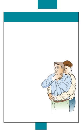
110
CPR
Obstructed Airway: Conscious Adult
or Child (1 yr or older)
Signs and Symptoms
■
Grabbing at the throat with one or both hands
■
Inability to speak; high-pitched crowing sounds
■
Wheezing, gagging, ineffective coughing
1. Determine that airway is obstructed. Ask, “Are you
choking? Can you speak?”
2. Let person know you are going to help.
3. Stand behind choking person and wrap your arms
around his or her waist. For someone who is obese or
pregnant, wrap arms around chest.
4. Make a fist. Place thumb side of fist in middle of
abdomen just above navel. Locate middle of sternum for
obese or pregnant persons.
5. Grasp fist with your other hand.
6. Press fist abruptly into
abdomen using an
upward, inward thrust.
Use a straight thrust back for
someone who is obese or
pregnant.
7. Continue thrusts until object
is dislodged or person loses
consciousness.
8. If person loses
consciousness, treat as
unconscious adult or child
with an obstructed airway
(pp 112–113).
Heimlich maneuver for adult or child.
05ECG-Tab 05 2/4/05 4:01 PM Page 110
Copyright © 2005 F. A. Davis.
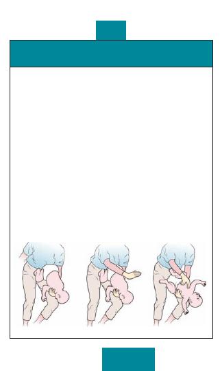
111
CPR
Obstructed Airway: Conscious
Infant (younger than 1 yr)
Signs and Symptoms
■
Inability to breathe or cry
■
High-pitched crowing sounds
■
Sudden wheezing or noisy breathing
1. Determine that airway is obstructed.
2. Lay infant down on your forearm, with the chest in your
hand and the jaw between your thumb and index finger.
3. Using your thigh or lap for support, keep infant’s head
lower than his or her body.
4. Give five quick, forceful blows between shoulder
blades with your palm.
5. Turn infant over to be face up on your other arm. Using
your thigh or lap for support, keep infant’s head lower than
his or her body.
6. Place two fingers on center of sternum just below nipple
line.
7. Give five quick thrusts down, depressing chest
1
/
2
–1
in. each time.
8. Continue sequence of five back blows and five chest
thrusts until object is dislodged or infant loses
consciousness. If infant loses consciousness, treat as
unconscious infant with an obstructed airway (p 114).
Heimlich maneuver for infant.
05ECG-Tab 05 2/4/05 4:01 PM Page 111
Copyright © 2005 F. A. Davis.

112
Obstructed Airway: Unconscious
Adult (older than 8 yr)
Signs and Symptoms
■
Failure to breathe
■
Inability to move air into lungs with rescue breaths
■
Cyanosis
1. Establish unresponsiveness. Gently shake or tap person.
Shout, “Are you OK?”
2. If no response, call for an AED, summon help, call a
code, or call 911. Send second rescuer, if available, for
help.
3. Position person supine on a hard, flat surface. Support
head and neck, loosen clothing, and expose chest.
4. Open airway by the head tilt–chin lift method or, if spinal
injury is suspected, use the jaw thrust method.
5. Look, listen, and feel for breathing for up to 10 sec.
6. If person is not breathing, begin rescue breaths. If the
chest does not rise, reposition the head and the chin and jaw,
and attempt to ventilate.
7. If ventilation is unsuccessful and chest still does not rise,
begin abdominal thrusts. Straddle thighs or kneel to side
for someone who is obese or pregnant. Place heel of hand in
middle of abdomen just above umbilicus (middle of sternum
if person is obese or pregnant).
8. Place other hand on top of first hand and give five quick
thrusts inward and upward.
9. Open mouth by placing thumb over tongue and index finger
under chin. Perform a finger sweep to try to remove
object.
10. Repeat steps 6 through 9 until rescue breaths are effective.
Then continue steps for CPR.
♥
Clinical Tip: The most common cause of airway obstruction
is the tongue.
CPR
05ECG-Tab 05 2/4/05 4:01 PM Page 112
Copyright © 2005 F. A. Davis.
113
Obstructed Airway: Unconscious Child (1–8 yr)
Signs and Symptoms
■
Failure to breathe
■
Inability to move air into lungs with rescue breaths
■
Cyanosis
1. Check for unresponsiveness. Gently shake or tap child.
Shout, “Are you OK?”
2. If no response send a second rescuer, if available, for help.
3. Position child supine on a hard, flat surface. Support head
and neck, loosen clothing, and expose chest.
4. Open airway by the head tilt–chin lift method or, if spinal
injury is suspected, use the jaw thrust method.
5. Look, listen, and feel for breathing for up to 10 sec.
6. If child is not breathing, begin rescue breaths. If the chest
does not rise, reposition the head and the chin and jaw, and
attempt to ventilate.
7. If ventilation is unsuccessful and chest still does not rise,
begin abdominal thrusts. Straddle child’s thighs. Place
heel of hand in middle of abdomen just above umbilicus.
8. Place other hand on top of first hand and give five quick
thrusts inward and upward.
9. Open child’s mouth by placing thumb over tongue and index
finger under chin. If object is visible and loose, perform
a finger sweep and remove it. Do not perform a blind
finger sweep.
10. If airway obstruction is not relieved after 1 min and rescuer is
alone, call for an AED, summon help, call a code, or
call 911.
11. Repeat steps 6 through 9 until rescue breaths are effective.
Then continue steps for CPR.
♥
Clinical Tip: Avoid compression of the xiphoid process.
CPR
05ECG-Tab 05 2/4/05 4:01 PM Page 113
Copyright © 2005 F. A. Davis.

114
Obstructed Airway: Unconscious
Infant (younger than 1 yr)
Signs and Symptoms
■
Inability to breathe, high-pitched noises
■
Inability to move air into lungs with rescue breaths
■
Cyanosis
1. Check for unresponsiveness. Gently rub infant’s back or
sternum. Never shake an infant.
2. If no response send a second rescuer, if available, for help.
3. Position infant supine on a hard, flat surface. Support head
and neck, loosen clothing, and expose chest.
4. Open airway by the head tilt–chin lift method, or, if spinal
injury is suspected, use the jaw thrust method.
5. Look, listen, and feel for breathing for up to 10 sec.
6. If infant is not breathing, begin rescue breaths. If the chest
does not rise, reposition the head and the chin and jaw, and
attempt to ventilate.
7. If ventilation is unsuccessful and chest still does not rise, begin
back blows.
8. Lay infant down on your forearm, with the chest in your hand
and the jaw between your thumb and index finger.
9. Using your thigh or lap for support, keep infant’s head lower
than his or her body. Give five quick, forceful blows
between shoulder blades with your palm.
10. Turn infant over to be face up on your other arm. Using your
thigh or lap for support, keep infant’s head lower than his or
her body. Place two fingers on center of sternum just below
nipple line. Give five quick thrusts down, depressing chest
1
/
2
–1 in. each time.
11. Open infant’s mouth by placing thumb over tongue and index
finger under chin. If object is visible and loose, perform a
finger sweep and remove it. Do not perform a blind
finger sweep.
12. If airway obstruction is not relieved after 1 min and rescuer is
alone, call for an AED, summon help, call a code, or call
911.
13. Repeat steps 6 through 11 until rescue breaths are effective.
Then continue steps for CPR.
CPR
05ECG-Tab 05 2/4/05 4:01 PM Page 114
Copyright © 2005 F. A. Davis.
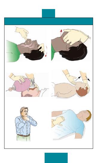
115
CPR and Obstructed Airway Positions
Head tilt–chin lift (adult or child).
Jaw thrust maneuver.
Bag-valve-mask.
Head tilt–chin lift (infant).
Universal choking sign.
Abdominal thrusts.
CPR
05ECG-Tab 05 2/4/05 4:01 PM Page 115
Copyright © 2005 F. A. Davis.
116
ACLS
Ventricular Fibrillation or
Pulseless Ventricular Tachycardia
Signs and Symptoms
■
Unresponsive state
■
No respiration, pulse, or BP
1. Establish unresponsiveness with no respiration or pulse.
2. Deliver a precordial thump if cardiac arrest is witnessed
and a defibrillator is not immediately available.
3. Begin CPR with high-flow oxygen.
4. Defibrillate at 200 J (or equivalent biphasic energy).
5. Defibrillate at 200–300 J (or equivalent biphasic energy).
6. Defibrillate at 360 J (or equivalent biphasic energy).
7. Intubate and establish IV.
8. Administer epinephrine 1 mg (10 mL of 1:10,000) IVP
(follow with 20 mL IV flush), repeat every 3–5 min; give
2.0–2.5 mg diluted in 10 mL normal saline if administering
via ET tube; or administer a single dose of vasopressin 40
U IVP.
9. Defibrillate at 360 J (or equivalent biphasic energy) within
30–60 sec after each dose of medication. Pattern should be
drug, shock; drug, shock. Consider the following anti-
arrhythmics for shock-refractory VF or VT:
10. Administer amiodarone 300 mg (diluted in 20-30 mL D5W)
IVP; or lidocaine 1.0–1.5 mg/kg IVP, 2-4 mg/kg by ET tube.
11. Repeat initial antiarrhythmic for shock-refractory VF or VT:
amiodarone 150 mg IVP; or lidocaine 0.5–0.75 mg/kg IVP,
repeat lidocaine every 5–10 min, max. 3 mg/kg.
12. Administer magnesium sulfate 1–2 g (2–4 mL of a 50%
solution) diluted in 10 mL of D5W IVP in polymorphic VT,
torsade de pointes, or suspected hypomagnesemia.
13. If no response, consider procainamide 30–50 mg/min IV
infusion, max. 17 mg/kg; or sodium bicarbonate 1 mEq/kg
IVP, may repeat 0.5 mEq/kg every 10 min.
♥
Clinical Tip: Do not delay defibrillation.
♥
Clinical Tip: If vasopressin is used, wait 10–20 min before
administering epinephrine.
06ECG-Tab 06 2/4/05 4:01 PM Page 116
Copyright © 2005 F. A. Davis.
117
ACLS
Thrombosis (pulmonary)
Thrombosis (coronary)
Tension pneumothorax
Tamponade (cardiac)
Tablets (drug overdose)
Pulseless Electrical Activity
Signs and Symptoms
■
Unresponsive state
■
No respiration, pulse, or BP
■
Identifiable electrical rhythm on monitor but no pulse
1. Establish unresponsiveness with no respiration or pulse.
2. Begin CPR with high-flow oxygen.
3. Intubate and establish IV.
4. Consider and treat possible causes: pulmonary embolism,
MI, acidosis, tension pneumothorax, hyper- or hypokalemia,
cardiac tamponade, hypovolemia, hypoxia, hypothermia,
drug overdose (e.g., cyclic antidepressants, beta blockers,
calcium channel blockers, digoxin).
5. Administer epinephrine 1 mg (10 mL of 1:10,000) IVP, repeat
every 3–5 min; give 2.0–2.5 mg diluted in 10 mL normal
saline if administering by ET tube.
6. Administer atropine 1 mg IVP if ECG rate is
60 bpm.
Repeat every 3–5 min as needed to a total dose of 0.03–0.04
mg/kg. May be given by ET tube at 2–3 mg diluted in 10 mL
normal saline.
7. Consider fluid challenge of 500 mL normal saline, especially
in suspected hypovolemia.
8. If no response, consider sodium bicarbonate 1 mEq/kg IVP,
may repeat 0.5 mEq/kg every 10 min.
♥
Clinical Tip: Sodium bicarbonate may be harmful in
hypercarbic acidosis.
♥
Clinical Tip: Memory aid for causes of PEA:
Five “H” Causes
Five “T” Causes
Hypothermia
Hyperkalemia/hypokalemia
Hydrogen ion (acidosis)
Hypoxia
Hypovolemia
06ECG-Tab 06 2/4/05 4:01 PM Page 117
Copyright © 2005 F. A. Davis.

118
Asystole
Signs and Symptoms
■
Unresponsive state
■
No respiration, pulse, or BP
■
ECG shows flat line; no electrical activity
1. Establish unresponsiveness with no respiration or pulse.
2. Begin CPR with high-flow oxygen.
3. Intubate and establish IV.
4. Consider and treat possible causes: pulmonary embolism,
MI, acidosis, tension pneumothorax, hyper- or
hypokalemia, cardiac tamponade, hypovolemia, hypoxia,
hypothermia, drug overdose (e.g., cyclic antidepressants,
beta blockers, calcium channel blockers, digoxin).
5. If condition remains unchanged, begin immediate
transcutaneous pacing if equipment is available.
6. Administer epinephrine 1 mg (10 mL of 1:10,000) IVP,
repeat every 3–5 min; give 2.0–2.5 mg diluted in 10 mL
normal saline if administering by ET tube.
7. Administer atropine 1 mg IVP, repeat every 3–5 min as
needed, to a total dose of 0.03–0.04 mg/kg. May be given
by ET tube at 2–3 mg diluted in 10 mL normal saline.
8. If no response, consider sodium bicarbonate 1 mEq/kg IVP,
may repeat 0.5 mEq/kg every 10 min.
9. If asystole persists, consider quality of resuscitation,
identification of reversible causes, and support for
termination protocols.
♥
Clinical Tip: Do not delay transcutaneous pacing; it takes
priority over medication.
♥
Clinical Tip: Always confirm asystole by checking the ECG in
two different leads. Also, search to identify underlying VF.
♥
Clinical Tip: Study local policy to learn established criteria
for stopping resuscitation efforts.
ACLS
06ECG-Tab 06 2/4/05 4:01 PM Page 118
Copyright © 2005 F. A. Davis.

119
Ischemic Chest Pain
Signs and Symptoms
■
History of acute MI or angina
■
Chest pain or discomfort
■
Pain spreading to neck, shoulders, arms, or jaw
■
Nausea, diaphoresis, shortness of breath
1. Establish responsiveness.
2. Measure vital signs, including oxygen saturation.
3. Supply oxygen, begin cardiac monitoring, start IV, and
obtain 12-lead ECG.
4. Administer aspirin 162–325 mg.
5. Administer nitroglycerin by sublingual route 0.3–0.4 mg (1
tablet), repeat every 5 min, max. 3 doses/15 min; or
administer aerosol spray for 0.5–1.0 sec at 5-min intervals
(provides 0.4 mg per dose).
6. Nitroglycerin administration requires BP >100 mm Hg
systolic.
7. Repeat nitroglycerin (see step 5) until chest pain is relieved,
systolic BP falls below 100 mm Hg, or signs of ischemia or
infarction are resolved.
8. If chest pain is not relieved by nitroglycerin, administer
morphine 2–4 mg IVP (over 1–5 min) every 5–30 min. Do
not administer morphine if systolic BP is
100 mm Hg.
♥
Clinical Tip: Patients should not be given nitroglycerin if they
have taken sildenafil (Viagra), tadalafil (Cialis), or vardenafil
(Levitra) in the last 24 hr. The use of nitroglycerin with these
medications may cause irreversible hypotension.
♥
Clinical Tip: Diabetic patients and women frequently present
with atypical symptoms (e.g., weakness, fatigue, complaints of
indigestion).
ACLS
06ECG-Tab 06 2/4/05 4:01 PM Page 119
Copyright © 2005 F. A. Davis.
120
Bradycardia
Signs and Symptoms
■
Pulse rate
60 bpm
■
AV block
■
Hypotension, altered mental status, pulmonary edema, shock
1. Establish responsiveness.
2. Measure vital signs, including oxygen saturation.
3. Supply oxygen, begin cardiac monitoring, and start IV.
4. In 2nd-degree (Mobitz type II) or 3rd-degree AV block,
proceed directly to step 5, transcutaneous pacing;
otherwise administer atropine 0.5–1.0 mg IVP every 3–5
min, max. 0.03–0.04 mg/kg.
5. If patient remains symptomatic or has 2nd-degree (Mobitz
type II) or 3rd-degree AV block, sedate patient and begin
transcutaneous pacing, if available.
6. If no response, consider dopamine with continuous
infusions (titrate to patient response) of 5–20
g/kg/min.
Mix 400 mg/250 mL in normal saline, lactated Ringer’s
solution, or D5W.
7. If patient is still hypotensive with severe bradycardia,
consider epinephrine infusion, 2–10
g/min IV (add 1 mg of
1:1000 to 500 mL normal saline and infuse at 1–5 mL/min).
8. If still no response, consider isoproterenol, IV infusion: mix
1 mg in 250 mL normal saline, lactated Ringer’s solution, or
D5W with rate of 2–10
g/min, titrate to patient response.
♥
Clinical Tip: If patient is symptomatic, do not delay
transcutaneous pacing while waiting for atropine to take effect
or for IV access.
♥
Clinical Tip: Use atropine with caution in a suspected acute
MI; atropine may induce rate-related ischemia.
♥
Clinical Tip: If patient is asymptomatic but has 2nd-degree
(Mobitz type II) or 3rd-degree AV block, use transcutaneous
pacemaker until transvenous pacer is placed.
ACLS
06ECG-Tab 06 2/4/05 4:01 PM Page 120
Copyright © 2005 F. A. Davis.
121
Tachycardia—Unstable
Signs and Symptoms
■
Altered level of consciousness
■
Chest pain or discomfort, palpitations
■
Shortness of breath, diaphoresis
■
Hypotension, pulmonary edema, crackles, rhonchi, jugular
vein distention, peripheral edema
1. Establish responsiveness.
2. Measure vital signs, including oxygen saturation.
3. Supply oxygen, begin cardiac monitoring, and start IV.
4. Establish that serious signs and symptoms are related to
the tachycardia.
5. If ventricular rate is
150 bpm, prepare for immediate
synchronized cardioversion.
6. Premedicate with a sedative plus an analgesic whenever
possible.
7. Administer synchronized cardioversion at 100 J (or
equivalent biphasic energy).
8. If no response, administer synchronized cardioversion at
200 J (or equivalent biphasic energy).
9. If no response, administer synchronized cardioversion at
300 J (or equivalent biphasic energy).
10. If no response, administer synchronized cardioversion at
360 J (or equivalent biphasic energy).
11. If the unstable tachycardia converts to VF or pulseless VT,
treat with immediate defibrillation and follow algorithm for
VF and pulseless VT.
♥
Clinical Tip: Reactivate sync mode before next attempted
cardioversion.
♥
Clinical Tip: If a tachycardia is VT or torsade de pointes, it
may rapidly deteriorate to VF.
♥
Clinical Tip: A-flutter and PSVT may respond to lower energy
levels such as 50 J (or equivalent biphasic energy).
ACLS
06ECG-Tab 06 2/4/05 4:01 PM Page 121
Copyright © 2005 F. A. Davis.
122
Wide-Complex Tachycardia—Stable
Monomorphic VT
1. Establish responsiveness.
2. Measure vital signs, including oxygen saturation.
3. Supply oxygen, begin cardiac monitoring, and start IV.
4. May go directly to step 8, cardioversion.
For Impaired Cardiac Function
5. Administer amiodarone 150 mg IVP over 10 min (15 mg/min),
may repeat infusion of 150 mg IVP every 10 min as needed; or
administer lidocaine 0.5–0.75 mg/kg IVP (may use up to
1.0–1.5 mg/kg), repeat 0.5–0.75 mg/kg IVP every 5–10 min,
max. 3 mg/kg.
6. If rhythm converts to sinus rhythm, begin infusion of rhythm-
converting agent: amiodarone, slow infusion of 360 mg IV
over the next 6 hr (1 mg/min) with maintenance infusion of
540 mg over the next 18 hr (0.5 mg/min); or start lidocaine
infusion of 1–4 mg/min (30–50
g/kg/min).
7. If rhythm does not convert, prepare for immediate
cardioversion.
8. Premedicate with sedative plus analgesic agent whenever
possible.
9. Administer synchronized cardioversion incrementally at 100 J,
200 J, 300 J, then 360 J (or equivalent biphasic energy).
For Normal Cardiac Function
5. Follow steps 1–4 above.
6. Otherwise, consider procainamide or sotalol.
7. Other acceptable medication is amiodarone or lidocaine.
Notes:
ACLS
06ECG-Tab 06 2/4/05 4:01 PM Page 122
Copyright © 2005 F. A. Davis.
123
Wide-Complex Tachycardia—Stable
Polymorphic VT
1. Establish responsiveness.
2. Measure vital signs, including oxygen saturation.
3. Supply oxygen, begin cardiac monitoring, and start IV.
4. May go directly to step 8, cardioversion.
For Impaired Cardiac Function
5. Administer amiodarone 150 mg IVP over first 10 min (15
mg/min), may repeat infusion of 150 mg IVP every 10 min as
needed; or administer lidocaine 0.5–0.75 mg/kg IVP (may use
up to 1.0–1.5 mg/kg), repeat 0.5–0.75 mg/kg IVP every 5–10 min,
max. 3 mg/kg.
6. If rhythm converts to sinus rhythm, begin infusion of rhythm-
converting agent: amiodarone, slow infusion of 360 mg IV over
the next 6 hr (1 mg/min) with maintenance infusion of 540 mg
over the next 18 hr (0.5 mg/min); or start lidocaine infusion of
1–4 mg/min (30–50
g/kg/min).
7. If rhythm does not convert, prepare for immediate cardiover-
sion.
8. Premedicate with sedative plus analgesic agent whenever
possible.
9. Administer synchronized cardioversion incrementally at 100 J,
200 J, 300 J, then 360 J (or equivalent biphasic energy).
For Normal Cardiac Function
If possible, measure QT interval before onset of VT; it cannot be
obtained in sustained VT. Torsade de pointes is an example of
polymorphic VT with an abnormally prolonged QT interval.
Normal QT Interval
Prolonged QT Interval
Correct electrolytes
Treat ischemia
Consider (any one): beta
blockers, lidocaine,
amiodarone, procainamide,
or sotalol.
ACLS
Correct electrolytes
Treat ischemia
Consider (any one):
magnesium, overdrive
pacing, isoproterenol,
phenytoin, or lidocaine.
06ECG-Tab 06 2/4/05 4:01 PM Page 123
Copyright © 2005 F. A. Davis.

124
Narrow-Complex Tachycardia—Stable
Paroxysmal Supraventricular Tachycardia
Signs and Symptoms
■
If present, hypotension, syncope, or limited ability to exercise
■
Patient may be asymptomatic.
1. Establish responsiveness.
2. Measure vital signs, including oxygen saturation.
3. Supply oxygen, begin cardiac monitoring, and start IV.
4. Attempt vagal maneuvers (e.g., carotid sinus massage,
Valsalva maneuver).
5. If rhythm has not converted to sinus rhythm, administer
adenosine 6 mg rapid IVP over 1–3 sec followed by a 20-mL
bolus of normal saline.
6. If rhythm still has not converted, repeat adenosine 12 mg IVP
in 1–2 min. A third dose of 12 mg IVP may be given after
another 1–2 min, max. 30 mg.
For Impaired Cardiac Function
7. If still no response and patient has serious signs and
symptoms with ventricular rate
150 bpm, prepare for
immediate cardioversion.
8. Premedicate with sedative plus analgesic agent whenever
possible.
9. Administer synchronized cardioversion incrementally at 100
J, 200 J, 300 J, then 360 J (or equivalent biphasic energy).
10. If rhythm still has not converted, consider digoxin,
amiodarone, or diltiazem.
For Normal Cardiac Function
7. Follow steps 1–6 above.
8. Consider in order of priority an AV blocker (beta blocker,
calcium channel blocker, digoxin), cardioversion, and an
antiarrhythmic (procainamide, amiodarone, sotalol).
ACLS
06ECG-Tab 06 2/4/05 4:01 PM Page 124
Copyright © 2005 F. A. Davis.
125
Narrow-Complex Tachycardia—Stable
Junctional Tachycardia
1. Establish responsiveness.
2. Measure vital signs, including oxygen saturation.
3. Supply oxygen, begin cardiac monitoring, and start IV.
4. Attempt vagal maneuvers (e.g., carotid sinus massage,
Valsalva maneuver).
5. If rhythm has not converted to sinus rhythm, administer
adenosine 6 mg rapid IVP over 1–3 sec followed by a 20-mL
bolus of normal saline.
6. If rhythm still has not converted, repeat adenosine 12 mg IVP
in 1–2 min. A third dose of 12 mg IVP may be given after
another 1–2 min, max. 30 mg.
For Impaired Cardiac Function
7. If still no response consider amiodarone, 150 mg IVP over 10
min (15 mg/min), may repeat infusion of 150 mg IVP every 10
min as needed.
8. Do not attempt cardioversion.
For Normal Cardiac Function
7. Follow steps 1–6 above.
8. Consider a beta blocker, calcium channel blocker, or
amiodarone.
9. Do not attempt cardioversion.
♥
Clinical Tip: Avoid carotid massage in patients at risk for
carotid atherosclerosis.
Notes:
ACLS
06ECG-Tab 06 2/4/05 4:01 PM Page 125
Copyright © 2005 F. A. Davis.
126
Narrow-Complex Tachycardia—Stable
Ectopic or Multifocal Atrial Tachycardia
1. Establish responsiveness.
2. Measure vital signs, including oxygen saturation.
3. Supply oxygen, begin cardiac monitoring, and start IV.
4. Attempt vagal maneuvers (e.g., carotid sinus massage,
Valsalva maneuver).
5. If rhythm has not converted to sinus rhythm, administer
adenosine 6 mg rapid IVP over 1–3 sec followed by a 20-mL
bolus of normal saline.
6. If rhythm still has not converted, repeat adenosine 12 mg IVP
in 1–2 min. A third dose of 12 mg IVP may be given after
another 1–2 min, max. 30 mg.
For Impaired Cardiac Function
7. If still no response, consider amiodarone 150 mg IVP over 10
min (15 mg/min), may repeat infusion of 150 mg IVP every 10
min as needed.
8. Consider diltiazem 15–20 mg (0.25 mg/kg) IVP over 2 min.
May repeat in 15 min at 20–25 mg (0.35 mg/kg) IVP over 2
min. Start maintenance drip at 5–15 mg/hr and titrate to HR.
9. Do not attempt cardioversion.
For Normal Cardiac Function
7. Follow steps 1–6 above.
8. Consider a beta blocker, calcium channel blocker, or
amiodarone.
9. Do not attempt cardioversion.
Notes:
ACLS
06ECG-Tab 06 2/4/05 4:01 PM Page 126
Copyright © 2005 F. A. Davis.
127
ACLS
To control rate
Diltiazem (or another calcium channel
blocker) or metoprolol (or another beta
blocker)
To convert rhythm
Urgent cardioversion (
24 hr): IV heparin,
transesophageal echocardiography to
exclude atrial clot, cardioversion (within
24 hr), then anticoagulation (4 wk); or
delayed cardioversion (
3 wk):
anticoagulation (3 wk), then
cardioversion, then anticoagulation (4 wk)
To control rate
Diltiazem, digoxin, or amiodarone
To convert rhythm
Urgent cardioversion (
24 hr): IV heparin ,
transesophageal echocardiography to
exclude atrial clot, cardioversion (within
24 hr), then anticoagulation (4 wk); or
delayed cardioversion (
3 wk):
anticoagulation (3 wk), then
cardioversion, then anticoagulation (4 wk)
Narrow-Complex Tachycardia—Stable
Atrial Fibrillation or Atrial Flutter
1. Establish responsiveness.
2. Measure vital signs, including oxygen saturation.
3. Supply oxygen, begin cardiac monitoring, and start IV.
4. If rate or rhythm has not converted, proceed to the following tables:
Agents Used in Normal Cardiac Function
Duration
≤ 48 hr
Duration
48 hr
To control rate
Diltiazem (or another
calcium channel
blocker) or metoprolol
(or another beta blocker)
To convert rhythm
Recommended:
cardioversion
Or consider:
procainamide,
amiodarone, ibutilide,
flecainide, propafenone
Agents Used in Impaired Cardiac Function
Duration
≤ 48 hr
Duration
48 hr
To control rate
Diltiazem, digoxin, or
amiodarone
To convert rhythm
Recommended:
cardioversion
Or consider:
amiodarone
06ECG-Tab 06 2/4/05 4:01 PM Page 127
Copyright © 2005 F. A. Davis.
128
Narrow-Complex Tachycardia—Stable
Atrial Fibrillation or Atrial Flutter with
Wolff-Parkinson-White Syndrome
1. Establish responsiveness.
2. Measure vital signs, including oxygen saturation.
3. Supply oxygen, begin cardiac monitoring, and start IV.
4. If rate or rhythm has not converted, proceed to the following
tables:
To Control Rate and Rhythm
Agents Used in Normal Cardiac Function
Duration
≤ 48 hr
Duration
48 hr
Recommended:
cardioversion
Or consider:
amiodarone,
procainamide,
flecainide, pro-
pafenone, sotalol
Agents Used in Impaired Cardiac Function
Duration
≤ 48 hr
Duration
48 hr
Recommended:
cardioversion
Or consider:
amiodarone
♥
Clinical Tip: Do not use adenosine, beta blockers, calcium
channel blockers, or digoxin with A-fib or A-flutter associated
with WPW.
ACLS
Urgent cardioversion (
24 hr): IV
heparin, transesophageal
echocardiography to exclude atrial
clot, cardioversion (within 24 hr), then
anticoagulation (4 wk); or delayed
cardioversion (
3 wk):
anticoagulation (3 wk), then cardio-
version, then anticoagulation (4 wk)
Urgent cardioversion (
24 hr): IV heparin,
transesophageal echocar-diography to
exclude atrial clot, cardioversion (within
24 hr), then anticoagulation (4 wk); or
delayed cardioversion (
3 wk): anticoa-
gulation (3 wk), then cardioversion,
then anticoagulation (4 wk)
06ECG-Tab 06 2/4/05 4:01 PM Page 128
Copyright © 2005 F. A. Davis.
129
Notes:
ACLS
06ECG-Tab 06 2/4/05 4:01 PM Page 129
Copyright © 2005 F. A. Davis.
ECG Test Strip 1
Note: All ECG strips in this tab were recorded in lead II.
ECG Test Strip 2
130
TEST
STRIPS
07ECG-Tab 07 2/4/05 4:02 PM Page 130
Copyright
©
2005
F.
A.
Davis.
131
ECG Test Strip 3
ECG Strip 1
ECG Strip 2
ECG Strip 3
Rate:
Rate:
Rate:
Rhythm:
Rhythm:
Rhythm:
P Waves:
P Waves:
P Waves:
PR Interval:
PR Interval:
PR Interval:
QRS:
QRS:
QRS:
Interpretation:
Interpretation:
Interpretation:
TEST
STRIPS
07ECG-Tab 07 2/4/05 4:02 PM Page 131
Copyright
©
2005
F.
A.
Davis.
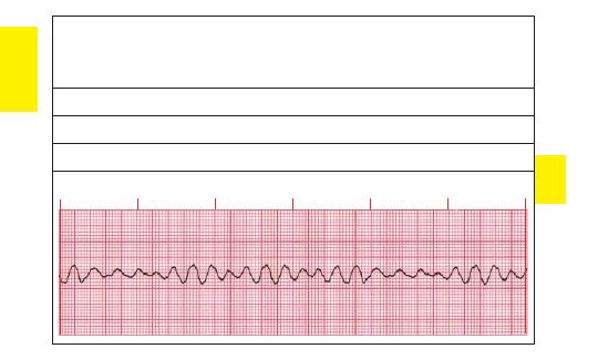
132
Case Study One: A 66-year-old woman with a history of heart disease is found unresponsive.
This is an unwitnessed cardiac arrest with the initial rhythm shown in ECG strip 4. CPR is
initiated while the defibrillator is charged. Strip 5 shows the rhythm following defibrillation.
Because the first defibrillation was unsuccessful, the machine is charged a second time. The
next rhythm is shown in strip 6.
ECG Strip 4 Interpretation:
ECG Strip 5 Interpretation:
ECG Strip 6 Interpretation:
ECG Test Strip 4
TEST
STRIPS
07ECG-Tab 07 2/4/05 4:02 PM Page 132
Copyright
©
2005
F.
A.
Davis.
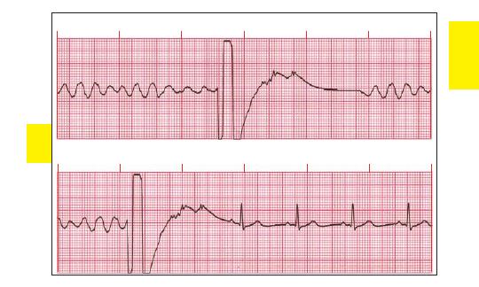
133
ECG Test Strip 5
ECG Test Strip 6
TEST
STRIPS
07ECG-Tab 07 2/4/05 4:02 PM Page 133
Copyright
©
2005
F.
A.
Davis.
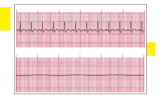
134
ECG Test Strip 7
ECG Test Strip 8
TEST
STRIPS
07ECG-Tab 07 2/4/05 4:02 PM Page 134
Copyright
©
2005
F.
A.
Davis.
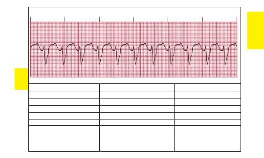
ECG Test Strip 9
ECG Strip 7
ECG Strip 8
ECG Strip 9
Rate:
Rate:
Rate:
Rhythm:
Rhythm:
Rhythm:
P Waves:
P Waves:
P Waves:
PR Interval:
PR Interval:
PR Interval:
QRS:
QRS:
QRS:
Interpretation:
Interpretation:
Interpretation:
135
TEST
STRIPS
07ECG-Tab 07 2/4/05 4:02 PM Page 135
Copyright
©
2005
F.
A.
Davis.
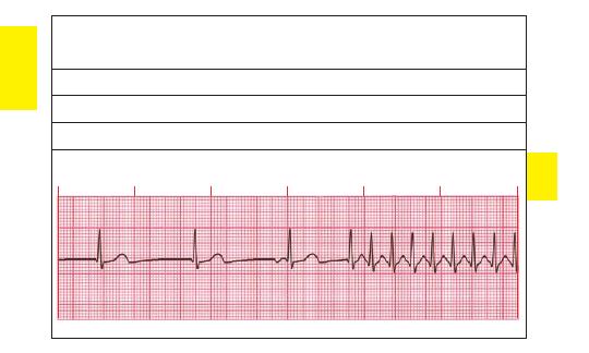
136
Case Study Two: A 72-year-old man is complaining of dizziness and anxiety. Strip 10 shows his
initial rhythm. An IV is started and the patient is given oxygen, but his vital signs become
unstable (strip 11). An IVP of adenosine is given and his condition stabilizes with the final
rhythm, shown in strip 12.
ECG Strip 10 Interpretation:
ECG Strip 11 Interpretation:
ECG Strip 12 Interpretation:
ECG Test Strip 10
TEST
STRIPS
07ECG-Tab 07 2/4/05 4:02 PM Page 136
Copyright
©
2005
F.
A.
Davis.
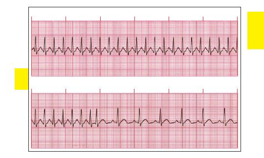
137
ECG Test Strip 11
ECG Test Strip 12
TEST
STRIPS
07ECG-Tab 07 2/4/05 4:02 PM Page 137
Copyright
©
2005
F.
A.
Davis.
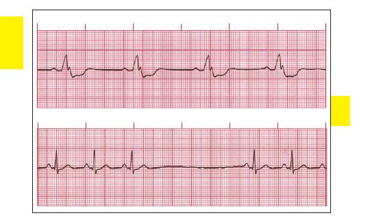
138
ECG Test Strip 13
ECG Test Strip 14
TEST
STRIPS
07ECG-Tab 07 2/4/05 4:02 PM Page 138
Copyright
©
2005
F.
A.
Davis.
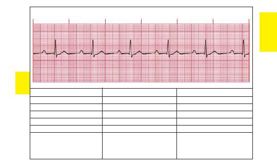
139
TEST
STRIPS
ECG Test Strip 15
ECG Strip 13
ECG Strip 14
ECG Strip 15
Rate:
Rate:
Rate:
Rhythm:
Rhythm:
Rhythm:
P Waves:
P Waves:
P Waves:
PR Interval:
PR Interval:
PR Interval:
QRS:
QRS:
QRS:
Interpretation:
Interpretation:
Interpretation:
07ECG-Tab 07 2/4/05 4:02 PM Page 139
Copyright
©
2005
F.
A.
Davis.
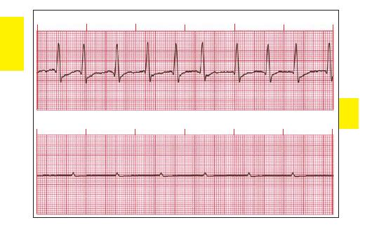
140
ECG Test Strip 16
ECG Test Strip 17
TEST
STRIPS
07ECG-Tab 07 2/4/05 4:02 PM Page 140
Copyright
©
2005
F.
A.
Davis.
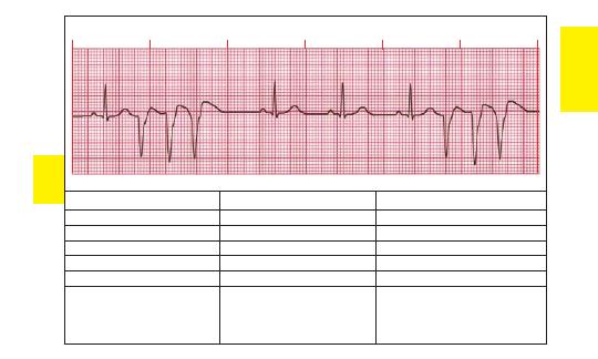
141
TEST
STRIPS
ECG Test Strip 18
ECG Strip 16
ECG Strip 17
ECG Strip 18
Rate:
Rate:
Rate:
Rhythm: Rhythm:
Rhythm:
P Waves:
P Waves:
P Waves:
PR Interval:
PR Interval:
PR Interval:
QRS:
QRS:
QRS:
Interpretation:
Interpretation:
Interpretation:
07ECG-Tab 07 2/4/05 4:02 PM Page 141
Copyright
©
2005
F.
A.
Davis.
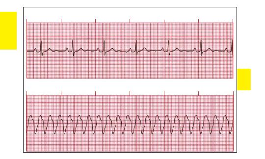
142
ECG Test Strip 19
ECG Test Strip 20
TEST
STRIPS
07ECG-Tab 07 2/4/05 4:02 PM Page 142
Copyright
©
2005
F.
A.
Davis.
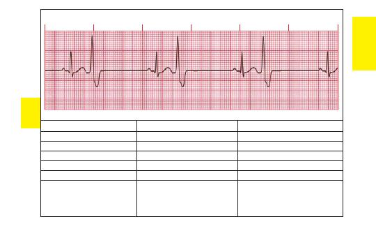
143
TEST
STRIPS
ECG Test Strip 21
ECG Strip 19
ECG Strip 20
ECG Strip 21
Rate:
Rate:
Rate:
Rhythm:
Rhythm:
Rhythm:
P Waves:
P Waves:
P Waves:
PR Interval:
PR Interval:
PR Interval:
QRS:
QRS:
QRS:
Interpretation:
Interpretation:
Interpretation:
07ECG-Tab 07 2/4/05 4:02 PM Page 143
Copyright
©
2005
F.
A.
Davis.
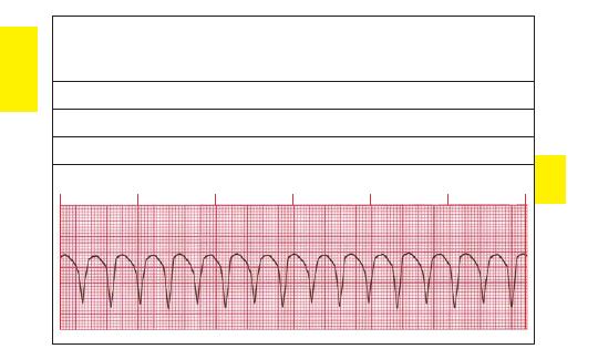
144
Case Study Three:
A 44-year-old man complains of severe chest pain. He has diaphoresis, a
BP of 80/60, and 24 respirations per min. The initial rhythm, recorded by the paramedics, is
shown in strip 22. An IV is started and the patient is given oxygen. Because his condition is
unstable, he receives sedation and cardioversion (strip 23). There is no change, and
cardioversion is performed a second time (strip 24).
ECG Strip 22 Interpretation:
ECG Strip 23 Interpretation:
ECG Strip 24 Interpretation:
ECG Test Strip 22
TEST
STRIPS
07ECG-Tab 07 2/4/05 4:02 PM Page 144
Copyright
©
2005
F.
A.
Davis.
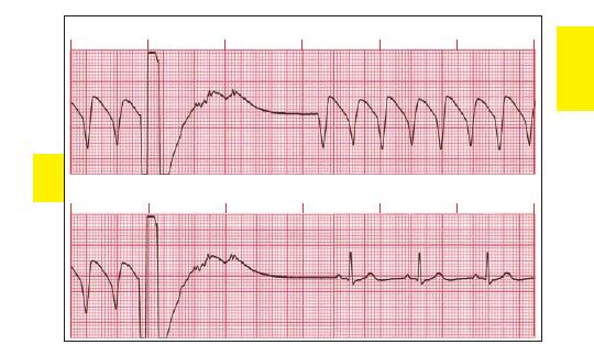
145
ECG Test Strip 23
ECG Test Strip 24
TEST
STRIPS
07ECG-Tab 07 2/4/05 4:02 PM Page 145
Copyright
©
2005
F.
A.
Davis.
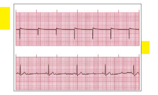
146
ECG Test Strip 25
ECG Test Strip 26
TEST
STRIPS
07ECG-Tab 07 2/4/05 4:02 PM Page 146
Copyright
©
2005
F.
A.
Davis.
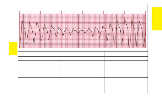
147
TEST
STRIPS
ECG Test Strip 27
ECG Strip 25
ECG Strip 26
ECG Strip 27
Rate:
Rate:
Rate:
Rhythm:
Rhythm:
Rhythm:
P Waves:
P Waves:
P Waves:
PR Interval:
PR Interval:
PR Interval:
QRS:
QRS:
QRS:
Interpretation:
Interpretation:
Interpretation:
07ECG-Tab 07 2/4/05 4:02 PM Page 147
Copyright
©
2005
F.
A.
Davis.
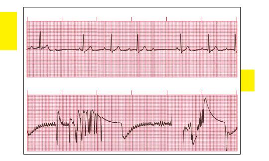
148
ECG Test Strip 28
ECG Test Strip 29
TEST
STRIPS
07ECG-Tab 07 2/4/05 4:02 PM Page 148
Copyright
©
2005
F.
A.
Davis.
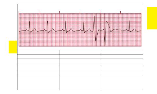
149
TEST
STRIPS
ECG Test Strip 30
ECG Strip 28
ECG Strip 29
ECG Strip 30
Rate:
Rate:
Rate:
Rhythm:
Rhythm:
Rhythm:
P Waves:
P Waves:
P Waves:
PR Interval:
PR Interval:
PR Interval:
QRS:
QRS:
QRS:
Interpretation:
Interpretation:
Interpretation:
07ECG-Tab 07 2/4/05 4:02 PM Page 149
Copyright
©
2005
F.
A.
Davis.
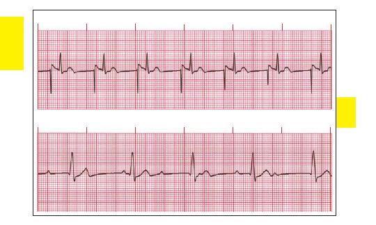
150
ECG Test Strip 31
ECG Test Strip 32
TEST
STRIPS
07ECG-Tab 07 2/4/05 4:02 PM Page 150
Copyright
©
2005
F.
A.
Davis.
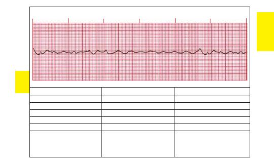
151
ECG Test Strip 33
ECG Strip 31
ECG Strip 32
ECG Strip 33
Rate:
Rate:
Rate:
Rhythm:
Rhythm:
Rhythm:
P Waves:
P Waves:
P Waves:
PR Interval:
PR Interval:
PR Interval:
QRS:
QRS:
QRS:
Interpretation:
Interpretation:
Interpretation:
TEST
STRIPS
07ECG-Tab 07 2/4/05 4:02 PM Page 151
Copyright
©
2005
F.
A.
Davis.
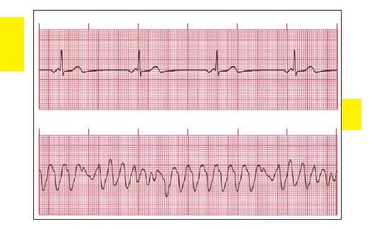
152
ECG Test Strip 34
ECG Test Strip 35
TEST
STRIPS
07ECG-Tab 07 2/4/05 4:02 PM Page 152
Copyright
©
2005
F.
A.
Davis.

153
TEST
STRIPS
ECG Test Strip 36
ECG Strip 34
ECG Strip 35
ECG Strip 36
Rate:
Rate:
Rate:
Rhythm:
Rhythm:
Rhythm:
P Waves:
P Waves:
P Waves:
PR Interval:
PR Interval:
PR Interval:
QRS:
QRS:
QRS:
Interpretation:
Interpretation:
Interpretation:
07ECG-Tab 07 2/4/05 4:02 PM Page 153
Copyright
©
2005
F.
A.
Davis.
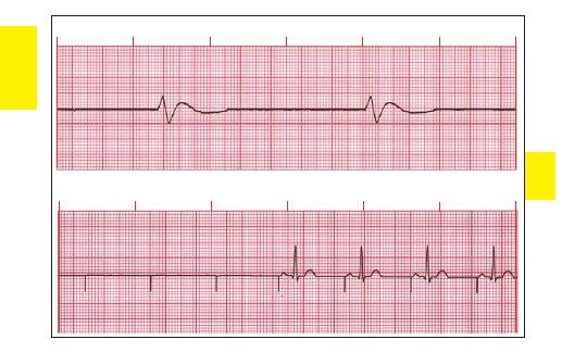
154
ECG Test Strip 37
ECG Test Strip 38
TEST
STRIPS
07ECG-Tab 07 2/4/05 4:02 PM Page 154
Copyright
©
2005
F.
A.
Davis.
155
TEST
STRIPS
ECG Test Strip 39
ECG Strip 37 Interpretation:
ECG Strip 38 Interpretation:
ECG Strip 39 Interpretation:
07ECG-Tab 07 2/4/05 4:02 PM Page 155
Copyright
©
2005
F.
A.
Davis.
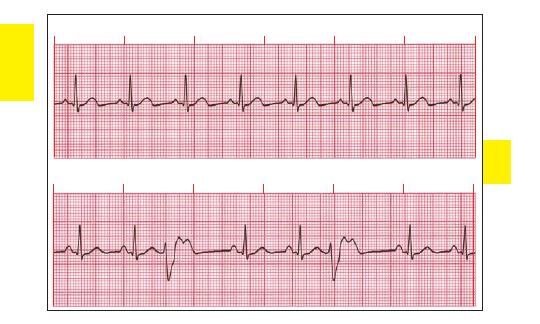
156
ECG Test Strip 40
ECG Test Strip 41
TEST
STRIPS
07ECG-Tab 07 2/4/05 4:02 PM Page 156
Copyright
©
2005
F.
A.
Davis.

157
ECG Test Strip 42
ECG Strip 40
ECG Strip 41
ECG Strip 42
Rate:
Rate:
Rate:
Rhythm: Rhythm:
Rhythm:
P Waves:
P Waves:
P Waves:
PR Interval:
PR Interval:
PR Interval:
QRS:
QRS:
QRS:
Interpretation:
Interpretation:
Interpretation:
TEST
STRIPS
07ECG-Tab 07 2/4/05 4:02 PM Page 157
Copyright
©
2005
F.
A.
Davis.
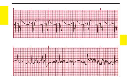
158
ECG Test Strip 43
ECG Test Strip 44
TEST
STRIPS
07ECG-Tab 07 2/4/05 4:02 PM Page 158
Copyright
©
2005
F.
A.
Davis.
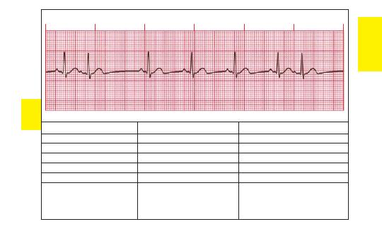
159
ECG Test Strip 45
ECG Strip 43
ECG Strip 44
ECG Strip 45
Rate:
Rate:
Rate:
Rhythm: Rhythm:
Rhythm:
P Waves:
P Waves:
P Waves:
PR Interval:
PR Interval:
PR Interval:
QRS:
QRS:
QRS:
Interpretation:
Interpretation:
Interpretation:
TEST
STRIPS
07ECG-Tab 07 2/4/05 4:02 PM Page 159
Copyright
©
2005
F.
A.
Davis.
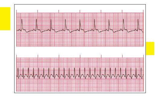
160
ECG Test Strip 46
ECG Test Strip 47
TEST
STRIPS
07ECG-Tab 07 2/4/05 4:02 PM Page 160
Copyright
©
2005
F.
A.
Davis.

161
ECG Test Strip 48
ECG Strip 46
ECG Strip 47
ECG Strip 48
Rate:
Rate:
Rate:
Rhythm: Rhythm:
Rhythm:
P Waves:
P Waves:
P Waves:
PR Interval:
PR Interval:
PR Interval:
QRS:
QRS:
QRS:
Interpretation:
Interpretation:
Interpretation:
TEST
STRIPS
07ECG-Tab 07 2/4/05 4:02 PM Page 161
Copyright
©
2005
F.
A.
Davis.
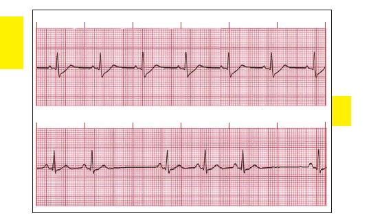
162
ECG Test Strip 49
ECG Test Strip 50
TEST
STRIPS
07ECG-Tab 07 2/4/05 4:02 PM Page 162
Copyright
©
2005
F.
A.
Davis.

163
TEST
STRIPS
ECG Test Strip 51
ECG Strip 49
ECG Strip 50
ECG Strip 51
Rate:
Rate:
Rate:
Rhythm: Rhythm:
Rhythm:
P Waves:
P Waves:
P Waves:
PR Interval:
PR Interval:
PR Interval:
QRS:
QRS:
QRS:
Interpretation:
Interpretation:
Interpretation:
07ECG-Tab 07 2/4/05 4:02 PM Page 163
Copyright
©
2005
F.
A.
Davis.
164
TEST
STRIPS
Rate: 34 bpm
Rhythm: Regular
P Waves: None
PR Interval: None
QRS: 0.20 sec
Interpretation:
Idioventricular
rhythm
Rate: Ventricular 150 bpm,
atrial 280 bpm
Rhythm: Regular
P Waves: Flutter waves
PR Interval: Variable
QRS: 0.08 sec
Interpretation: Atrial flutter
with 2:1 conduction
Answers to ECG Test Strips
ECG Strip 1
ECG Strip 2
ECG Strip 3
Rate: 35 bpm
Rhythm: Regular
P Waves: Normal
PR Interval: 0.16 sec
QRS: 0.10 sec
Interpretation: Sinus
bradycardia
ECG Strip 4 Interpretation: Ventricular fibrillation
ECG Strip 5 Interpretation: VF with defibrillation converting back to same rhythm
ECG Strip 6 Interpretation: VF with defibrillation converting to sinus rhythm at 68 bpm
Notes:
07ECG-Tab 07 2/4/05 4:02 PM Page 164
Copyright
©
2005
F.
A.
Davis.

165
ECG Strip 7
ECG Strip 8
ECG Strip 9
Rate: 115 bpm
Rhythm: Regular
P Waves: Normal
PR Interval: 0.12 sec
QRS: 0.10 sec
Interpretation: Sinus
tachycardia
ECG Strip 10 Interpretation: Paroxysmal supraventricular tachycardia—initial junctional rhythm
at 48 bpm converting to supraventricular tachycardia at 250 bpm
ECG Strip 11 Interpretation: SVT at 250 bpm
ECG Strip 12 Interpretation: SVT at 250 bpm converting to a sinus rhythm at 100 bpm
Notes:
TEST
STRIPS
Rate: None
Rhythm: None
P Waves: None
PR Interval: None
QRS: None
Interpretation:
Asystole
Rate: 115 bpm
Rhythm: Regular
P Waves: None
PR Interval: None
QRS: Wide (
0.12 sec), bizarre
Interpretation: Ventricular
tachycardia—monomorphic
07ECG-Tab 07 2/4/05 4:02 PM Page 165
Copyright
©
2005
F.
A.
Davis.
ECG Strip 13
ECG Strip 14
ECG Strip 15
Rate: 41 bpm
Rhythm: Regular
P Waves: Normal
PR Interval: 0.20 sec
QRS: 0.24 sec
Interpretation: Sinus brady-
cardia with a bundle branch
block
ECG Strip 16
ECG Strip 17
ECG Strip 18
Rate: Atrial
350 bpm,
ventricular 88–115 bpm
Rhythm: Irregular
P Waves: None
PR Interval: None
QRS: 0.12 sec
Interpretation: Atrial
fibrillation
166
TEST
STRIPS
Rate: Basic rate 79 bpm
Rhythm: Irregular
P Waves: Normal
PR Interval: 0.16 sec
QRS: 0.08 sec
Interpretation: Sinus rhythm
with sinus pause/arrest
Rate: 58 bpm
Rhythm: Regular
P Waves: Normal
PR Interval: 0.32 sec
QRS: 0.08 sec
Interpretation: Sinus
bradycardia with
1st-degree AV block
Rate: Atrial 60 bpm
Rhythm: Atrial regular
P Waves: Normal
PR Interval: None
QRS: None
Interpretation: P Wave
asystole
Rate: Basic rate 68 bpm
Rhythm: Irregular
P Waves: Normal
PR Interval: 0.16 sec
QRS: 0.08 sec
Interpretation: Sinus rhythm
with premature ventricular
contractions—triplets
07ECG-Tab 07 2/4/05 4:02 PM Page 166
Copyright
©
2005
F.
A.
Davis.
167
ECG Strip 19
ECG Strip 20
ECG Strip 21
Rate: 65 bpm
Rhythm: Regular
P Waves: Normal
PR Interval: 0.20 sec
QRS: 0.08 sec
Interpretation: Normal
sinus rhythm with U wave
ECG Strip 22 Interpretation: VT—monomorphic
ECG Strip 23 Interpretation: VT—monomorphic with cardioversion converting to same rhythm
ECG Strip 24 Interpretation: VT—monomorphic with cardioversion converting to a sinus rhythm
at 65 bpm
Notes:
TEST
STRIPS
Rate: 214 bpm
Rhythm: Regular
P Waves: None
PR Interval: None
QRS: Wide (
0.12 sec),
bizarre
Interpretation: VT—
monomorphic
Rate: Basic rate 35 bpm
Rhythm: Regular
P Waves: Normal
PR Interval: 0.16 sec
QRS: 0.08 sec
Interpretation: Sinus
bradycardia with ventricular
bigeminy
07ECG-Tab 07 2/4/05 4:02 PM Page 167
Copyright
©
2005
F.
A.
Davis.
168
ECG Strip 25
ECG Strip 26
ECG Strip 27
Rate: Pacing spikes 68 bpm
Rhythm: Regular pacing
spikes
P Waves: None
PR Interval: None
QRS: None
Interpretation: Pacemaker—
100% failure to capture,
underlying rhythm asystole
ECG Strip 28
ECG Strip 29
ECG Strip 30
Rate: 50–75 bpm
Rhythm: Irregular
P Waves: Normal
PR Interval: 0.12–0.28 sec
QRS: 0.08 sec
Interpretation: 2nd-degree
AV block Type I
TEST
STRIPS
Rate: Atrial 125 bpm,
ventricular 44 bpm
Rhythm: Regular
P Waves: Normal
PR Interval: 0.16 sec
QRS: 0.10 sec
Interpretation: 2nd-degree AV
block Type II with 3:1
conduction
Rate: 200–250 bpm
Rhythm: Irregular
P Waves: None
PR Interval: None
QRS: Wide (
0.12 sec), bizarre
Interpretation: VT—torsade de
pointes
Rate: None
Rhythm: None
P Waves: None
PR Interval: None
QRS: None
Interpretation: Loose
electrodes
Rate: Basic rate 68 bpm
Rhythm: Irregular
P Waves: Normal
PR Interval: 0.16 sec
QRS: 0.10 sec
Interpretation: Sinus rhythm
with multiform PVCs—
couplets
07ECG-Tab 07 2/4/05 4:02 PM Page 168
Copyright
©
2005
F.
A.
Davis.
169
TEST
STRIPS
ECG Strip 31
ECG Strip 32
ECG Strip 33
Rate: 68 bpm
Rhythm: Regular
P Waves: Upright with pacing
spikes
PR Interval: 0.16 sec
QRS: 0.10 sec
Interpretation: Atrial
pacemaker with 100%
capture
ECG Strip 34
ECG Strip 35
ECG Strip 36
Rate: 48 bpm
Rhythm: Regular
P Waves: Inverted
PR Interval: 0.12 sec
QRS: 0.08 sec
Interpretation: Junctional
rhythm
Rate: Atrial 75 bpm,
ventricular 48 bpm
Rhythm: Regular
P Waves: Normal,
superimposed on QRS and
T waves
PR Interval: Varies
QRS: 0.16 sec
Interpretation: 3rd-degree AV
block
Rate: 250 bpm
Rhythm: Irregular
P Waves: None
PR Interval: None
QRS: Wide (
0.12 sec),
bizarre
Interpretation: VT—
polymorphic
Rate: Indeterminate
Rhythm: Irregular
P Waves: None
PR Interval: None
QRS: None
Interpretation: VF
Rate: Atrial
≥350 bpm,
ventricular 94–167 bpm
Rhythm: Irregular
P Waves: None
PR Interval: None
QRS: 0.10 sec
Interpretation: A-fib
07ECG-Tab 07 2/4/05 4:02 PM Page 169
Copyright
©
2005
F.
A.
Davis.
170
ECG Strip 37 Interpretation: Agonal rhythm at 22 bpm
ECG Strip 38 Interpretation: Pacemaker failure to capture. When the pacemaker voltage is
increased, there is capture at pacemaker spike 4.
ECG Strip 39 Interpretation: Junctional bradycardia at 38 bpm converting to sinus bradycardia
at 38 bpm
Notes:
ECG Strip 40
ECG Strip 41
ECG Strip 42
Rate: 75 bpm
Rhythm: Regular
P Waves: Normal
PR Interval: 0.16 sec
QRS: 0.08 sec
Interpretation: Normal
sinus rhythm
TEST
STRIPS
Rate: Basic rate 79 bpm
Rhythm: Irregular
P Waves: Normal
PR Interval: 0.20 sec
QRS: 0.10 sec
Interpretation: Sinus
rhythm with ventricular
trigeminy
Rate: Basic rate 68 bpm
Rhythm: Irregular
P Waves: Normal; none
associated with premature
junctional contraction
PR Interval: 0.16 sec
QRS: 0.10 sec
Interpretation: Sinus rhythm
with PJCs at beats 4 and 6
07ECG-Tab 07 2/4/05 4:02 PM Page 170
Copyright
©
2005
F.
A.
Davis.
171
ECG Strip 43
ECG Strip 44
ECG Strip 45
Rate: 75 bpm
Rhythm: Regular
P Waves: Upright with
pacing spike
PR Interval: 0.20 sec
QRS: 0.16 sec
Interpretation: Atrial-
ventricular pacemaker
ECG Strip 46
ECG Strip 47
ECG Strip 48
Rate: 88 bpm
Rhythm: Regular
P Waves: Normal
PR Interval: 0.12 sec
QRS: 0.12 sec
Interpretation: Sinus
rhythm with ST segment
elevation
TEST
STRIPS
Rate: 75 bpm
Rhythm: Regular
P Waves: Not visible
PR Interval: Not measurable
QRS: Not measurable
Interpretation: Sinus
rhythm with muscle
artifact
Rate: 68 bpm
Rhythm: Irregular
P Waves: Normal
PR Interval: 0.16 sec
QRS: 0.10 sec
Interpretation: Sinus rhythm
with two premature atrial
contractions (beats 2 and 7)
Rate: 250 bpm
Rhythm: Regular
P Waves: Buried in T waves
PR Interval: Not measurable
QRS: 0.08 sec
Interpretation: SVT
Rate: 136 bpm
Rhythm: Regular
P Waves: Not visible
PR Interval: Not measurable
QRS: 0.10 sec
Interpretation: Sinus
tachycardia with muscle
artifact
07ECG-Tab 07 2/4/05 4:02 PM Page 171
Copyright
©
2005
F.
A.
Davis.
172
ECG Strip 49
ECG Strip 50
ECG Strip 51
Rate: 71 bpm
Rhythm: Regular
P Waves: Normal
PR Interval: 0.16 sec
QRS: 0.10 sec
Interpretation: Sinus rhythm
with ST segment depression
Notes:
TEST
STRIPS
Rate: Basic rate 79 bpm
Rhythm: Irregular
P Waves: Normal
PR Interval: 0.16 sec
QRS: 0.10 sec
Interpretation: Sinus rhythm
with two SA blocks
Rate: 107 bpm
Rhythm: Regular
P Waves: Notched (P prime)
PR Interval: 0.20 sec
QRS: 0.12 sec
Interpretation: Sinus
tachycardia with P
′ wave
07ECG-Tab 07 2/4/05 4:02 PM Page 172
Copyright
©
2005
F.
A.
Davis.

173
Notes:
TEST
STRIPS
07ECG-Tab 07 2/4/05 4:02 PM Page 173
Copyright
©
2005
F.
A.
Davis.
174
TOOLS
Troubleshooting ECG Problems
■
Place leads in the correct position. Incorrect placement can give
false readings.
■
Avoid placing leads over bony areas.
■
In patients with large breasts, place the electrodes under the
breast. Accurate tracings are obtained through the least amount
of fat tissue.
■
Apply tincture of benzoin to the electrode sites if the patient is
diaphoretic. The electrodes will adhere to the skin better.
■
Shave hair at the electrode site if it interferes with contact
between the electrode and skin.
■
Discard old electrodes and use new ones if the gel on the back of
the electrode dries.
Cable Connections
It is important to know if you are using an American or European cable
for ECG monitoring. The colors of the wires differ as shown below.
Monitoring Cable Connections
U.S.A.
Connect to
Europe
White
Right arm
Red
Black
Left arm
Yellow
Red
Left leg
Green
Green
Right leg
Black
Brown
Chest
White
Patient Cable
Monitoring cables contain varying numbers of wires.
3- and 4-wire cables: Allow a choice of limb and augmented leads.
5-wire cable: Allows a choice of limb and augmented leads plus a
chest lead.
10-wire cable: Records a 12-lead ECG.
08ECG-Tab 08 2/4/05 4:03 PM Page 174
Copyright © 2005 F. A. Davis.
175
TOOLS
Patient ECG Record
Patient Name: _______________________________________________
Sex
F
M
Heart rate: ________ bpm
■
Normal (60–100 bpm)
Y
N
■
Bradycardia (
60 bpm)
Y
N
■
Tachycardia (
100 bpm)
Y
N
Rhythm
■
Regular
Y
N
■
Irregular
Y
N
■
P waves
Y
N
P Waves (form)
■
Normal (upright and uniform)
Y
N
■
Inverted
Y
N
P wave associated with QRS
Y
N
PR interval normal (0.12–0.20 sec)
Y
N
P waves and QRS complexes associated with one another
Y N
QRS Interval
■
Normal (0.6–0.10 sec)
Y
N
■
Wide (
0.10 sec)
Y
N
Are the QRS complexes grouped or not grouped?
Are there any dropped beats?
Is there a compensatory or noncompensatory pause?
QT interval:
Interpretation:
08ECG-Tab 08 2/4/05 4:03 PM Page 175
Copyright © 2005 F. A. Davis.
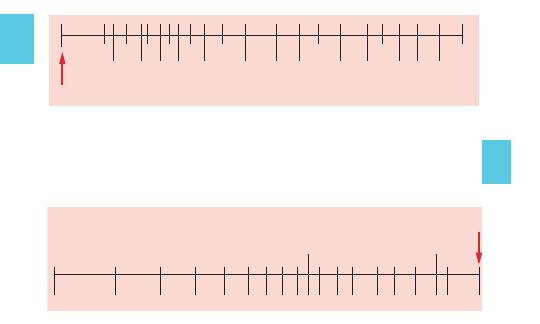
176
TOOLS
HEART RATE 1 Cycle from reference arrow (25 mm/s)
150 100
75 60
50
40
25
20
17
18
19
21
23
27
30
35
45
55
65
80
125
16
HEART RATE 2 Cycles from reference arrow (25 mm/s)
400
200150
125100
90
80
70
75
300
60
65
55
50
45
40
35
30
08ECG-Tab 08 2/4/05 4:03 PM Page 176
Copyright
©
2005
F.
A.
Davis.
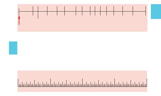
177
TOOLS
HEART RATE 3 Cycles from reference arrow (25 mm/s)
50
45
55
60
65
70
75
80
90
100
125
150
200
300
400
Inches
4
3
2
1
08ECG-Tab 08 2/4/05 4:03 PM Page 177
Copyright
©
2005
F.
A.
Davis.

178
Abbreviations
ACE
angiotensin-converting enzyme
AED
automatic external defibrillator
A-fib
atrial fibrillation
A-flutter
atrial flutter
ARDS
acute respiratory distress syndrome
AV
atrioventricular
BBB
bundle branch block
BP
blood pressure
bpm
beats per min
BUN
blood urea nitrogen
CAD
coronary artery disease
CHF
congestive heart failure
CO
cardiac output
COPD
chronic obstructive pulmonary disease
CPR
cardiopulmonary resuscitation
CVA
cerebrovascular accident
ECG
electrocardiogram
EMD
electromechanical dissociation
ET
endotracheal
FAB
fragment antigen binding
HR
heart rate
HTN
hypertension
IHSS
idiopathic hypertrophic subaortic stenosis
IM
intramuscular
IV
intravenous
IVP
intravenous push
LA
left arm
LL
left leg
LOC
level of consciousness
MAT
multifocal atrial tachycardia
MCL
modified chest lead
MI
myocardial infarction
NSR
normal sinus rhythm
PAC
premature atrial contraction
PAT
paroxysmal atrial tachycardia
PEA
pulseless electrical activity
TOOLS
08ECG-Tab 08 2/4/05 4:03 PM Page 178
Copyright © 2005 F. A. Davis.

179
PJC
premature junctional contraction
PO
by mouth
PSVT
paroxysmal supraventricular tachycardia
PVC
premature ventricular contraction
RA
right arm
RL
right leg
SA
sinoatrial
SC
subcutaneous
SV
stroke volume
SVT
supraventricular tachycardia
VF
ventricular fibrillation
VT
ventricular tachycardia
WAP
wandering atrial pacemaker
WPW
Wolff-Parkinson-White
TOOLS
08ECG-Tab 08 2/4/05 4:03 PM Page 179
Copyright © 2005 F. A. Davis.

180
Selected References
1. Cummins, RO (ed.): ACLS Provider Manual. American Heart
Association, Dallas, 2002.
2. Deglin, JH, Vallerand, AH: Davis’s Drug Guide for Nurses, ed
8. FA Davis, Philadelphia, 2003.
3. Deglin, JH, Vallerand, AH: Med Notes. FA Davis, Philadelphia,
2004.
4. Myers, E: RNotes. FA Davis, Philadelphia, 2003.
5. Myers, E, Hopkins, T: MedSurg Notes. FA Davis, Philadelphia,
2004.
6. Physicians’ Desk Reference, ed 59. Thomson Healthcare,
Montvale, NJ, 2005.
7. Scanlon, VC, Sanders, T: Essentials of Anatomy and
Physiology, ed 4. FA Davis, Philadelphia, 2003.
8. Stapleton, ER, et al. (eds.): BLS for Healthcare Providers.
American Heart Association, Dallas, 2001.
9. Taber’s Cyclopedic Medical Dictionary, ed 19. FA Davis,
Philadelphia, 2001.
TOOLS
09ECG-Ref 2/4/05 4:03 PM Page 180
Copyright © 2005 F. A. Davis.

181
Illustration Credits
Pages 2, 3, 4, 5, 6, 7, 8, from: Scanlon, Essentials of Anatomy and
Physiology, ed 4. F.A. Davis, Philadelphia, 2003.
Pages 10, 11, 16, 19, 73, 100, 111, 115 from: Myers, MedSurg
Notes. F.A. Davis, Philadelphia, 2004.
Pages 28–64, 66–70, 130–163 from: Armstrong Medical
Industries, Inc. Lincolnshire, IL.
TOOLS
10ECG-Cre 2/4/05 4:04 PM Page 181
Copyright © 2005 F. A. Davis.

182
TOOLS
A
Accelerated idioventricular
rhythm, 49, 49f
Accelerated junctional rhythm,
44, 44f
ACE (angiotensin-converting
enzyme) inhibitors, as emer-
gency cardiac medications,
86
Acute MI. See Infarction,
myocardial.
Adenosine, as emergency
cardiac medication, 86–87
Adult, cardiopulmonary resus-
citation of, 106t, 107
conscious, emergency
management of obstructed
airway in, 110
Heimlich maneuver in, 110f
unconscious, emergency
management of obstructed
airway in, 112
AED (automated external
defibrillator), 101
A-fib (atrial fibrillation), 41, 41f,
140f, 153f, 166, 169
management of, 127
in Wolff-Parkinson-White
syndrome, 128
A-flutter (atrial flutter), 40, 40f,
131f, 164
management of, 127
in Wolff-Parkinson-White
syndrome, 128
Agonal rhythm, 154f, 170
Airway obstruction, emergency
management of, 110–114,
115f
in conscious adult, 110
in conscious child, 110
in conscious infant, 111
in unconscious adult,
112
in unconscious child, 113
in unconscious infant, 114
via Heimlich maneuver,
110f, 111f
universal sign of, 115f
Amiodarone, as emergency
cardiac medication, 87
Angiotensin-converting
enzyme (ACE) inhibitors, as
emergency cardiac medica-
tions, 86
Anterior myocardial infarction,
79, 79f
Arrest, sinus, 32, 32f, 138f, 166
Arrhythmia(s), 28–59, 29f–59f
atrial, 34–42, 34f–42f
junctional, 43–47, 43f–47f
sinoatrial node, 28–33,
29f–33f
sinus, 31, 31f
ventricular, 48–59, 48f–59f
Arterial circulation, 7f
coronary, 5f
Artifacts, 69, 69f, 70, 70f
muscle, 70f, 158f, 161f, 171
Index
Note: Page numbers followed by f refer to illustrations and
pages followed by t refer to tables.
11ECG-Index 2/4/05 4:04 PM Page 182
Copyright © 2005 F. A. Davis.

183
TOOLS
Aspirin, as emergency cardiac
medication, 88
Asystole, 59, 59f, 134f, 140f,
165, 166
management of, 118
Atrial arrhythmias, 34–42,
34f–42f
Atrial contraction(s), prema-
ture, 36, 36f, 159f, 171
Atrial fibrillation (A-fib), 41,
41f, 140f, 153f, 166, 169
management of, 127
in Wolff-Parkinson-White
syndrome, 128
Atrial flutter (A-flutter), 40, 40f,
131f, 164
management of, 127
in Wolff-Parkinson-White
syndrome, 128
Atrial pacemaker, wandering,
34, 34f
Atrial tachycardia, 37, 37f
multifocal, 35, 35f
Atrioventricular block(s),
60–64, 60f–64f
first-degree, 60, 60f, 139f,
166
Mobitz I, 61, 61f, 148f, 168
Mobitz II, 62, 62f, 146f, 168
second-degree, 61, 61f, 62,
62f, 146f, 148f, 168
third-degree, 63, 63f, 150f,
169
Wenckebach, 61, 61f
Atrioventricular node, 10
Atropine, as emergency
cardiac medication, 88
Augmented limb leads, in elec-
trocardiography, 15, 15f
Automated external defibrilla-
tor (AED), 101
Automaticity, of cardiac cells,
9
AV. See Atrioventricular
entries.
B
Bag-valve mask, in airway
management, 115f
BBB (bundle branch block), 64,
64f, 138f, 166
left, 84, 84f
right, 85, 85f
Beta blockers, as emergency
cardiac medications, 88–89
Bicarbonate, as emergency
cardiac medication, 98
Bigeminy, ventricular, 52, 52f,
143f, 167
Block(s), atrioventricular,
60–64, 60f–64f
first-degree, 60, 60f, 139f,
166
Mobitz I, 61, 61f, 148f, 168
Mobitz II, 62, 62f, 146f, 168
second-degree, 61, 61f, 62,
62f, 146f, 148f, 168
third-degree, 63, 63f, 150f,
169
Wenckebach, 61, 61f
bundle branch, 64, 64f, 138f,
166
left, 84, 84f
right, 85, 85f
sinoatrial, 33, 33f, 162f, 172
Blood vessel structures, 6, 6f
Bradycardia, 120
junctional, 155f, 170
11ECG-Index 2/4/05 4:04 PM Page 183
Copyright © 2005 F. A. Davis.

184
TOOLS
Bradycardia (Continued)
management of, 120
sinus, 29, 29f, 130f, 138f,
139f, 143f, 164, 166, 167
junctional bradycardia
converting to, 155f, 170
with muscle artifact, 161f,
171
Bundle branch(es), 10
Bundle branch block (BBB), 64,
64f, 138f, 166
left, 84, 84f
right, 85, 85f
Bundle of His, 10
C
Cable connections, in electro-
cardiography, 17, 17f, 174
Calcium chloride, as emer-
gency cardiac medication,
89
Cardiac anatomy, 1, 1f–5f, 3
Cardiac electrophysiology,
9–11, 10f–11f, 74, 74f. See
also Electrocardiography
(ECG).
Cardiac ischemia, 76, 76f. See
also Infarction, myocardial.
chest pain due to, manage-
ment of, 119
Cardiopulmonary resuscitation
(CPR), 106t, 107–109, 115f
by one rescuer, 106t
by two rescuers, 106t
of adult, 106t, 107
of child, 106t, 108
of infant, 106t, 109
of newborn, 106t
Cardiovascular system, 6, 6f–8f
Cardioversion, 102
Carotid sinus massage,
104–105, 105f
Chambers of heart, 4f
Chest compressions, in CPR,
106t, 107, 108, 109
Chest leads, in electrocardiog-
raphy, 16, 16f, 18, 18f
Chest pain, ischemic, manage-
ment of, 119
Child, cardiopulmonary resus-
citation of, 106t, 108
conscious, emergency
management of
obstructed airway in, 110
Heimlich maneuver in, 110f
unconscious, emergency
management of
obstructed airway in, 113
Chin lift, in airway manage-
ment, 115f
Choking. See Airway obstruc-
tion.
Circulation, arterial, 7f
coronary, 5f
venous, 8f
Complex, 21
QRS, 22, 27
Complex width, tachycardia
management in relation to,
122, 123, 124–128
Conduction system, of heart,
10–11, 10f–11f. See also
Electrocardiography (ECG).
Conductivity, of cardiac cells, 9
Conscious patient, emergency
management of obstructed
airway in, 110, 111
Contractility, of cardiac cells, 9
11ECG-Index 2/4/05 4:04 PM Page 184
Copyright © 2005 F. A. Davis.

185
TOOLS
Contraction(s), atrial, prema-
ture, 36, 36f, 159f, 171
junctional, premature, 47,
47f, 157f, 170
ventricular, premature, 50,
50f
couplet (paired), 53, 53f,
149f, 168
every 2nd beat (bigemi-
nal), 52, 52f, 147f,
163
every 3rd beat (trigemi-
nal), 52, 52f, 156f, 170
every 4th beat
(quadrigeminal), 53,
53f
multiform, 51, 51f
paired (couplet), 53, 53f,
149f, 168
triplet, 141f, 166
uniform, 51, 51f
Coronary arterial circulation,
5f
Couplet PVCs (paired PVCs),
53, 53f, 149f, 168
CPR. See Cardiopulmonary
resuscitation (CPR).
D
Defibrillation, 99–101, 100f
automated external, 101
manual, 100
Depolarization, 11
Digoxin, as emergency cardiac
medication, 90
Digoxin toxicity, antidote for,
89
Diltiazem, as emergency
cardiac medication, 90
Dopamine, as emergency
cardiac medication,
91
Dropped beats, 26
E
ECG. See Electrocardiography
(ECG).
Electrical activity, pulseless,
58, 58f
management of, 117
mnemonics for causes of,
117
Electrical axis deviation, of
heart, 74, 74f
Electrical conduction system,
of heart, 10–11, 10f–11f. See
also Electrocardiography
(ECG).
Electrocardiography (ECG),
12–13, 21f
cable connections in, 17, 17f,
174
electrode placement in,
13–20, 13f–20f
loose, artifact due to, 69f,
148f, 168
fifteen-lead, 20, 20f
heart rate calculation via,
23–24, 23f, 25f, 176f,
177f
interpretation of results of,
22, 26–27
as abnormal, 28–64,
29f–64f. See also
Arrhythmia(s) and
specific patterns.
as artifactual. See
Artifacts.
11ECG-Index 2/4/05 4:04 PM Page 185
Copyright © 2005 F. A. Davis.

186
TOOLS
Electrocardiography (Cont’d)
as diagnostic yield in
suspected MI, 79–83,
79f–83f
as pacemaker rhythms, 66,
66f, 67f, 150f, 158f, 169,
171
on patient’s record, 175
leads used in, 13–20, 13f–20f
monitoring cables in, 174
troubleshooting in, 174
twelve-lead, 19, 19f, 72, 78,
78f
Emergency medical skills,
99–115
Emergency medications, 86–99
Endocardium, 2f
Epinephrine, as emergency
cardiac medication, 91–92
Escape beat, junctional, 46,
46f
Excitability, of cardiac cells, 9
External defibrillator, auto-
mated, 101
F
Fibrillation, atrial, 41, 41f, 140f,
153f, 166, 169
management of, 127
in Wolff-Parkinson-White
syndrome, 128
ventricular, 57, 57f, 132f,
133f, 151f, 164, 169
management of, 116
Fibrinolytic agents, as emer-
gency cardiac medications,
92
Fifteen-lead electrocardiogra-
phy, 20, 20f
First-degree atrioventricular
block, 60, 60f, 139f, 166
Flutter, atrial, 40, 40f, 131f,
164
management of, 127
in Wolff-Parkinson-White
syndrome, 128
Furosemide, as emergency
cardiac medication, 92–93
G
Great vessels of heart, 4f
H
H mnemonic, for causes of
pulseless electrical activity,
117
Head tilt, in airway manage-
ment, 115f
Heart, anatomy of, 1, 1f–5f, 3
cells of, properties of, 9
chambers of, 4f
coronary arteries of, 5f
electrical axis deviation of,
74, 74f
electrical conduction system
of, 10–11, 10f–11f. See also
Electrocardiography
(ECG).
great vessels of, 4f
ischemia of, 76, 76f. See also
Infarction, myocardial.
chest pain due to,
management of, 119
layers of, 2f
infarct sites in. See
Infarction, myocardial.
physiology of, 9
valves of, 3, 3f
11ECG-Index 2/4/05 4:04 PM Page 186
Copyright © 2005 F. A. Davis.

187
TOOLS
Heart rate, methods of calcu-
lating, 23–24, 23f, 25f, 176f,
177f
Heimlich maneuver, 110f, 111f
I
Ibutilide, as emergency cardiac
medication, 93
Idioventricular rhythm, 48, 48f,
130f, 164
accelerated, 49, 49f
Infant, cardiopulmonary resus-
citation of, 106t, 109
conscious, emergency
management of
obstructed airway in, 111
Heimlich maneuver in, 111f
unconscious, emergency
management of
obstructed airway in, 114
Infarction, myocardial, 75, 76,
79–83
anterior, 79, 79f
electrocardiographic
evidence for, 79–83,
79f–83f
inferior, 80, 80f
lateral, 81, 81f
posterior, 83, 83f
propagation of, 76, 76f
septal, 82, 82f
Inferior myocardial infarction,
80, 80f
Interval(s), 21
PR, 22, 26
QRS, 26
QT, 22, 26
Ischemia, cardiac, 76, 76f. See
also Infarction, myocardial.
chest pain due to, manage-
ment of, 119
Isoproterenol, as emergency
cardiac medication, 93–94
J
Jaw thrust, in airway manage-
ment, 115f
Junctional arrhythmias, 43–47,
43f–47f
Junctional bradycardia, 155f,
170
Junctional contraction(s),
premature, 47, 47f, 157f, 170
Junctional escape beat, 46, 46f
Junctional rhythm, 43, 43f
accelerated, 44, 44f
Junctional tachycardia, 45, 45f
management of, 125
L
Lateral myocardial infarction,
81, 81f
Left bundle branch, 10
Left bundle branch block, 84,
84f
Lidocaine, as emergency
cardiac medication, 94
Limb leads, in electrocardiog-
raphy, 13–15, 13f–15f
Loose electrode(s), artifact due
to, 69f, 148f, 168
M
Magnesium sulfate, as emer-
gency cardiac medication, 95
Manual defibrillation, 100
Massage, carotid sinus,
104–105, 105f
11ECG-Index 2/4/05 4:04 PM Page 187
Copyright © 2005 F. A. Davis.

188
TOOLS
MAT (multifocal atrial tachycar-
dia), 35, 35f
Medical skills, emergency,
99–115
Medications, emergency,
86–99
MI. See Myocardial infarction.
Mobitz I atrioventricular block,
61, 61f, 148f, 168
Mobitz II atrioventricular block,
62, 62f, 146f, 168
Monitoring cables, in electro-
cardiography, 174
Monomorphic ventricular
tachycardia, 54, 54f, 135f,
142f, 143f, 145f, 165, 167
management of, 122
Morphine, as emergency
cardiac medication, 95
Multifocal atrial tachycardia
(MAT), 35, 35f
Multiform premature ventricu-
lar contraction, 51, 51f
Muscle artifacts, 70f, 158f, 161f,
171
Myocardial infarction, 75, 76,
79–83
anterior, 79, 79f
electrocardiographic
evidence for, 79–83,
79f–83f
inferior, 80, 80f
lateral, 81, 81f
posterior, 83, 83f
propagation of, 76, 76f
septal, 82, 82f
Myocardium, 2f
acute infarction of. See
Myocardial infarction.
N
Narrow-complex tachycardia,
management of, 124–128
Newborn, cardiopulmonary
resuscitation of, 106t
Nitroglycerin, as emergency
cardiac medication, 96
O
Obstructed airway, emergency
management of, 110–114,
115f
in conscious adult, 110
in conscious child, 110
in conscious infant, 111
in unconscious adult, 112
in unconscious child, 113
in unconscious infant,
114
via Heimlich maneuver,
110f, 111f
universal sign of, 115f
One-rescuer cardiopulmonary
resuscitation, 106t
Oxygen, as emergency cardiac
medication, 96–97
P
P wave, 22, 26
PAC(s) (premature atrial
contraction[s]), 36, 36f, 159f,
171
Pacemaker, atrial, wandering,
34, 34f
Pacemaker device(s), 65
malfunction of, 68, 68f, 146f,
154f, 168, 170
Pacemaker rhythm, 66, 66f,
67f, 150f, 158f, 169, 171
11ECG-Index 2/4/05 4:04 PM Page 188
Copyright © 2005 F. A. Davis.

189
TOOLS
Pacing, transcutaneous, 102,
103f
Pain, ischemic, management
of, 119
Paired PVCs (couplet PVCs),
53, 53f, 149f, 168
Paroxysmal supraventricular
tachycardia (PSVT), 39, 39f,
136f, 165
management of, 124
Pause, sinus, 32, 32f, 138f, 166
PEA (pulseless electrical activ-
ity), 58, 58f
management of, 117
mnemonics for causes of,
117
Pediatric patient, cardiopul-
monary resuscitation of,
106t, 108, 109
conscious, emergency
management of
obstructed airway in, 110,
111
Heimlich maneuver in, 110f,
111f
unconscious, emergency
management of
obstructed airway in, 113,
114
Pericardium, 2f
PJC(s) (premature junctional
contraction[s]), 47, 47f, 157f,
170
Polymorphic ventricular tachy-
cardia, 55, 55f, 152f, 169
management of, 123
Posterior myocardial infarc-
tion, 83, 83f
PR interval, 22, 26
Precordial thump, 104
Premature atrial contraction(s)
(PAC[s]), 36, 36f, 159f, 171
Premature junctional contrac-
tion(s) (PJC[s]), 47, 47f, 157f,
170
Premature ventricular contrac-
tion(s) (PVC[s]), 50, 50f
couplet (paired), 53, 53f,
149f, 168
every 2nd beat (bigeminal),
52, 52f, 143f, 167
every 3rd beat (trigeminal),
52, 52f, 156f, 170
every 4th beat (quadrigemi-
nal), 53, 53f
multiform, 51, 51f
paired (couplet), 53, 53f,
149f, 168
triplet, 141f, 166
uniform, 51, 51f
Procainamide, as emergency
cardiac medication, 97
PSVT (paroxysmal supraven-
tricular tachycardia), 39, 39f,
136f, 165
management of, 124
Pulseless electrical activity
(PEA), 58, 58f
management of, 117
mnemonics for causes of,
117
Pulseless ventricular tachycar-
dia, management of, 116
Purkinje system, 10
PVC(s) (premature ventricular
contraction[s]), 50, 50f
couplet (paired), 53, 53f,
149f, 168
11ECG-Index 2/4/05 4:04 PM Page 189
Copyright © 2005 F. A. Davis.

190
TOOLS
PVC(s) (Continued)
every 2nd beat (bigeminal),
52, 52f, 143f, 157
every 3rd beat (trigeminal),
52, 52f, 156f, 170
every 4th beat (quadrigemi-
nal), 53, 53f
multiform, 51, 51f
paired (couplet), 53, 53f,
149f, 168
triplet, 141f, 166
uniform, 51, 51f
Q
Q wave, 22
QRS complex, 27
QRS interval, 22, 26
QT interval, 22, 26
Quadrigeminy, ventricular, 53,
53f
R
R wave, propagation of, 73, 73f
Rate component, in rhythm
evaluation, 26
Regularity component, in
rhythm evaluation, 26
Repolarization, 11
Resuscitation, cardiopul-
monary, 106t, 107–109, 115f
by one rescuer, 106t
by two rescuers, 106t
of adult, 106t, 107
of child, 106t, 108
of infant, 106t, 109
of newborn, 106t
Rhythm. See also
Arrhythmia(s).
agonal, 154f, 170
idioventricular, 48, 48f, 130f,
164
accelerated, 49, 49f
junctional, 43, 43f
accelerated, 44, 44f
pacemaker, 66, 66f, 67f, 158f,
159f, 169, 171
sinus, 28, 28f, 156f, 170
with muscle artifact, 158f,
171
with premature atrial
contractions, 159f,
171
with premature junctional
contractions, 157f,
170
with premature ventricular
contractions, 141f, 149f,
156f, 166, 168, 170
with sinoatrial block, 162f,
172
with sinus pause/arrest,
138f, 166
with ST segment depres-
sion, 162f, 172
with ST segment eleva-
tion, 160f, 171
with U wave, 142f, 167
Right bundle branch, 10
Right bundle branch block, 85,
85f
Right-sided twelve-lead elec-
trocardiography, 19, 19f
S
SA. See Sinoatrial entries.
Second-degree atrioventricular
block, 61, 61f, 62, 62f, 146f,
148f, 168
11ECG-Index 2/4/05 4:04 PM Page 190
Copyright © 2005 F. A. Davis.

191
TOOLS
Segment(s), 21
ST, 22
depression of, 77, 77f, 162f,
172
elevation of, 77, 77f, 160f,
171
Septal myocardial infarction,
82, 82f
Sinoatrial block, 33, 33f, 162f,
172
Sinoatrial node, 10
Sinoatrial node arrhythmias,
28–33, 29f–33f
Sinus arrest (sinus pause), 32,
32f, 138f, 166
Sinus arrhythmia, 31, 31f
Sinus bradycardia, 29, 29f,
130f, 138f, 139f, 143f, 164,
166, 167
junctional bradycardia
converting to, 155f, 170
with muscle artifact, 161f,
171
Sinus pause (sinus arrest), 32,
32f, 138f, 166
Sinus rhythm, 28, 28f, 156f,
170
with muscle artifact, 158f,
171
with premature atrial
contractions, 159f,
171
with premature junctional
contractions, 157f, 170
with premature ventricular
contractions, 141f, 149f,
156f, 166, 168, 170
with sinoatrial block, 162f,
172
with sinus pause/arrest, 138f,
166
with ST segment depression,
162f, 172
with ST segment elevation,
160f, 171
with U wave, 142f, 167
Sinus tachycardia, 30, 30f,
134f, 163f, 165, 172
Sodium bicarbonate, as emer-
gency cardiac medication,
98
ST segment, 22
depression of, 77, 77f, 162f,
172
elevation of, 77, 77f, 160f,
171
Stable narrow-complex tachy-
cardia, management of,
124–128
Stable wide-complex tachycar-
dia, management of, 122,
123
Supraventricular tachycardia
(SVT), 38, 38f, 137f, 160f,
165, 171
paroxysmal, 39, 39f, 136f,
165
management of, 124
T
T mnemonic, for causes of
pulseless electrical activity,
117
T wave, 22
Tachycardia, atrial, 37, 37f
multifocal, 35, 35f
junctional, 45, 45f
management of, 125
11ECG-Index 2/4/05 4:04 PM Page 191
Copyright © 2005 F. A. Davis.

192
TOOLS
Tachycardia (Continued)
monomorphic ventricular,
54, 54f, 135f, 142f, 143f,
145f, 165, 167
management of, 122
multifocal atrial, 35, 35f
narrow-complex, manage-
ment of, 124–128
paroxysmal supraventricular,
39, 39f, 136f, 165
management of, 124
polymorphic ventricular, 55,
55f
management of, 123
sinus, 30, 30f, 134f, 163f, 165,
172
stable, management of,
122–128
supraventricular, 38, 38f,
137f, 160f, 165, 171
paroxysmal, 39, 39f, 136f,
165
management of, 124
unstable, management of,
121
ventricular, 54, 54f, 55,
55f
monomorphic, 54, 54f,
135f, 142f, 143f, 145f,
165, 167
management of, 122
polymorphic, 55, 55f, 152f,
169
management of, 123
pulseless, management of,
116
wide-complex, management
of, 122, 123
Third-degree atrioventricular
block, 63, 63f, 150f, 169
Thump, precordial, 104
Torsade de pointes, 56, 56f,
147f, 168
Transcutaneous pacing, 102,
103f
Trigeminy, ventricular, 52, 52f,
156f, 170
Triplet PVCs, 141f, 166
Troubleshooting, in electrocar-
diography, 174
Twelve-lead electrocardiogra-
phy, 19, 19f, 72, 78, 78f
Two-rescuer cardiopulmonary
resuscitation, 106t
U
U wave, 22, 142f, 167
Unconscious patient, emer-
gency management of
obstructed airway in, 112,
113, 114
Uniform premature ventricular
contraction, 51, 51f
Universal sign, of airway
obstruction, 115f
Unstable tachycardia, manage-
ment of, 121
V
Vagal maneuver(s), 105
carotid sinus massage as,
104–105, 105f
Valves of heart, 3, 3f
Vasopressin, as emergency
cardiac medication, 98
Venous circulation, 8f
11ECG-Index 2/4/05 4:04 PM Page 192
Copyright © 2005 F. A. Davis.

193
TOOLS
Ventricular arrhythmias, 48–59,
48f–59f
Ventricular bigeminy, 52, 52f,
143f, 167
Ventricular contraction(s),
premature, 50, 50f
couplet (paired), 53, 53f,
149f, 168
every 2nd beat (bigeminal),
52, 52f, 143f, 167
every 3rd beat (trigeminal),
52, 52f, 156f, 170
every 4th beat (quadrigemi-
nal), 53, 53f
multiform, 51, 51f
paired (couplet), 53, 53f,
149f, 168
triplet, 141f, 166
uniform, 51, 51f
Ventricular fibrillation (VF), 57,
57f, 132f, 133f, 151f, 164, 169
management of, 116
Ventricular quadrigeminy, 53,
53f
Ventricular tachycardia (VT),
54, 54f, 55, 55f
monomorphic, 54, 54f, 135f,
142f, 143f, 145f, 165, 167
management of, 122
polymorphic, 55, 55f, 152f,
169
management of, 123
pulseless, management of,
116
Ventricular trigeminy, 52, 52f,
156f, 170
Verapamil, as emergency
cardiac medication, 98–99
VF (ventricular fibrillation), 57,
57f, 132f, 133f, 151f, 164, 169
management of, 116
VT (ventricular tachycardia),
54, 54f, 55, 55f
monomorphic, 54, 54f, 135f,
142f, 143f, 145f, 165, 167
management of, 122
polymorphic, 55, 55f, 152f,
169
management of, 123
pulseless, management of,
116
W
Wandering atrial pacemaker,
34, 34f
Wave(s), 21
P, 22, 26
Q, 22
R, propagation of, 73, 73f
T, 22
U, 22, 142f, 167
Wenckebach atrioventricular
block, 61, 61f
Wide-complex tachycardia,
management of, 122, 123
Wolff-Parkinson-White (WPW)
syndrome, 42, 42f
management of, 128
11ECG-Index 2/4/05 4:04 PM Page 193
Copyright © 2005 F. A. Davis.

194
TOOLS
Notes
11ECG-Index 2/4/05 4:04 PM Page 194
Copyright © 2005 F. A. Davis.

195
TOOLS
Notes
11ECG-Index 2/4/05 4:04 PM Page 195
Copyright © 2005 F. A. Davis.

196
TOOLS
Notes
11ECG-Index 2/4/05 4:04 PM Page 196
Copyright © 2005 F. A. Davis.

197
TOOLS
Notes
11ECG-Index 2/4/05 4:04 PM Page 197
Copyright © 2005 F. A. Davis.

198
TOOLS
Notes
11ECG-Index 2/4/05 4:04 PM Page 198
Copyright © 2005 F. A. Davis.

199
TOOLS
Notes
11ECG-Index 2/4/05 4:04 PM Page 199
Copyright © 2005 F. A. Davis.

200
TOOLS
Notes
11ECG-Index 2/4/05 4:04 PM Page 200
Copyright © 2005 F. A. Davis.

Notes
11ECG-Index 2/4/05 4:04 PM Page 201
Copyright © 2005 F. A. Davis.