1
Fifth stage
Ophthalmology
Lec-1
د
.
نزار
8/11/2016
-
:
GROSS ANATOMY
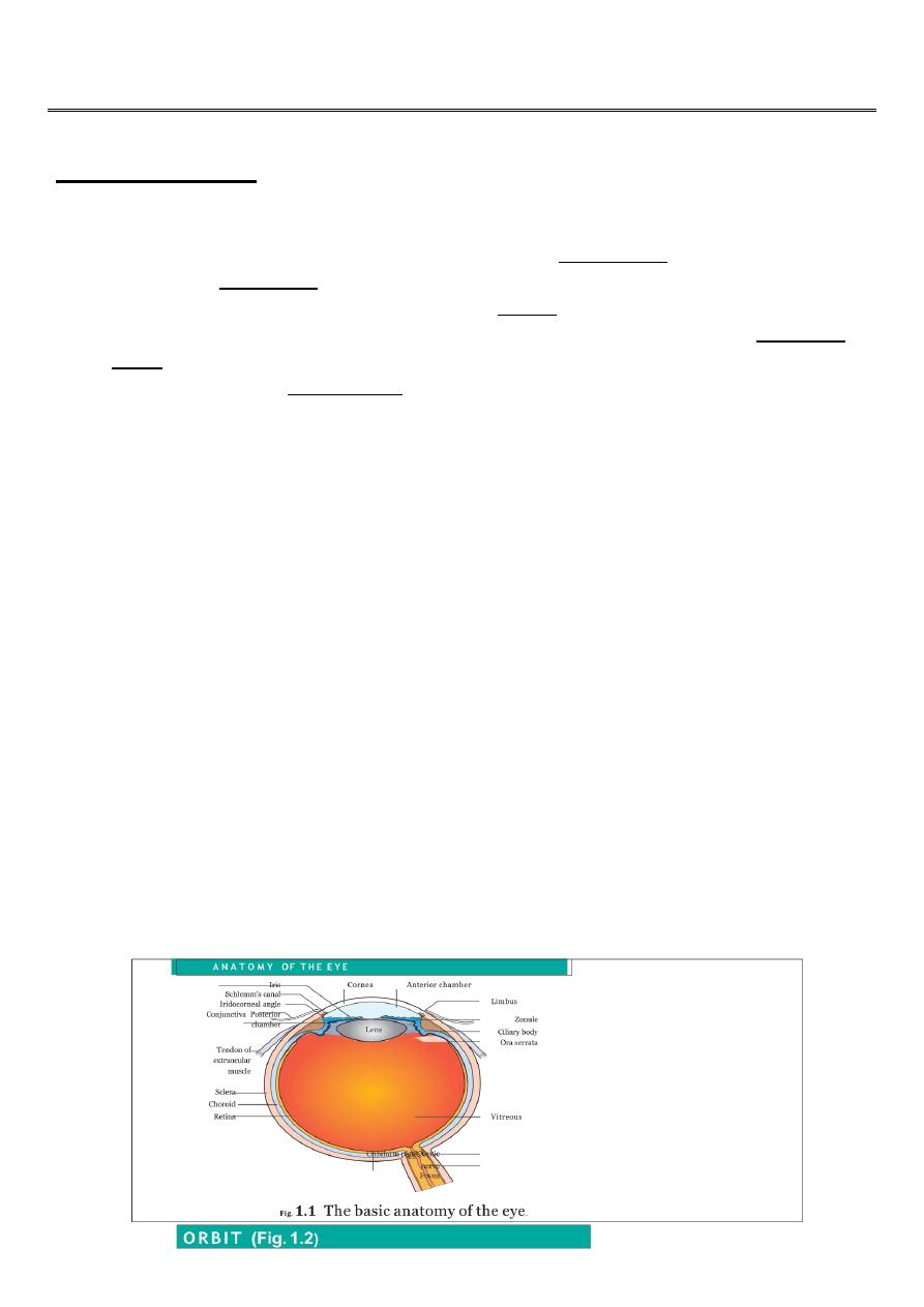
The eye comprises
o A tough outer coat which is transparent anteriorly (the cornea) and opaque
posteriorly (the sclera).
o The junction between the two is called the limbus. The extraocular muscles attach to
the sclera while the optic nerve leaves the sclera posteriorly through the cribriform
plate.
o A rich vascular coat (the choroid) lines the posterior segment of the eye and
nourishes the retina at its inner surface.
o The ciliary body lies anteriorly. It contains the smooth ciliary muscle whose
contraction alters lens shape and enables the focus of the eye to be changed. The
ciliary epithelium secretes aqueous humour and maintains the ocular pressure. The
ciliary body provides attachment for the iris.
o The lens lies behind the iris and is supported by fine fibrils (the zonule) running
between the lens and the ciliary body.
o The angle formed by the iris and cornea (the iridocorneal angle) is lined by a
meshwork of cells and collagen beams (the trabecular meshwork). In the sclera
outside this, Schlemm?s canal conducts the aqueous humour from the anterior
chamber into the venous system, permitting aqueous drainage. This region is
termed the drainage angle.
o Between the cornea anteriorly and the lens and iris posteriorly lies the anterior
chamber.
o Between the iris, the lens and the ciliary body lies the ( posterior chamber).
o Both these chambers are filled with aqueous humour.
o Between the lens and the retina lies the vitreous body.
o Anteriorly, the conjunctiva is reflected from the sclera onto the underside of the
upper and lower eyelids. A connective tissue layer (Tenon?s capsule) separates the
conjunctiva from the sclera and is prolonged backwards as a sheath around the
rectus muscles.
2
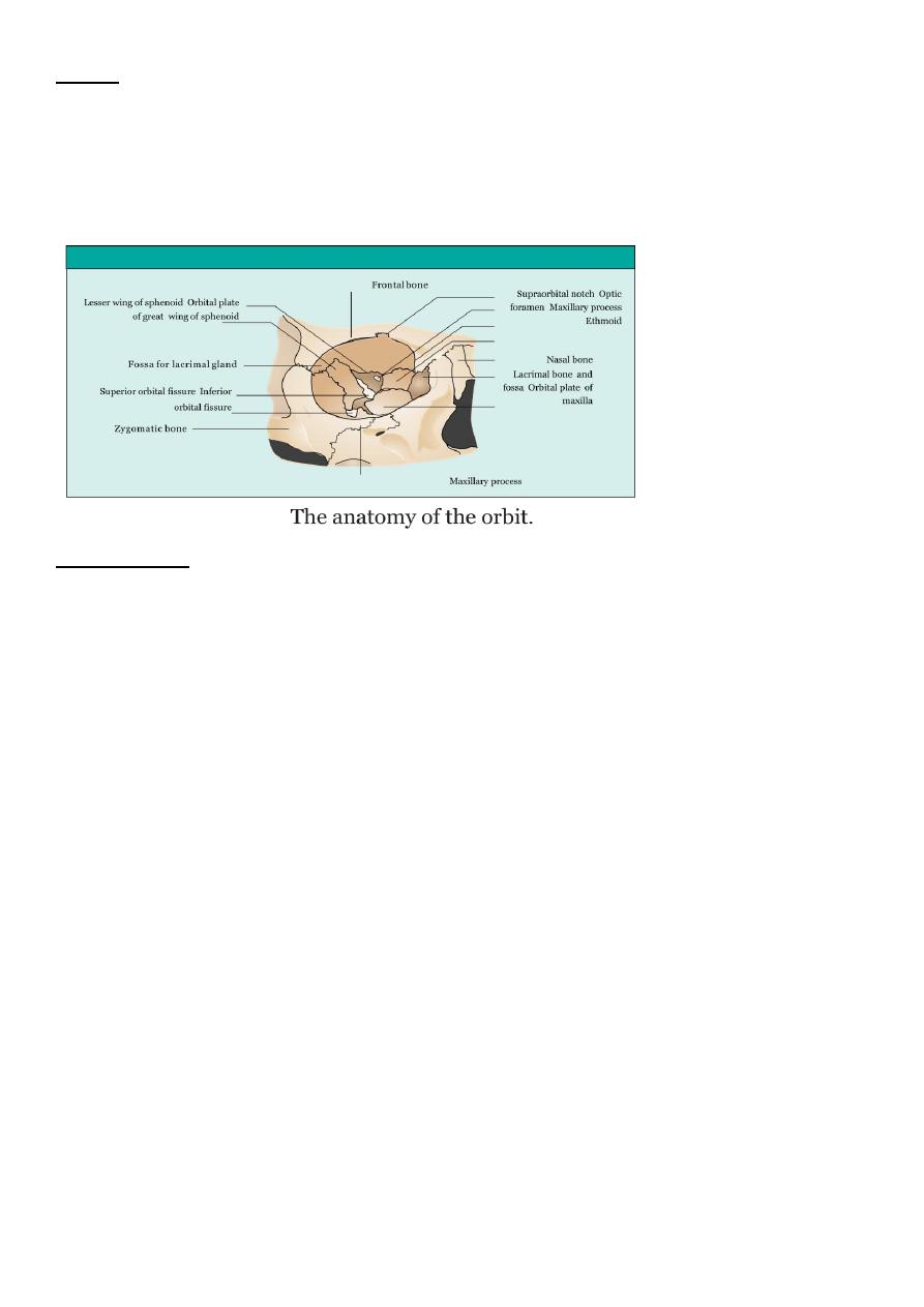
ORBIT
The eye lies within the bony orbit whose structure .The orbit has the shape of a four-sided
pyramid. At its posterior apex is the optic canal which transmits the optic nerve to the
brain. The superior and inferior orbital fissures allow the passage of blood vessels and
cranial nerves which supply orbital structures. On the anterior medial wall lies a fossa for
the lacrimal sac. The lacrimal gland lies anteriorly in the superolateral aspect of the orbit.
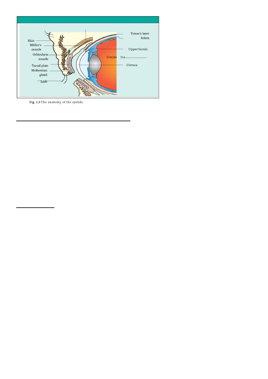
THE EYELIDS
Function:-
1-provide mechanical protection to the anterior globe.
2-secrete the oily part of the tear film.
3-spread the tear film over the conjunctiva and corneaprevent drying of the eyes
4-contain the puncta through which the tears drain into the lacrimal drainage system.
Components:-
1. A surface layer of skin.
2. The orbicularis muscle.
3. A tough collagenous layer (the tarsal plate).
4. An epithelial lining, the conjunctiva, reflected onto the globe.
The levator muscle passes forwards to the upper lid and inserts into the tarsal plate. It is
innervated by the third nerve.
Damage to the nerve or changes in old age result in drooping of the eyelid (ptosis). A flat
smooth muscle arising from the sympathetic supply is damaged (as in Horner?s syndrome)
a slight ptosis results.
The margin of the eyelid is the site of the mucocutaneous junction. It contains the
openings of the
meibomian oil glands which are located in the tarsal plate. These secrete the lipid
component of the tear
film. Medially, on the upper and lower lids, two small puncta form the initial part of the
lacrimal drainage system.
3
THE LACRIMAL DRAINAGE SYSTEM :-
Tears drain into the upper and lower puncta and then into the lacrimal sac via the upper
and lower canaliculi.
They form a common canaliculus before entering the lacrimal sac.
The nasolacrimal duct passes from the sac to the nose.
Failure of the distal part of the nasolacrimal duct to fully canalize at birth is the usual cause
of a watering, sticky eye in a baby.
Tear drainage is an active process.
Each blink of the lids helps to pump tears through the system.
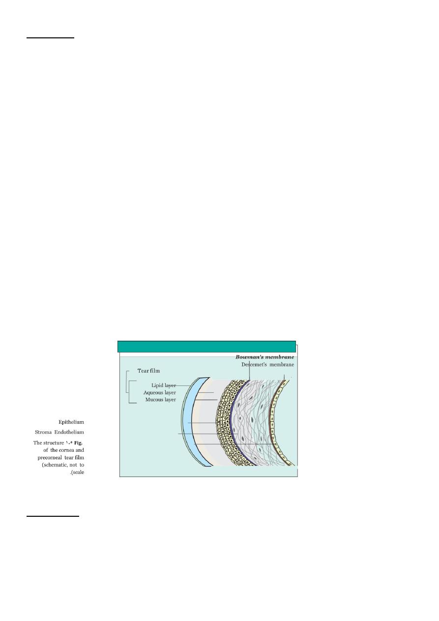
The tear film
The tear film (10 ?m thick) covers the external ocular surface.
Compositions:- 3 layers
1. a thin mucin layer in contact with the ocular surface and produced mainly by the
conjunctival goblet cells;
2. an aqueous layer produced by the lacrimal gland;
3. a surface oil layer produced by the tarsal meibomian glands and delivered to the lid
margins.
functions of the tear film:-
1. it provides a smooth air/tear interface for distortion free refraction of light at the
cornea;
2. it provides oxygen anteriorly to the avascular cornea;
3. it removes debris and foreign particles from the ocular surface through the flow of
tears;
4. it has antibacterial properties through the action of lysozyme, lactoferrin and the
immunoglobulins, particularly secretory IgA.
4
The cornea
The cornea is 0.5 mm thick and comprises:
1.The epithelium, an anterior squamous layer thickened peripherally at the limbus where it
is continuous with the conjunctiva. The limbus houses its germinative?or stem?cells.
2.An underlying stroma of collagen fibrils, ground substance and fibro- blasts. The regular
packing and small diameter of the collagen fibrils accounts for corneal transparency.
3.The endothelium, a monolayer of non-regenerating cells which actively pumps ions and
water from the stroma to control
corneal hydration and transparency.
**The difference between the regenerative capacity of the epithelium and endothelium is
important. Damage to the epithelial
layer, by an abrasion for example, is rapidly repaired. Endothelium, damaged by disease or
surgery, cannot be regenerated.
Loss of its barrier and pumping functions leads to overhydration, distortion of the regular
packing of collagen fibres and
corneal clouding.
functions:-
1. it refracts light and together with the lens, focuses light onto the retina;
2. it protects the internal ocular structures.
The sclera:
is formed from interwoven collagen fibrils of different widths lying within a ground
substance and maintained by fibroblasts;is of variable thickness, 1 mm around the optic
nerve head and 0.3 mm just posterior to the muscle insertions.

5
The choroid:-
has a high blood flow
Function:-nourishes the deep, outer layers of the retina and may have a role in its
temperature homeostasis.
composition:-
arterioles, venules and a dense fenestrated capillary network
o *is loosely attached to the sclera;
o *Its basement membrane together with that of the retinal pigment epithelium (RPE)
forms the acellular, Bruch's membrane, which acts as a diffusion barrier between the
choroid and the retina.
The retinal pigment epithelium
o Composition:- single layer of cells.
o is loosely attached to the retina except at the periphery (ora serrata) and around the
optic disc;
o phagocytoses the redundant external segments of the rods and cones.
o Function:-
1. facilitates the passage of nutrients and metabolites between the retina and choroid.
2. takes part in the regeneration of rhodopsin and cone opsin, the photoreceptor visual
pigments recycling vitamin A; melanin granules absorb scattered light.
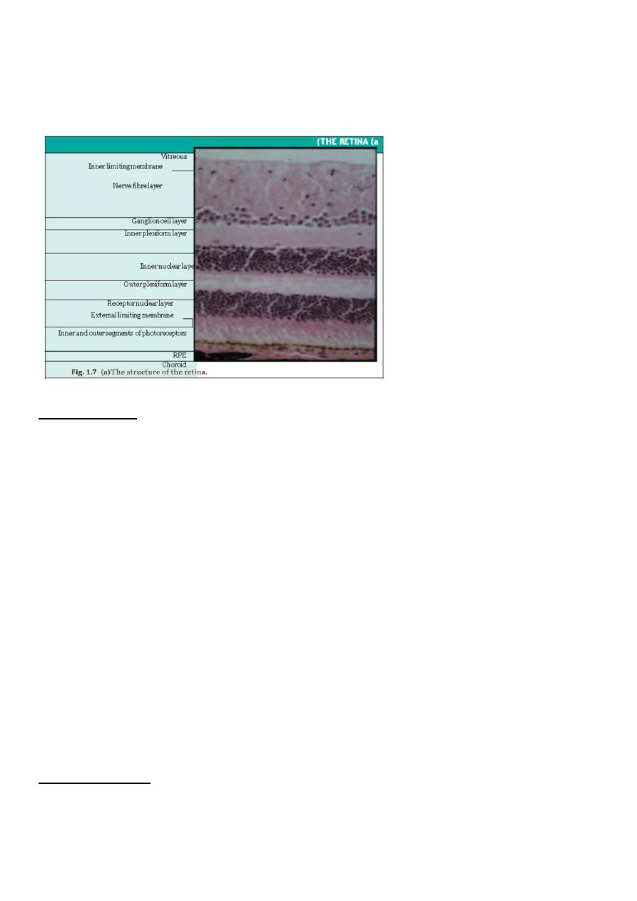
The retina
Composition:-
Is a highly complex structure divided into ten separate layers comprising photoreceptors
(rods and cones) and
neurones, some of which (the ganglion cells) give rise to the optic nerve fibres.
Function:-
o Is responsible for converting light into electrical signals. The initial integration of
these signals is also performed by
o the retina.
6
o Cones are responsible for daylight vision. Subgroups of cones are responsive to
different short, medium and long
o wavelengths (blue, green, red). They are concentrated at the fovea which is
responsible for detailed vision.
The vitreous:-
Is a clear gel occupying two-thirds of the globe.
Composition:-
98% water,the remainder consists of hyaluronic acid and a fine collagen network. There
are few cells.
Attachment:-
Is firmly attached anteriorly to the peripheral retina, pars plana and around the optic disc,
and less firmly to the
macula and retinal vessels.
Function:-
it Has a nutritive and supportive role.
**Detachment of the vitreous from the retina, which commonly occurs in later life,
increases traction on the points of firm attachment.
This may occasionally lead to a peripheral retinal break, when the vitreous pulls away a
piece of the underlying retina.
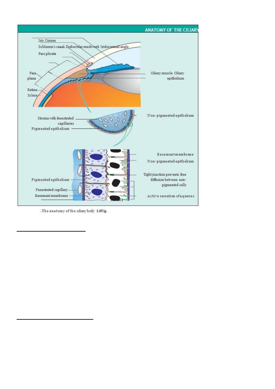
The ciliary body
subdivided into three parts
1. the ciliary muscle
2. the ciliary processes (pars plicata)
3. the pars plana.
7
THE CILIARY MUSCLE
smooth muscle arranged in a ring overlying the ciliary
processes.
Nerve supply:-the parasympathetic system via the third cranial nerve.
Action:-
Is responsible for changes in lens thickness and curvature during accommodation. The
zonular fibres supporting the lens are under tension
during distant viewing. Contraction of the muscle relaxes them and permits the lens to
increase its curvature and hence its refractive power.
THE CILIARY PROCESSES
(PARS PLICATA)
There are about 70 radial ciliary processes arranged in a ring around the posterior chamber.
Function:-They are responsible for the secretion of aqueous humour.
Composition:-Each ciliary process is formed by an epithelium two layers thick (the outer
pigmented and inner non-pigmented) with a vascular stroma.
The stromal capillaries are fenestrated, allowing plasma constituents ready access.
8
The tight junctions between the non-pigmented epithelial cells provide a barrier to free
diffusion into the posterior chamber. They are essential for the active secretion of aqueous
by the non-pigmental cells.
THE PARS PLANA:-
This comprises a relatively avascular stroma covered by an epithelial layer two cells thick.
It is safe to make surgical incisions through the scleral wall here to gain access to the
vitreous cavity.
The iris:
is attached peripherally to the anterior part of the ciliary body.
It forms the pupil at its centre, the aperture of which can be varied by the sphincter
and dilator muscles to control the amount of light entering the eye;
has an anterior border layer of fibroblasts and collagen and a cellular stroma in
which the sphincter muscle is embedded at the pupil margin.
The sphincter muscle is innervated by the parasympathetic system.
The smooth dilator muscle extends from the iris periphery towards the sphincter.
It is innervated by the sympathetic system.
Posteriorly the iris is lined with a pigmented epithelium two layers thick.
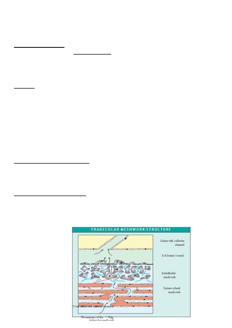
The iridocorneal (drainage) angle
Site :-lies between the iris, cornea and the ciliary body.
Function:-It is the site of aqueous drainage from the eye via the trabecular meshwork.
THE TRABECULAR MESHWORK
This overlies Schlemm's canal.
Composition:- collagen beams covered by trabecular cells.
Function:-This meshwork accounts for most of the resistance to aqueous outflow.
*Damage here is thought to be the cause of the raised intraocular pressure in primary
open angle glaucoma.
9
The lens
Is the second major refractive element of the eye; the cornea, with its tear film, is
the first.
Grows throughout life.
Is supported by zonular fibres running between the ciliary body and the lens capsule.
Comprises an outer collagenous capsule under whose anterior part lies a monolayer
of epithelial cells. Towards the equator the epithelium gives rise to the lens fibres.
The zonular fibres transmit changes in the ciliary muscle allowing the lens to change
its shape and refractive power.
The lens fibres make up the bulk of the lens. They are elongated cells arranged in
layers which arch over the lens equator.
Anteriorly and pos- teriorly they meet to form the lens sutures. With age the deeper
fibres lose their nuclei and intracellular organelles.
The oldest fibres are found centrally and form the lens nucleus; the peripheral fibres
make up the lens cortex.
The high refractive index of the lens arises from the high protein content of the
fibres.
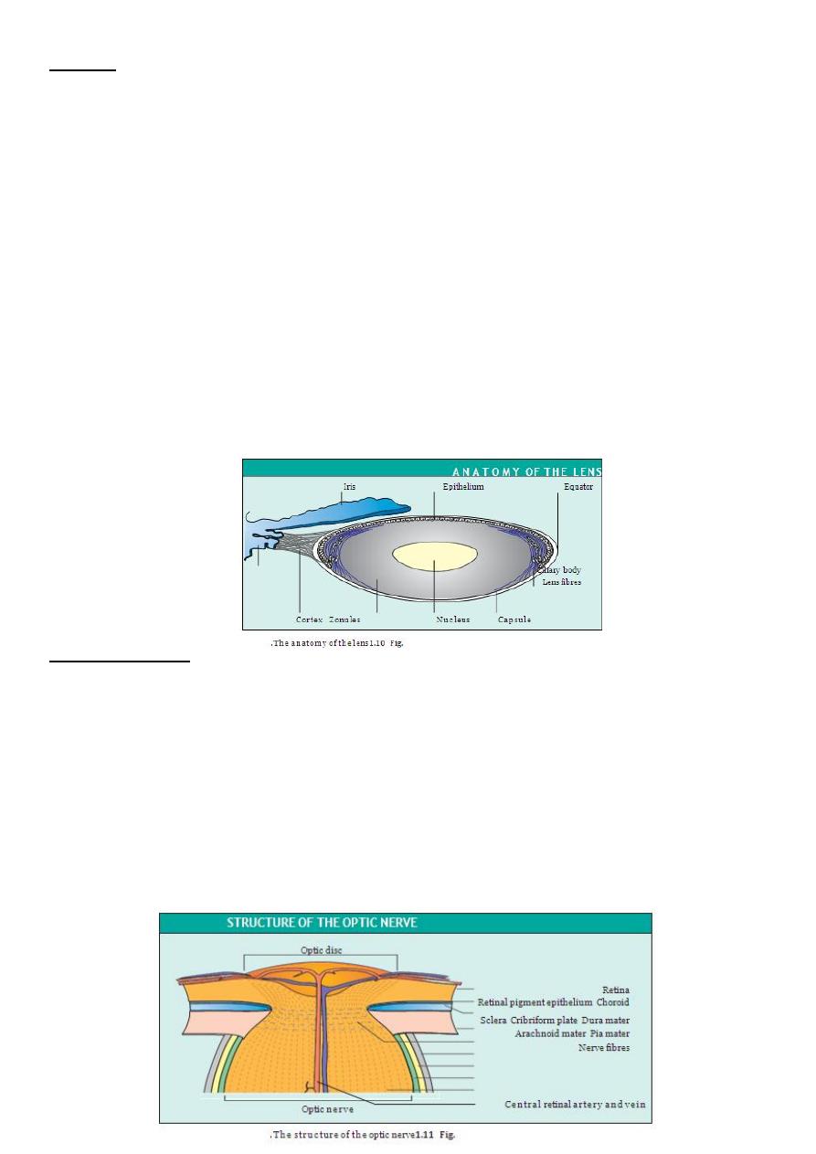
The optic nerve
This is formed by the axons arising from the retinal ganglion cell layer, which form
the nerve fibre layer, the innermost
layer of the retina.
Passes out of the eye through the cribriform plate of the sclera, a sieve- like
structure.
In the orbit the optic nerve is surrounded by a sheath formed by the dura, arachnoid
and pia mater continuous with that surrounding the brain. It is bathed in
cerebrospial fluid.
The central retinal artery and vein enter the eye in the centre of the optic nerve.
The extraocular nerve fibres are myelinated; those within the eye are not.
11
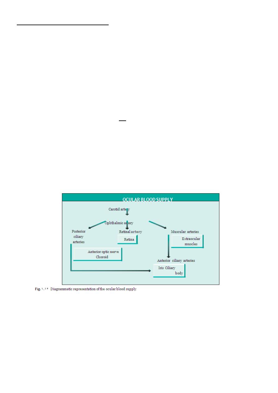
THE OCULAR BLOOD SUPPLY
The eye receives its blood supply from :-
1-the ophthalmic artery (a branch of the internal carotid artery) via the retinal artery.
2-ciliary arteries
3-muscular arteries .
The conjunctival circulation anastomoses anteriorly with branches from the external
carotid artery.
The anterior optic nerve is supplied by branches from the ciliary arteries. The retina is
supplied by arterioles branching from the central retinal artery.
These arterioles each supply an area of retina with little overlap.
Obstruction results in ischaemia of most of the area supplied by that arteriole.
The fovea is so thin that it requires no supply from the retinal circulation.
It is supplied indirectly, as are the outer layers of the retina, by diffusion of oxygen and
metabolites across the retinal pigment epithelium from the choroid.
The endothelial cells of the retinal capillaries are joined by tight junctions so that the
vessels are impermeable to small molecules. This forms an inner blood retinal barrier
The capillaries of the choroid, however, are fenestrated and leaky.
The retinal pigment epithelial cells are also joined by tight junctions and present an
external blood retinal barrier between the leaky choroid and the retina.
It is the breakdown of these barriers that causes the retinal signs seen in many vascular
diseases.
11
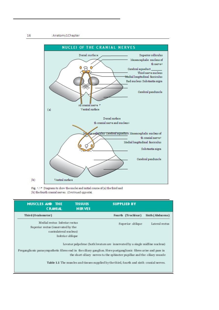
THE THIRD, FOURTH AND SIXTH CRANIAL NERVES
12
-
Peripheral course:
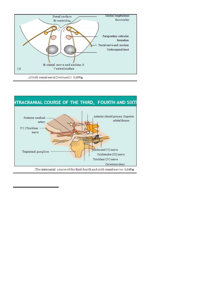
THIRD NERVE
It leaves the midbrain ventrally between the cerebral peduncles.
It then passes between the posterior cerebral and superior cerebellar arteries and
then lateral to the posterior
communicating artery. Aneurysms of this artery may cause a third nerve palsy.
The nerve enters the cavernous sinus in its lateral wall and enters the orbit through
the superior orbital fissure.
FOURTH NERVE
The nerve decussates and leaves the dorsal aspect of the midbrain below the inferior
colliculus.
It first curves around the midbrain before passing like the third nerve between the
posterior cerebral and superior cerebel- lar arteries to enter the
lateral aspect of the cavernous sinus inferior to the third nerve.
13
It enters the orbit via the superior orbital fissure.
SIXTH NERVE
Fibres leave from the inferior border of the pons.
It has a long intracranial course passing upwards along the pons to angle anteriorly
over the petrous bone and into the cavernous sinus where it lies infero-medial to
the fourth nerve in proximity to the internal carotid artery.
It enters the orbit through the superior orbital fissure.
This long course is important because the nerve can be involved in numerous
intracranial pathologies including base of skull fractures, invasion by nasopharyngeal
tumours, and raised intracranial pressure
.
CHAPTER-2 - History and examination
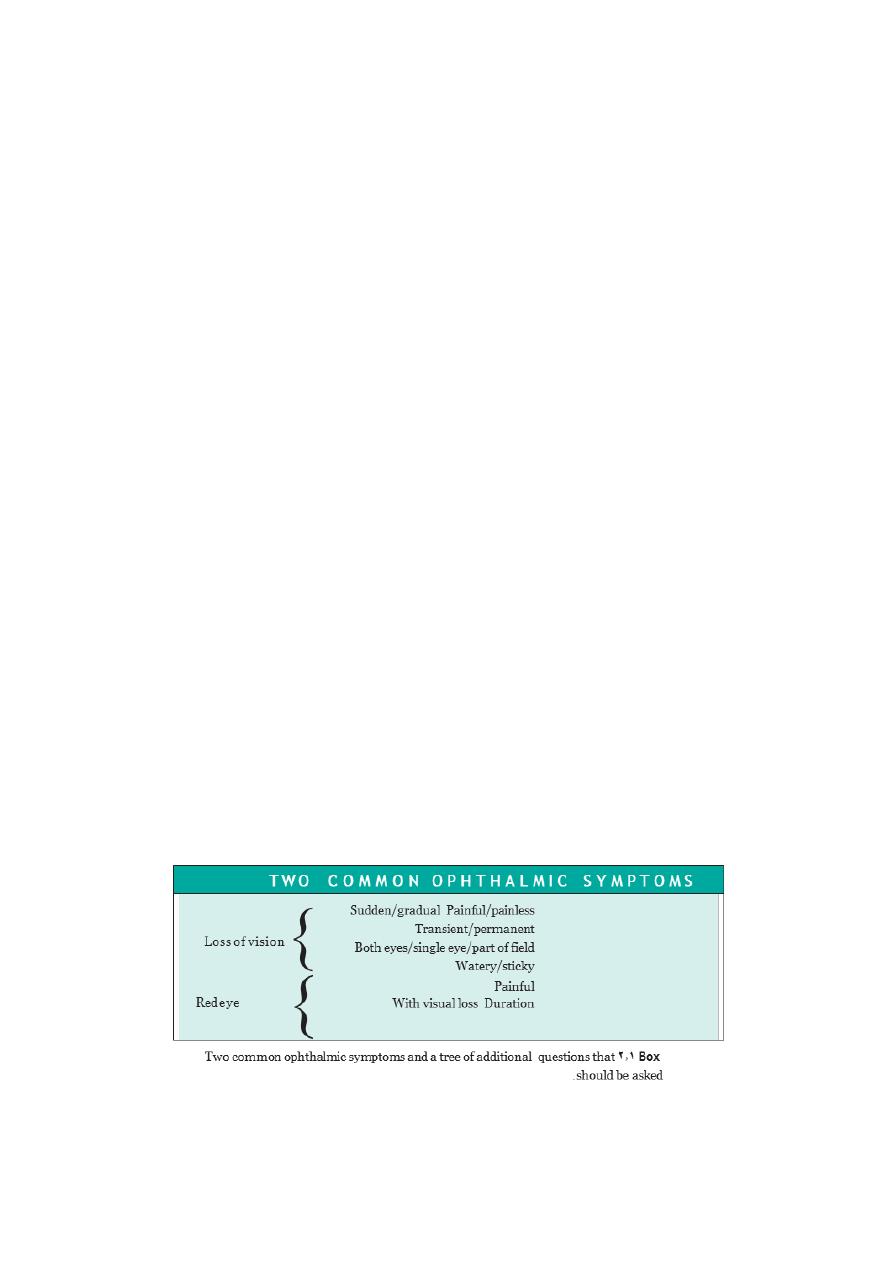
HISTORY
A good history must include details of:
• Ocular symptoms ,time of onset, eye affected, and associated non -
ocular
symptoms.
• Past ocular history
( e.g .poor vision in one eye since birth, recurrence of previous
disease, particularly inflammatory.)
• Past medical history
( e.g .of hypertension
which may be associated with some
vascular eye diseases such as central retinal vein occlusion ;diabetes
which may
cause retinopathy and systemic inflammatory
disease such as sarcoid which may
also cause ocular inflammation.)
• Drug history ,since some drugs such as isoniazid and chloroquine may be toxic to
the eye.steroid use is
• Family history
( e.g .of ocular diseases known to be inherited, such as retinitis
pigmentosa, or of disease where family history may be a risk factor ,such as
glaucoma.)
• Presence of allergies.
14
EXAMINATION
Physiological testing of the eye
VISUAL ACUITY
Adults
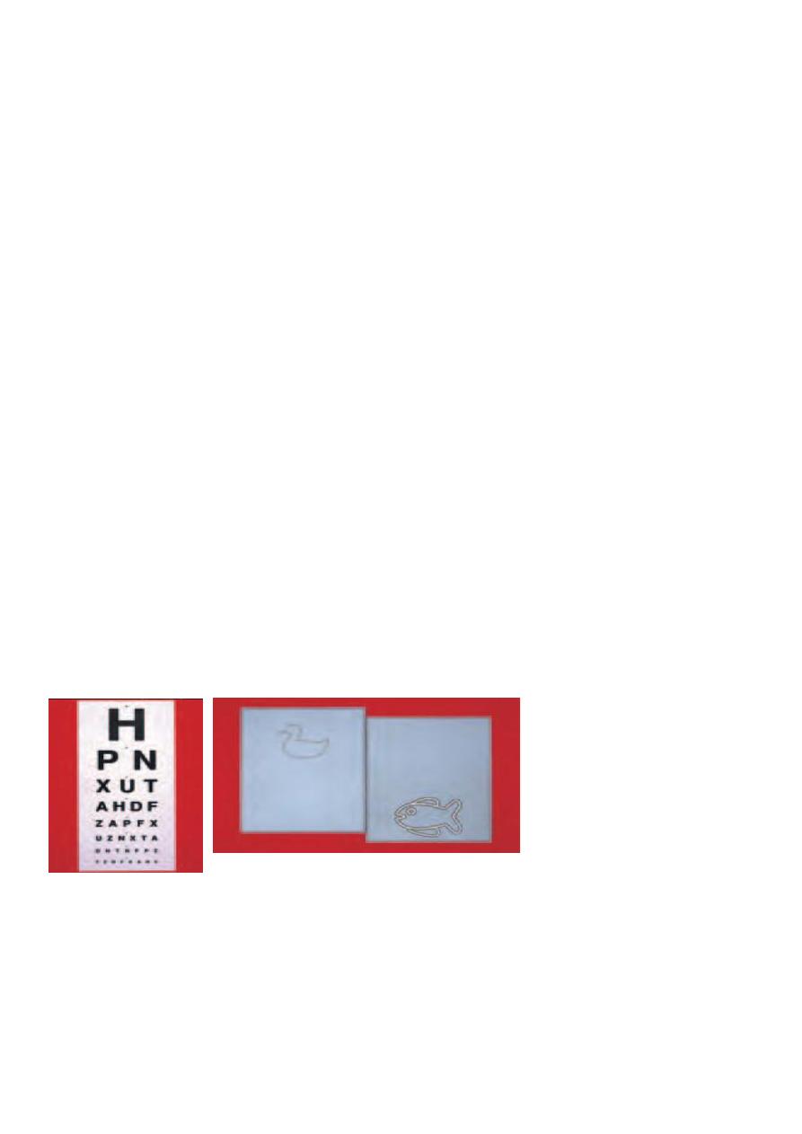
Visual acuity (VA) tests the resolving power of the eye. The standard test is the Snellen
chart ,consisting of rows of letters of decreasing size. Each row is numbered with the
distance in metres at which each letter width sub -
tends 1 minute of arc at the eye. Acuity
is recorded as the reading distance (e.g. 6 metres) over the row number, of the smallest
letter seen. If this is the 6 metre line, then VA is 6/6; if it is the 60 metre line then VA is
6/60 .Vision is tested with spectacles if worn, but a pinhole will correct for mod -
erate
refractive error.
Children
In children, various methods are used to assess visual acuity:
• Very young children are observed to see if they can follow objects or pick up ‘hundreds
and thousands ’cake decorations.
• The Cardiff AcuityTest can be used to assess vision in one to three year olds. This is a
preferential looking test
based on the finding that children prefer to look at complex
rather than plain targets.The grey cards present a variety of figures surrounded by a
white band bordered with two black bands. As the width of the bands decreases the
picture becomes harder to see against the grey background. The gaze of the child is
observed and the
examiner estimates whether the object seen is at the top or bottom of the card. When the
examiner is unable to identify the position of the object from the child’s gaze it is assumed
that the child cannot see the picture.
Older children are able to identify or match single pictures and letters of varying size
VISUAL FIELDS
The visual fields map the peripheral extent of the visual world. Each field can be
represented as a series of contours or isoptres ,demonstrating the ability to resolve a
target of given size and brightness. The field is not flat ;towards the centre the eye is able
to detect much smaller objects than at the periphery. This produces a ‘hill of vision ’in
which objects which are resolved in finest detail are at the peak of the hill (at the fovea )
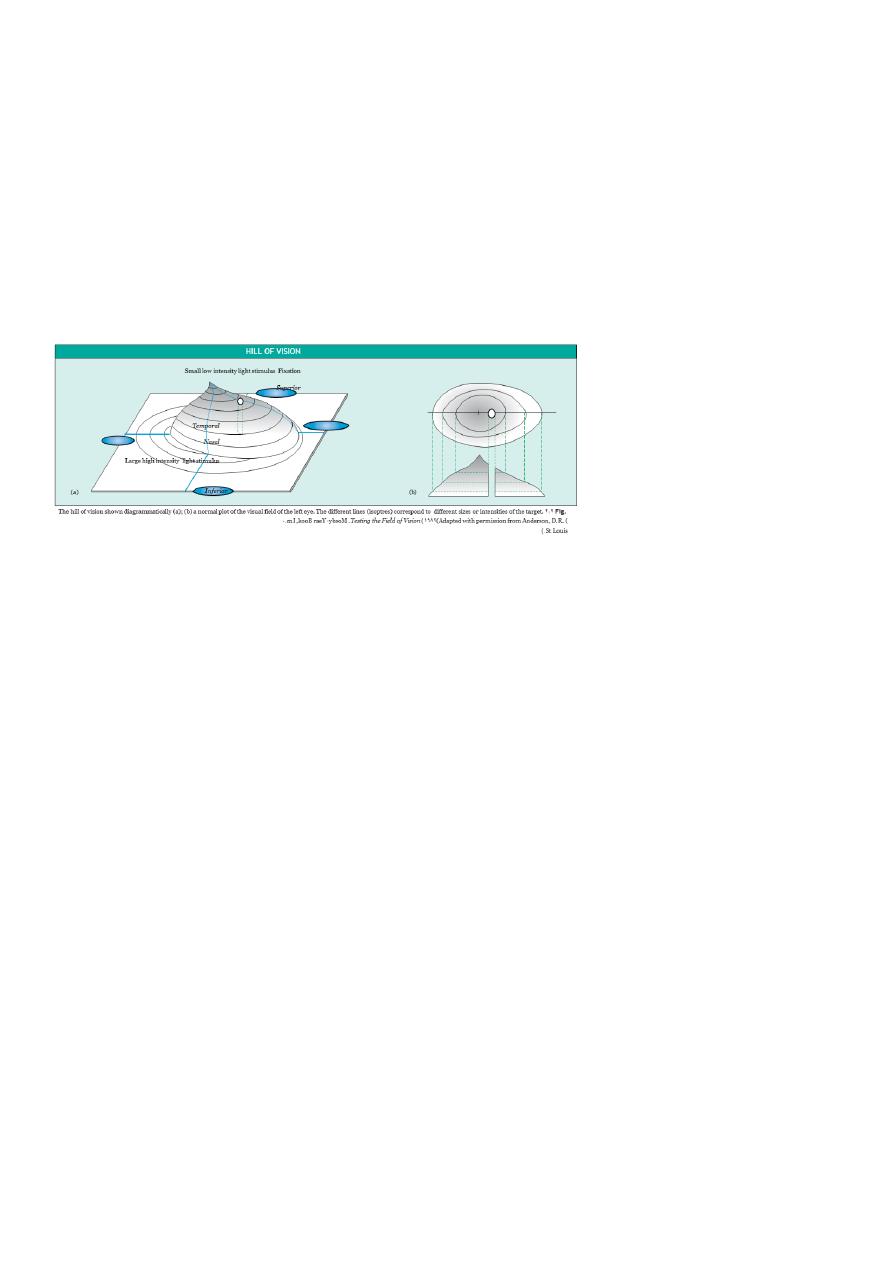
15
(Fig. 2.2 .)On the temporal side of the field is the blind spot.This corresponds to the optic
nerve head where there is an absence of photoreceptors.
The visual field may be tested in various ways.
CONFRONTATION TESTS
One eye of the patient is covered and the examiner sits opposite, closing his eye on the
same side. An object ,traditionally the head of a large hat pin ,is then brought into view
from the periphery and moved centrally .The patient is asked to say when he first sees the
test object. Each quadrant is tested and the location of the blind spot determined. The
patient’s field is thus compared with that of the examiner. With practice central sco
-
tomas
( a scotoma is a focal area of decreased sensitivity within the visual field,
surrounded by a more sensitive area) can also be identified.
Crude testing of the field can be performed as follows:
• Ask the patient to cover one eye. Sit facing the patient and hold up your hands in
front of the unoccluded eye ,palms facing the patient, one on either side of the
midline. Enquire if the two palms apear the same. Repeat the test with the fellow
eye .This can be useful in picking up a bitemporal hemianopia
( patients may also
miss the temporal letters on the Snellen chart when their visual acuity is measured.)
• Ask the patient to count the number of fingers which you show in each quadrant of
the visual field.
A useful test to identify a neurological field defect is to use a red object .The red field is
the most sensitive to optic nerve lesions .A red -
topped pin is used to perform a
confrontation test, the patient being asked to say when he first sees the pin top as red
(not when he first sees the pin top). More simply a red object can be held in each quadrant
or hemi -
field and the patient asked to compare the quality of red in each location .In a
hemianopic field defect the red would appear duller in the affected field.
PERIMETERS
These machines permit more accurate plotting of the visual field. They measure:
• The kinetic
visual field in which the patient indicates when he first sees a light of a
specific size and brightness brought in from the periphery .This is rather like the
moving pinhead of the confrontation test.
• The static
visual field in which the patient indicates when he first sees a stationary
light of increasing brightness.
These techniques are particularly useful in chronic ocular and neurological conditions to
monitor changes in the visual field (e.g. in glaucoma.)
16
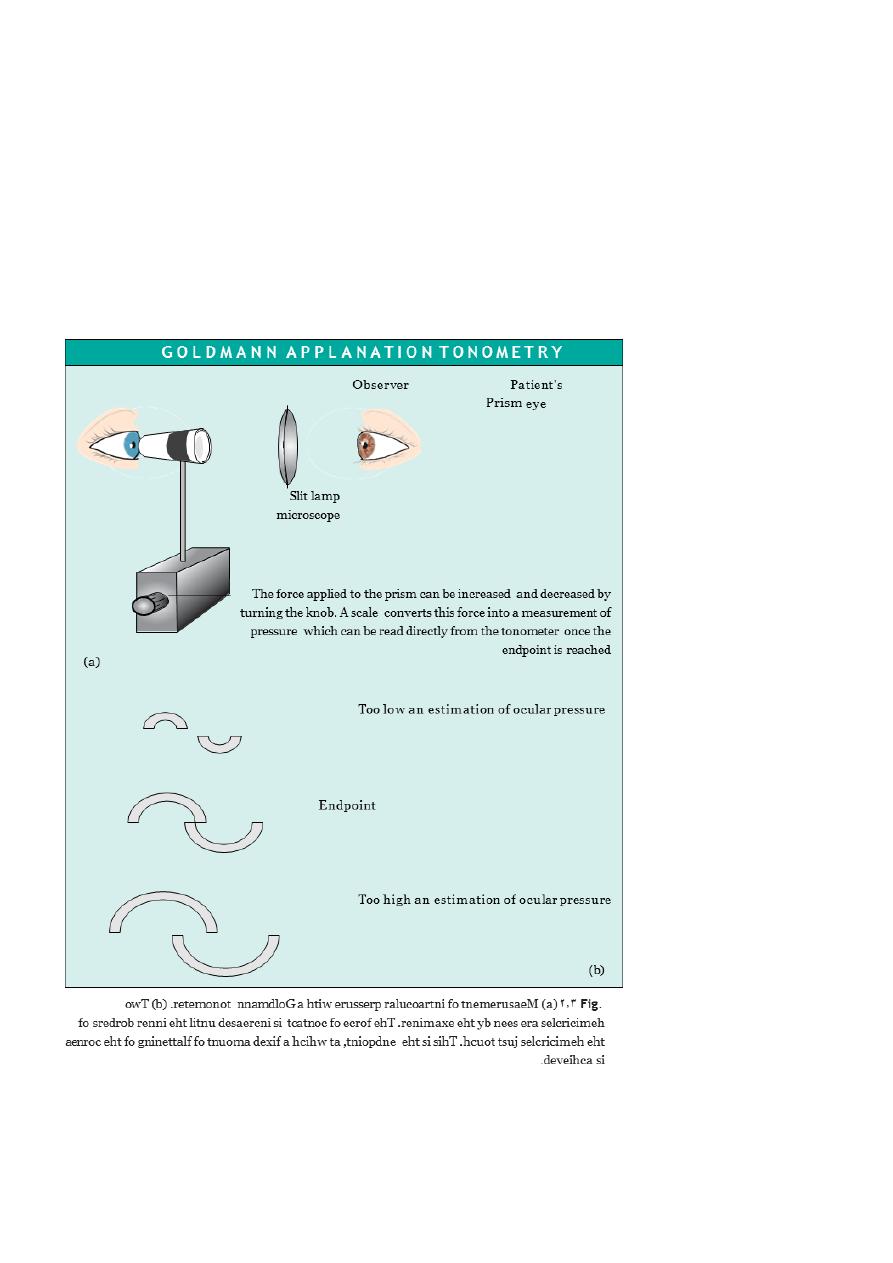
INTRAOCULAR PRESSURE
Intraocular pressure is measured with a Goldmann tonometer
. A clear plastic cylinder is
pressed against the anaesthetized cornea. The ring of flattening, viewed through the
cylinder ,is made visible by the presence of fluorescein in the tear film (see p. 27). A
horizontally disposed prism ,within the cylinder, splits the ring of contact into two
hemicircles. The force applied to the cylinder can be varied to alter the amount of corneal
flattening and thus the size of the ring. It is adjusted so that the two hemi -
circles just
interlock.This is the endpoint of the test, and the force applied ,converted into units of
ocular pressure (mmHg) can now be read from the tonometer.
Optometrists use a puff of air of varying intensity to produce corneal
flattening rather than the prism of the Goldmann tonometer. Various other tonometers
are also available including small hand held electronic devices.

17
PUPILLARY REACTIONS
The size of the pupils
( miosis ,constricted ;mydriasis ,dilated) and their response to light
and accommodation gives important information about:
• the function of the afferent pathway controlling the pupils (the optic nerve and
tract;)
• the function of the efferent pathway.
Examination of the pupils begins with an assessment of the size of the pupils in a uniform
light. If there is asymmetry
( anisocoria )it must be decided whether the small or large
pupil is abnormal .A pathologically
small pupil (after damage to the sympathetic
nervous
system) will be more
apparent in
dim illumination, since dilation of the normal pupil will
be
greater. A pathologically large pupil (seen
in disease of the parasympathetic nervous
system) will be more apparent in the
light
.
Patients with a history of inflammation of the anterior eye
( iritis ,)trauma or previous
ocular surgery may have structural iris changes which mechanically alter the shape of the
pupil. Some individuals have asymmetrical pupillary diameters unassociated with disease.
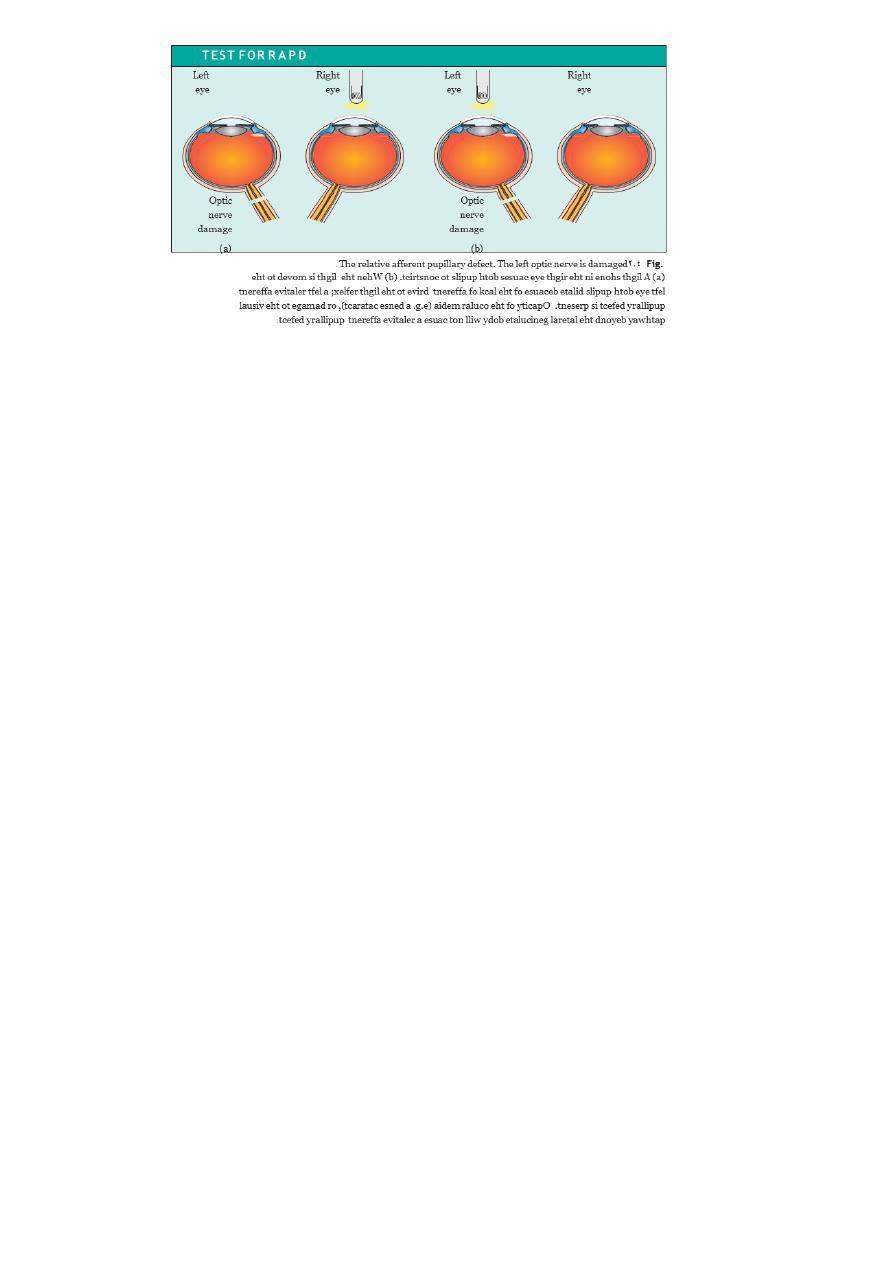
In a patient in whom the pupil sizes are equal, the next step is to look for a defect in optic
nerve function, using the ‘swinging flashlight test .’This is a sensitive index of an afferent
conduction defect. The patient is seated in a dimly illuminated room and views a distant
object. A torch is directed at each eye in turn while the pupils are observed. A unilateral
defect in optic nerve conduction is demonstrated as a relative afferent pupil defect
(RAPD( )see Fig. 2.4.)
In order to test the efferent limb of the pupil reflex, the patient is now asked to look at a
near object; the normal pupils constrict in conjunction with accommodation and
convergence. This is termed the near reflex.
EYE MOVEMENTS
These are assessed while sitting facing the patient. Note the following:
• the position of the eyes;
• the range of eye movements;
• the type of eye movements.
An abnormal direction of one of the eyes in the primary position of gaze (looking straight
ahead) may suggest a squint .This can be confirmed by performing a cover test
( see p.
173.)
The
range
of
eye
movements
is
assessed
by
asking
the
subject
to follow
a moving
object. Horizontal, vertical and oblique movements are
checked from the
primary
position
of gaze asking the patient to report
any
double vision
(
diplopia
.)
The
presence of
oscillating eye movements
(
nystagmus
)
(see p. 184) is also noted. Movement of the eyes
when
following an object is recorded. Such
movements
(
pursuit
movements
)
are
usually
smooth but may be altered in
disease. The ability to direct gaze
rapidly from one object to
another
(
saccadic
eye
movements) can be
tested by asking the
patient to look at targets
(such as the finger) held
at either side of the head. These movements
should be fast,
smooth and
accurate
(
that is they should not overshoot or undershoot the target
.)
18
EYELIDS
These are usually at a symmetrical height. The margin of the lid is applied closely to the
globe in the healthy eye. If the lid margin is turned away from the globe an ectropion
is
present; if the lid margin is turned in and the lashes are rubbing against the globe an
entropion
is present.
A drooping lid
( ptosis )may reflect:
• An anatomical disorder (e.g. a failure of the levator tendon to insert properly into
the lid.)
• An organic problem (e.g. weakness of the levator muscle in myasthenia gravis or
impairment of its nerve supply in third nerve palsy.)
In assessing ptosis, the distance between the upper and lower lid is measured with the
patient looking straight ahead. The excursion of the upper lid from extreme downgaze to
extreme upgaze is then recorded. In myasthenia, repeated up and down movement of the
lids will increase the ptosis by fatiguing the levator muscle (see p. 50).
Anatomical examination of the eye
LIDS AND ANTERIOR SEGMENT
Simple examination of the eye and adnexae can reveal a great deal about pathological
processes within the eye.
DIAGNOSTIC USE OF FLUORESCEIN
Fluorescein has the property of absorbing light in the blue wavelength and emitting a
green fluorescence. The application of fluorescein
to the eye can identify corneal
abrasions (where the surface epithelial cells have been lost) and leakage of aqueous
humour from the eye (Fig. 2.5).
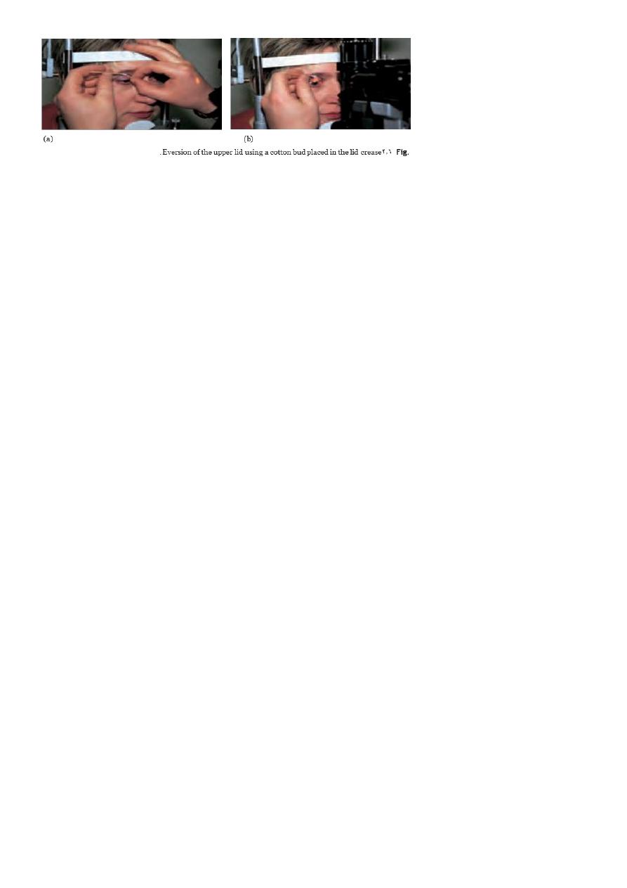
EVERSION OF THE UPPER LID
( Fig
.
2.6
)
The underside of the upper lid is examined by everting it over a small blunt ended object
(e.g. a cotton bud) placed in the lid crease. This is an important technique to master as
foreign bodies may often lodge under the upper lid causing considerable pain to the victim
.
19
RETINA
The retina is examined by:
• Direct ophthalmoscopy
( the conventional ophthalmoscope) (see Fig
.
2.7
.)
• Indirect ophthalmoscopy ,which allows the extreme retinal periphery to be viewed.
The examiner wears a head-mounted binocular microscope with a light source. A
lens placed between the examiner and the eye of the subject is used to produce an
inverted image of the retina.
A special contact lens
( e.g .a
3
- mirror lens )is also used at the slit lamp.
The latter two techniques are reserved for specialists; the technique that must be
mastered by the non-specialist is direct ophthalmoscopy.
The direct ophthalmoscope provides:
• an image of the red reflex;
• a magnified view of the optic nerve head, macula, retinal blood vessels and the
retina to the equator.
It comprises:
• a light source, the size and colour of which can be changed;
• a system of lenses which permits the refractive error of both observer and patient to
be corrected.
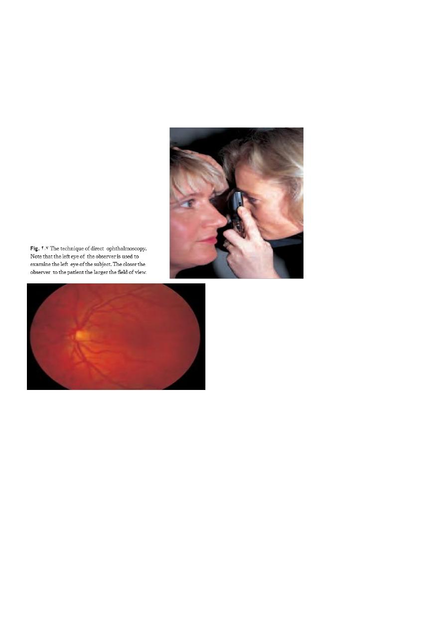
Confident use of the ophthalmoscope comes with practice. The best results are obtained if
the pupil is first dilated with tropicamide,a mydriatic with a short duration of action.
The patient and examiner must be comfortable and the patient looks straight ahead at a
distant object. The examiner’s right eye is used to examine the patient’s right eye and the
left eye to examine the left eye.
The examiner, with the ophthalmoscope about 30 cm away from the eye ,views the red
reflex through the pupil. The correct power of lens in the ophthalmoscope to produce a
clear image is found by ratcheting down from a high to a low hypermetropic (plus)
correction. Opacities in the cornea or lens of the eye will appear black against the red
reflex.The eye is then approached to within a couple of centimetres and the power of the
lenses is adjusted in the myopic (minus) direction, to achieve focus on the retina.
The examiner may find it helpful to place a hand on the subject’s fore -
head which can also
be used to hold the upper lid open. The retina should now be in view. It is important to try
and examine the retina in a logical sequence so that nothing is overlooked.
• First find the optic disc (Fig. 2.8), assess its margins (are they distinct?) ,assess the
colour of the disc (is it pale?) ,assess the optic cup (see p. 105).
• Examine the macular region. Is there a normal foveal reflex (in youth the foveal pit
appears as a bright pinpoint of light in the centre of the retina .)Are there any
abnormal lesions such as haemorrhages, exudates or cotton wool spots?
21
• Return to the optic disc and follow each major vessel branch of the vasculature out
to the periphery. Are the vessels of normal diameter, do the arteries nip the
veins where they cross
( A/V
nipping ,)are there any emboli in the arterioles ?
Also examine the surrounding retina for abnormalities.
• Examine the peripheral retina with a 360° sweep.
Fig
.
8.2
A normal left fundus. Note the optic disc with retinal veins and arteries passing
from it to branch over the retina. The large temporal vessels are termed arcades .The
macula lies temporal to the disc with the fovea at its centre.
Special examination techniques
DIAGNOSTIC LENSES
Ophthalmologists employ special lenses that can be used in conjunction with the slit lamp
to examine particular ocular structures.
A gonioscopy
lens is a diagnostic contact lens ,with a built-in mirror that permits
visualization of the iridocorneal angle .A larger lens with three
mirrors allows the peripheral retina to be seen. Both are applied to the anaesthetized
cornea with a lubricating medium .
Other lenses can be used to obtain a stereoscopic view of the retina+90D,,+78D.

21
RETINOSCOPY
The technique of retinoscopy allows the refractive state of the eye to be measured (i.e. the
required strength of a corrective spectacle lens .)
Investigative techniques
ULTRASOUND
provide information about the vitreous, retina and posterior coats of the eye, particularly
when they cannot be clearly visualized (if, for example, there is a dense cataract or
vitreous haemorrhage .)B-scan)
Ultrasound is also used to measure the length of the eyeball prior to cataract surgery to
estimate the power of the artificial lens that is implanted into the eye (A-scan
(
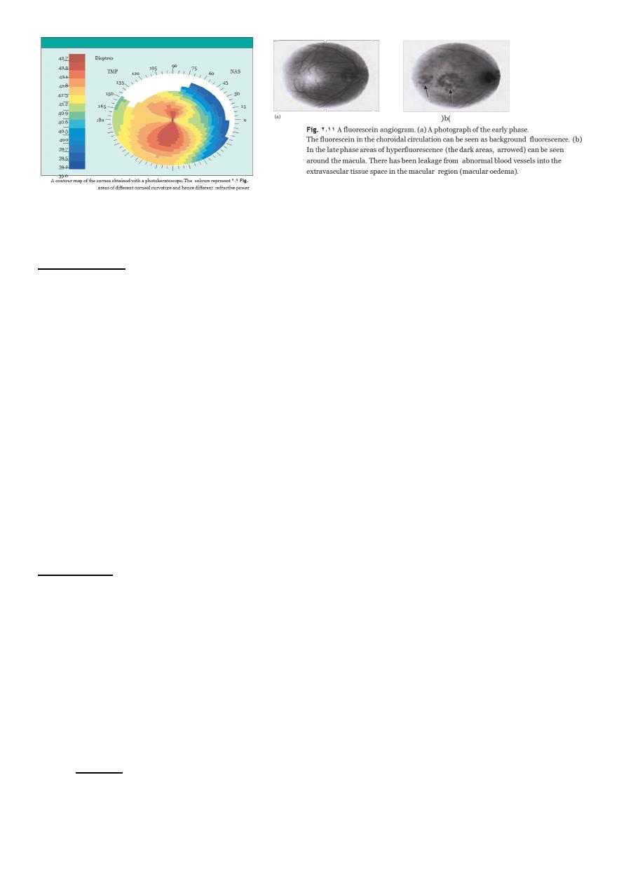
KERATOMETRY
The shape of the cornea (the radius of curvature) can be measured from the image of a
target reflected from its surface. This is important in contact lens assessment ,refractive
surgery
)( and in calculating the power of an artificial lens implant in cataract surgery
.)(
The technique of photokeratometry allows a very accurate contour map of the cornea.)
SYNOPTOPHORE
This machine permits the assessment of binocular single
vision ,the ability of the two
eyes to work together to produce a single image. It is also able to test the range over
which the eyes can move away from
( diverge )or towards each other
( converge )whilst
maintaining a single picture
( to
measure the range of fusion )
EXOPHTHALMOMETER
This device measures ocular protrusion
( proptosis.)
ELECTROPHYSIOLOGICAL TESTS
The electrical activity of the retina and visual cortex in response to specific visual stimuli,
for example a flashing light, can be used to assess the functioning of the retina
(electroretinogram ,)RPE
( electro-oculogram )and the visual pathway
( visually evoked
response or
potential.)
RADIOLOGICAL IMAGING TECHNIQUES
The CT and MRI scans have largely replaced skull and orbital X-rays in the imaging of the
orbit and visual pathway. The newer diagnostic techniques have enhanced the diagnosis
of orbital disease
( e.g. optic nerve sheath meningioma) and visual pathway lesions such as
pituitary tumours. They have also become the first line investigation in orbital trauma.
FLUORESCEIN ANGIOGRAPHY
)(
This technique provides detailed information about the retinal circulation.
22
CHAPTER-3-Clinical optics
Introduction
o Light :-is part of the electro-magnetic spectrum to which the eye is sensitive.
(waveband of 390 nm to 760inm).
o light must be correctly focused on the retina.
o The focus must be adjustable to allow equally clear vision of near and distant
objects.
o The cornea, or actually the air/tear interface is responsible for two-thirds and the
crystalline lens for one-third of the focusing power of the eye.
o These two refracting elements in the eye converge the rays of light because:
o The cornea has a higher refractive index than air; the lens has a higher refractive
index than the aqueous and vitreous humours that surround it.
o The velocity of light is reduced in a dense medium so that light is refracted towards
the normal.
o When passing from the air to the cornea or aqueous to lens the rays therefore
converge.
o The refracting surfaces of the cornea and lens are spherically convex.
AMETROPIA
o When parallel rays of light from a distant object are brought to focus on the retina
with the eye at rest (i.e. not accommodating)
o the refractive state of the eye is known as emmetropia .
o Such an individual can see sharply in the distance without accommodation.
o In ametropia, parallel rays of light are not brought to a focus on the retina in an eye
at rest.
o A change in refraction is required to achieve sharp vision.
Ametropia may be divided into:-
Myopia (short sightedness); the optical power of the eye is too high (usually due to
an elongated globe) and parallel rays of light are
brought to a focus in front of the retina (Fig. 3.2).
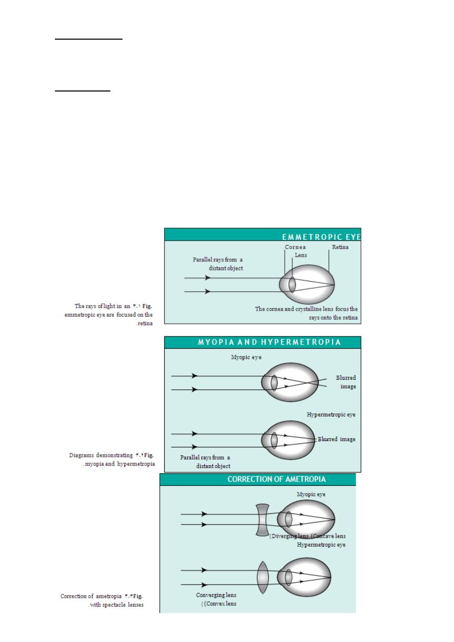
23
Hypermetropia (long sightedness); the optical power is too low (usually because the
eye is too short) and parallel rays of light converge
towards a point behind the retina.
Astigmatism; the optical power of the cornea in different planes is not equal.
Parallel rays of light passing through these different
planes are brought to different points of focus.
All three types of ametropia can be corrected by wearing spectacle lenses. These
diverge the rays in myopia, converge the rays in hypermetropia and correct for the
non-spherical shape of the cornea in astigmatism (Fig. 3.3). It should be noted that in
hypermetropia,accommodative effort will bring distant objects into focus by
increasing the power of the lens. This will use up the accommodative reserve for
near objects.

24
ACCOMMODATION AND PRESBYOPIA
o As an object is brought nearer to the eye
the power of the lens increases; this is
accommodation ,The eyes also converge.
o The ability to accommodate decreases with age, reaching a critical point at about 40
when the subject experiences difficulty with near
o vision (presbyopia).
o This occurs earlier in hypermetropes than myopes.
o The problem is overcome with convex reading lenses.
OPTICAL CORRECTION AFTER CATARACT EXTRACTION
The lens provides one-third of the refractive power of the eye so that after cataract
extraction (the removal of an opaque lens) the eye is rendered highly
hypermetropic, a condition termed aphakia. This can be corrected by:
1. the insertion of an intraocular lens at the time of surgery; 0% mag
2. contact lenses; 10% mag
3. aphakic spectacles. 33% mag.
Intraocular lenses give the best optical results.These mimic the natural lens position.
As they are unable to change shape the eye cannot accommodate.
An eye with an intraocular lens is said to be pseudophakic.
Contact lenses produce slight magnification of the retinal image
CONTACT LENSES
o These are made from rigid, gas permeable or soft hydrophilic materials.
o All contact lenses will retard the diffusion of oxygen to the cornea.
o Rigid gas permeable lenses are relatively more permeable to oxygen than soft
lenses.
o Although soft lenses are better tolerated, gas permeable lenses have certain
advantages:
1. their greater oxygen permeability reduces the risk of corneal damage from hypoxia.
2. their rigidity allows easier cleaning and offers less risk of infection.
3. their rigidity allows for a more effective correction of astigmatism.
4. proteinaceous debris is less likely to adhere to the lens and cause an allergic
conjunctivitis.

25
5. Plane soft contact lenses may also be used as ocular bandages, e.g. in the treatment
of some corneal diseases such as a persistent epithelial defect.
SPECTACLES
Spectacles are available to correct most refractive errors. Lenses can be made to
correct long and short sightedness and astigmatism.
They are simple and safe to use but may be lost or damaged. Some people find them
cosmetically unacceptable and prefer to wear
contact lenses. The correction of presbyopia requires additional lens power to
overcome the eye?s reduced accommodation for near focus. This can be achieved
with:
i.
Separate pairs of glasses for distance and near vision.
ii.
A pair of bifocal lenses where the near correction is added to the lower segment of
the distance lens.
iii.
Varifocal lenses where the power of the lens gradually changes from the distance
correction (in the upper part) to the near correction (in the lower part). This provides
sharper middle-distance vision but the lenses may be difficult to manage.
People with particular needs, such as musicians, may also need glasses for middle
distance.
REFRACTIVE SURGERY
o Although refractive errors are most commonly corrected by spectacles or
contact lenses, laser surgical correction is gaining popularity. The
o excimer laser precisely removes part of the superficial stromal tissue from
the cornea to modify its shape. Myopia is corrected by flattening the
cornea and hypermetropia by steepening it. In photorefractive
keratectomy (PRK), the laser is applied to the corneal surface.
o In laser assisted in situ keratomileusis (LASIK), a hinged partial thickness
corneal stromal flap is first created with a rapidly moving automated
blade.
o The flap is lifted and the laser applied onto the stromal bed. Unlike PRK,
LASIK provides a near instantaneous improvement in vision with minimal
discomfort.
26
o Serious complications during flap creation occur rarely. Intraocular lenses
can also be
placed in the eye but this carries all the risks of intraocular surgery and the
possibility of cataract formation.
CHAPTER-4-
The orbit
INTRODUCTION
The orbit provides:
1. protection to the globe.
2. attachments which stabilize the ocular movement
3. transmission of nerves and blood vessels.
Despite the number of different tissues present in the orbit the expression of
disease due to different pathologies is often
similar.
-
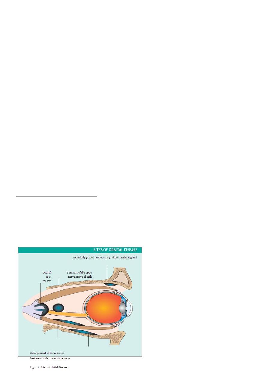
Proptosis(exophthalmos):
protrusion of the eye caused by a space-occupying lesion.
It can be measured with an exophthalmometer.
A difference of more than 3 mm between the two eyes is significant.
If the eye is displaced directly forwards ,,suggests
lesion that lies within the cone
formed by the extraocular muscles (an intra-conal
lesion)
ex:-optic nerve sheath meningioma.
27
*If the eye is displaced to one side a lesion outside the muscle cone is likely (an extra-conal
lesion). For example a tumour of the
lacrimal gland displaces the globe to the nasal side.
*A transient proptosis induced by increasing the cephalic venous pressure (by aValsalva
manoeuvre), is a sign of orbital varices.
* The speed of onset of proptosis may also give clues to the aetiology,,,ex:- slow
onsetbenign tumour// rapid onset inflammatory disorders, malignant tumours and
carotid-cavernous sinus fistula.
Note :-The presence of pain may suggest infection (e.g. orbital cellulitis).
backward displacement of the globe.
-
Enophthalmos:
*
This may be seen following an orbital fracture when orbital contents are displaced into an
adjacent sinus. It is also said to occur in Horner's syndrome but this is really a pseudo-
enophthalmos due to narrowing of the palpebral fissure.
Pain:-Inflammatory conditions, infective disorders and rapidly progressing
tumours cause pain. This is not usually present with benign tumours.
Conjunctival injection and swelling suggests an
-
:
Eyelid and conjunctival changes
inflammatory or infective process.
Infection is associated with:-
1. reduced eye movements
2. erythema
3. swelling of the lids (orbital cellulitis).
With more anterior lid inflammation (preseptal cellulitis) eye movements are full.

28
Florid engorgement of the conjunctival vessels suggests a vascular lesion caused by the
development of a fistula between the carotid artery and the cavernous sinus.
Diplopia
o This results from:-Direct involvement of the muscles in myositis and dysthyroid eye
disease.
o Sign :-Movement is restricted in a direction opposite to the field of action of the
affected muscle. The eye appears to be tethered (e.g. if the inferior rectus is thickened
in thyroid eye disease there will be Involvement of the nerve supply to the extraocular
muscles. Here diplopia occurs during gaze into the field of action of the muscle
(e.g.palsy of the right lateral rectus produces diplopia in right horizontal gaze)
.
Visual acuity:-
This may be reduced by:
1.exposure keratopathy from severe proptosis.
2.optic nerve involvement by compression or inflammation.
3.distortion of the macula due to posterior compression
INVESTIGATION OF ORBITAL DISEASE
The CT and MRI scans have greatly helped in the diagnosis of orbital disease; localizing the
site of the lesion, demonstrating enlarged
intraocular muscles in dysthyroid eye disease and myositis or visualizing fractures to the
orbit.
Additional systemic tests will be dictated by the differential diagnosis (e.g. tests to
determine the primary site of a secondary tumour).
DIFFERENTIAL DIAGNOSIS OF ORBITAL DISEASE
(Traumatic orbital disease is
discussed in Chapter 16.)
Disorders of the extraocular muscles:-
o Dysthyroid eye disease and ocular myositis present with symptoms and signs of
orbital disease.
o In children a rapidly developing proptosis may be caused by a rare
rhabdomyosarcoma arising from the extraocular muscles.
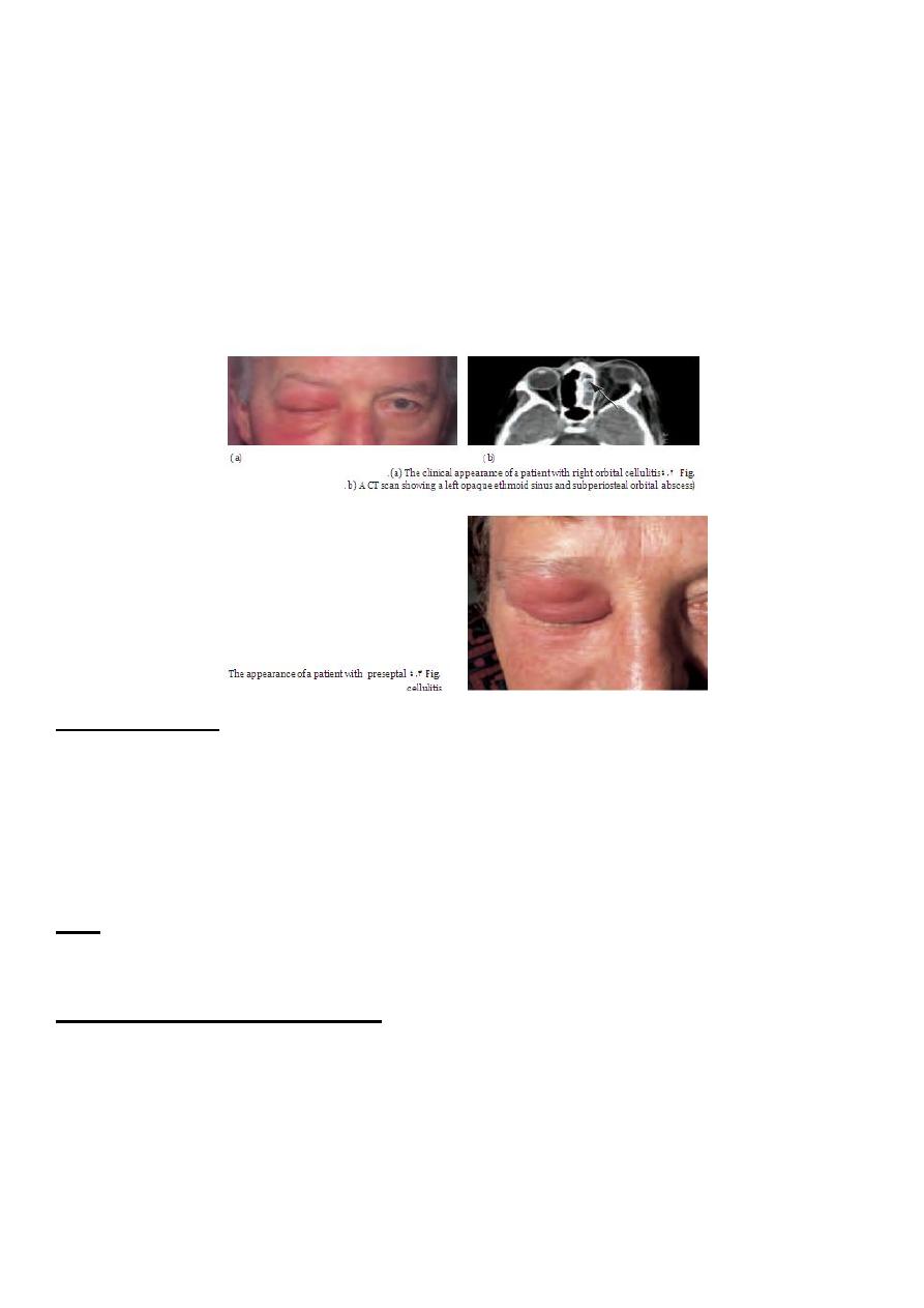
Infective disorders
o Orbital cellulitis is a serious condition which can cause blindness and may spread to
cause a brain abscess.
o The infection often arises from an adjacent ethmoid sinus.
o The commonest causative organism is Haemophilus influenzae.
Presentation:-
1. a painful eye.
2. periorbital inflammation and swelling; mild proptosis

29
3. reduced eye movements
4. conjunctival injection
5. possible visual loss
6. systemic illness and pyrexia.
Diagnosis & treatment :-
o An MRI or CT scan is helpful in diagnosis and in planning treatment .
o The condition usually responds to intravenous broad spectrum antibiotics.
o It may be necessary to drain an abscess or decompress the orbit particularly if the
optic nerve is compromised.
o Optic nerve function must be closely watched, monitoring acuity, colour vision and
testing for a relative afferent pupillary defect.
o Orbital decompression is usually performed with the help of an ENT specialist.
A preseptal cellulitis
involves only the lid.
It presents with periorbital inflammation and swelling but not the other ocular
features of orbital cellulitis.
Eye movement is not impaired.
Inflammatory disease
The orbit may become involved in various inflammatory disorders including
sarcoidosis and orbital pseudotumour, a non-specific lymphofibroblastic disorder.
Diagnosis of such conditions is difficult.The presence of other systemic signs of
sarcoidosis may be helpful. If an orbital pseudotumour is suspected it may be
necessary to biopsy the tissue to differentiate the lesion from a lymphoma.
Vascular abnormalities
(carotid-cavernous sinus fistula orbital varix) causing intermittent proptosis..
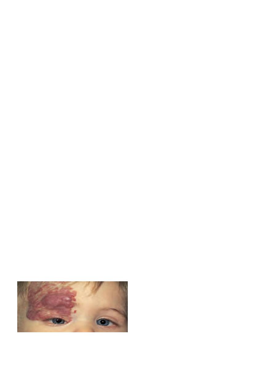
In infants, a capillary haemangioma may present as an extensive lesion of the orbit
and the surrounding skin .
Orbital tumours
The following tumours may produce signs of orbital disease:
1. lacrimal gland tumours.
2. optic nerve gliomas.
31
3. Meningiomas.
4. Lymphomas.
5. Rhabdomyosarcoma.
6. metastasis from other systemic cancers (neuroblastomas in children, the breast,
lung, prostate or gastrointestinal tract in the adult).
diagnosis :-A CT or MRI scan. Again systemic investigation, for example to determine the
site of a primary tumour, may
be required.
Malignant lacrimal gland tumours carry a poor prognosis.
Benign tumours still require complete excision to prevent malignant transformation.
Optic nerve gliomas may be associated with neurofibromatosis.
Treatment :-
They are difficult to treat but are often slow growing and thus may require no
intervention.
Meningiomas of the optic nerve are rare, and may also be difficult to excise.
Again they can be observed and some may benefit from treatment with
radiotherapy.
Meningiomas from the middle cranial fossa may spread through the optic canal into
the orbit.
The treatment of lymphoma requires a full systemic investigation to determine
whether the lesion is indicative of widespread disease or whether it is localized to
the orbit.
In the former case the patient is treated with chemotherapy, in the latter with
localized radiotherapy.
**In children the commonest orbital tumour is a rhabdomyosarcoma, a rapidly growing
tumour of striated muscle Chemotherapy is
effective if the disease is localized to the orbit.
(capillary hemangioma)
31
Dermoid cysts
Etiology:-
the continued growth of ectodermal tissue beneath the surface, which may
present in the medial or lateral aspectof the superior orbit.
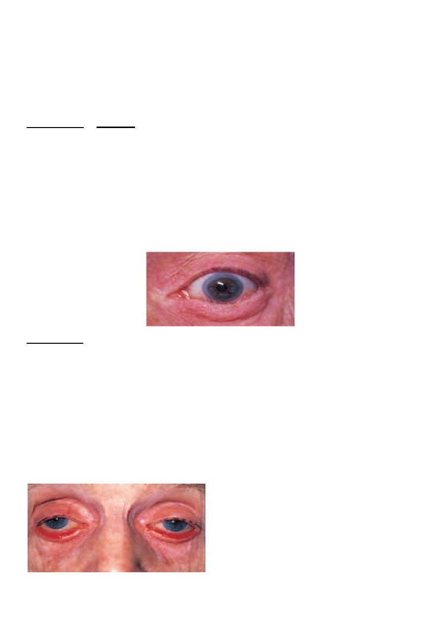
The commonest cause of bilateral proptosis is dysthyroid disease (Dysthyroid disease
may be associated with the serious complications of exposure keratopathy and optic
nerve compression).
Excision is usually performed for cosmetic reasons
Suspect orbital cellulitis in a patient with periorbital and conjunctival inflammation,
particularly when there is severe pain and the patient is systemically unwell.
Chapter-5-EYELIDS
INTRODUCTION
The eyelids are important because of :-
1-providing physical protection to the eyes.
2-in ensuring a normal tear film and tear drainage.
Diseases of the eyelids can be divided into those associated with:
1. abnormal lid position.
2. inflammation of the lid.
3. lid lumps.
4. abnormalities of the lashes.
ABNORMALITIES OF LID POSITION
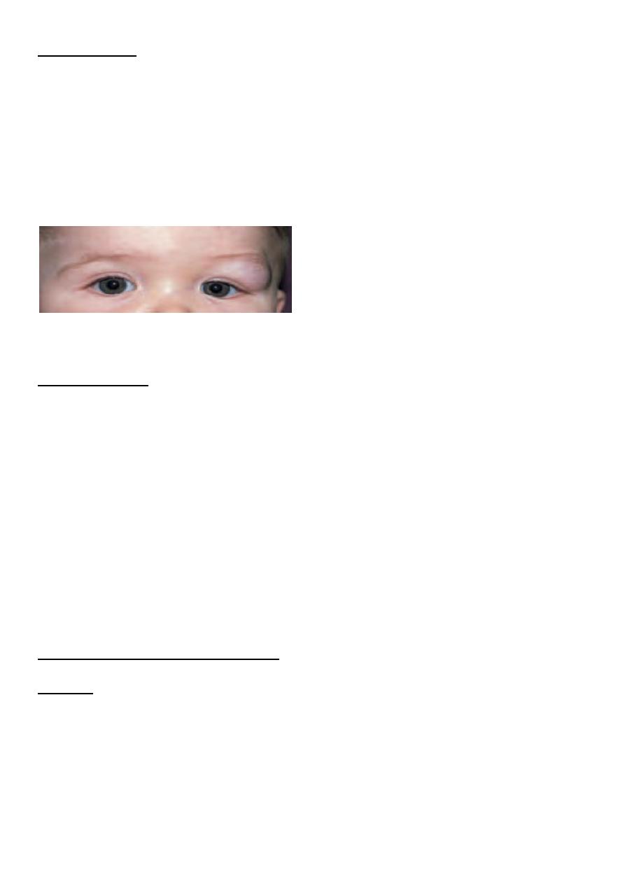
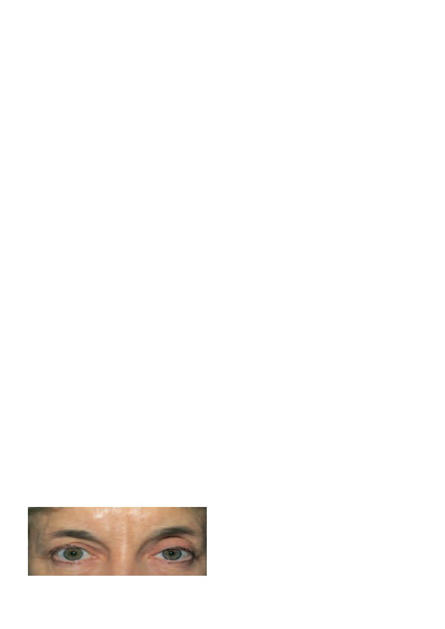
1-Ptosis:-
abnormally low position of the upper eyelid.
PATHOGENESIS
It may be caused by:
1. Mechanical factors
a)
Large lid lesions pulling down the lid.
b) Lid oedema.
c) Tethering of the lid by conjunctival scarring.
32
d) Structural abnormalities including a disinsertion of the aponeurosis of the levator
muscle, usually in elderly patients.
2. Neurological factors.
a) Third nerve palsy .
b) Horner's syndrome, due to a sympathetic nerve lesion.
c) Marcus Gunn jaw-winking syndrome. In this congenital ptosis there is a mis-wiring of
the nerve supply to the pterygoid muscle of the jaw and the levator of the eyelid so
that the eyelid moves in conjunc- tion with movements of the jaw.
3. Myogenic factors.
a) Myasthenia gravis .
b) Some forms of muscular dystrophy.
c) Chronic external ophthalmoplegia.
SYMPTOMS
Patients present because:
they object to the cosmetic effect
vision may be impaired
there are symptoms and signs associated with the underlying cause (e.g. asymmetric
pupils in Horner's syndrome,diplopia and reduced eye movements in a third nerve
palsy).
SIGNS
There is a reduction in size of the interpalpebral aperture. The upper lid margin, which
usually overlaps the upper limbus by
1-2 mm, may be partially covering the pupil.
The function of the levator muscle can be tested by measuring the maximum travel of the
upper lid from upgaze to downgaze (normally 15 -18 mm).
Pressure on the brow (frontalis muscle) during this test will prevent its contribution to lid
elevation.
If myasthenia is suspected the ptosis should be observed during repeated lid movement.
Increasing ptosis after repeated elevation and depression of the lid is suggestive of
myasthenia.
Other underlying signs, for example of Horner's syndrome or a third nerve palsy, may be
present.
(left ptosis)
33
MANAGEMENT
o It is important to exclude an underlying cause whose treatment could resolve the
problem (e.g. myasthenia gravis).
o Ptosis otherwise requires surgical correction.
o In very young children this is usually deferred but may be expedited if pupil cover
threatens to induce amblyopia.
Entropion :-
an inturning, usually of the lower lid.
o It is seen most commonly in elderly patients where the orbicularis muscle becomes
weakened.
o It may also be caused by conjunctival scarring distorting the lid (cicatricial
entropion).
o The inturned lashes cause irritation of the eye and may also abrade the cornea. The
eye may be red.
o Treatment:-Short-term treatment includes the application of lubricants to the eye or
taping of the lid to overcome the inturning. Permanent treatment requires surgery.
Ectropion :-
an eversion of the lid.
causes :-
1. involutional orbicularis muscle laxity.
2. scarring of the periorbital skin.
3. seventh nerve palsy.
**The malposition of the lids everts the puncta and prevents drainage of the tears, leading
to epiphora"excessive lacrimation".
It also exposes the conjunctiva ,, This again results in an irritable eye.
Treatment:- surgical.

34
INFLAMMATIONS OF THE EYELIDS
Blepharitis
chronic eyelid inflammation
.
very common condition.
It is sometimes associated with chronic staphylococcal infection.
The condition causes squamous debris, inflammation of the lid margin, skin and eyelash
follicles (anterior blepharitis).
The meibomian glands may be affected independently (meibomian gland disease or
posterior blepharitis).
SYMPTOMS:-tired, sore eyes, worse in the morning,crusting of the lid margin.
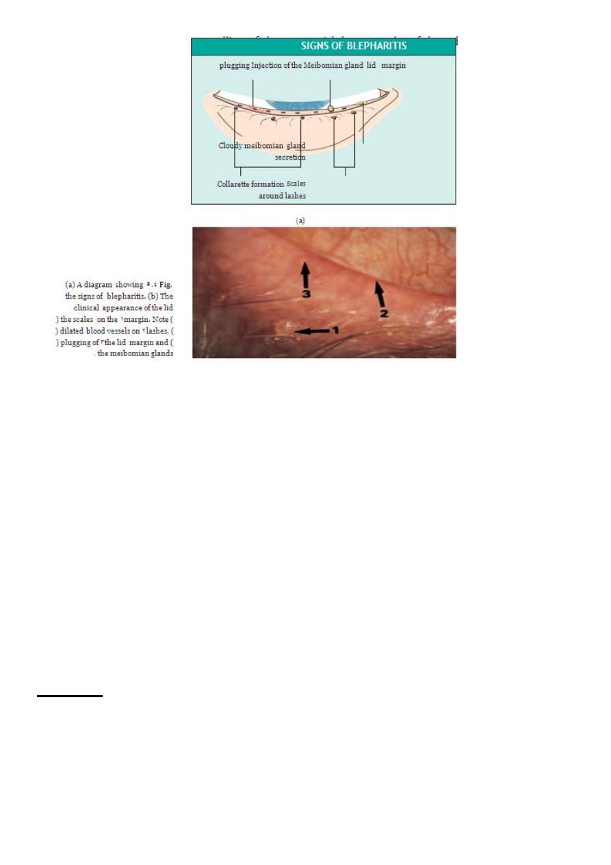
SIGNS :-There may be:-
1. scaling of the lid margins.
2. debris in the form of a rosette around the eyelash, the base of which may also be
ulcerated, a sign of staphylococcal infection.
3. A reduction in the number of eyelashes.
4. obstruction and plugging of the meibomian ducts.
5. cloudy meibomian secretions
6. injection of the lid margin
tear film abnormalities.
In severe disease the corneal epithelium is affected (blepharokeratitis).
Small ulcers may form in the peripheral cornea marginal ulceration secondary to
staphylococcal exotoxins.
The conjunctiva becomes injected.
Blepharitis is strongly associated with seborrhoeic dermatitis, atopic eczema and
acne rosacea.
In rosacea there is hyperaemia and telangiectasia of the facial skin and a rhinophima
(a bulbous irregular
swelling of the nose with hypertrophy of the sebaceous glands.
35
TREATMENT:-
This is often difficult and must be long term.
For anterior blepharitis
lid toilet with a cotton bud wetted with bicarbonate
solution or diluted baby shampoo helps to remove squamous debris from the eye.
Similarly, abnormal meibomian gland secretions can be expressed by lid massage
after hot bathing.
Staphylococcal lid disease may also require therapy with topical antibiotics (fusidic
acid gel) and, occasionally, with systemic antibiotics.
Meibomian gland function can be improved by oral tetracycline.
Topical steroids may improve an anterior blepharitis but frequent use is best
avoided.
Posterior blepharitis can be associated with a dry eye which requires treatment with
artificial tears.
PROGNOSIS
Although symptoms may be ameliorated by treatment, blepharitis may remain a chronic problem.
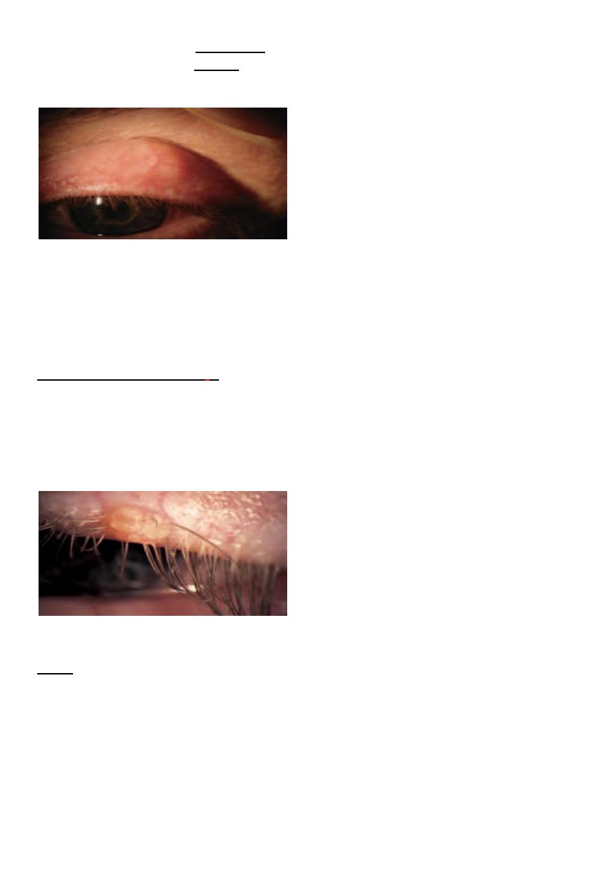
Chalazion
o common condition.
o painless
o An obstructed meibomian gland causes a granuloma within the tarsal plate.
o Symptoms :-are of an unsightly lid swelling which usually resolves within 6 months.
o If the lesion persists it can be incised and curetted from the conjunctival surface.
36
o An abscess (internal hordeolum) may also form within the meibomian gland, which
unlike a chalazion is painful. It may respond to topical anti- biotics but incision may
be necessary.
o A stye (external hordeolum) is a painful abscess of an eyelash follicle.
Treatment:-
1-removal of the associated eyelash
2-application of hot compresses.
3-Most cases are self-limiting.
4-Occasionally systemic antibiotics are required.
Molluscum contagiosum
:- is
umbilicated lesion found on the lid margin
caused by the pox virus.
It causes irritation of the eye.
The eye is red and small elevations of lymphoid tissue (follicles) are found on the
tarsal conjunctiva.
Treatment
excision of the lesion.
Cysts
o Various cysts may form on the eyelids.
o Sebaceous cysts are opaque.
o They rarely cause symptoms.
o They can be excised for cosmetic reasons.
A cyst of Moll :-a small translucent cyst on the lid margin caused by obstruction of a
sweat gland.
A cyst of Zeis :-an opaque cyst on the eyelid margin caused by blockage of an accessory
sebaceous gland.
37
Treatment:- These can be excised for cosmetic reasons.
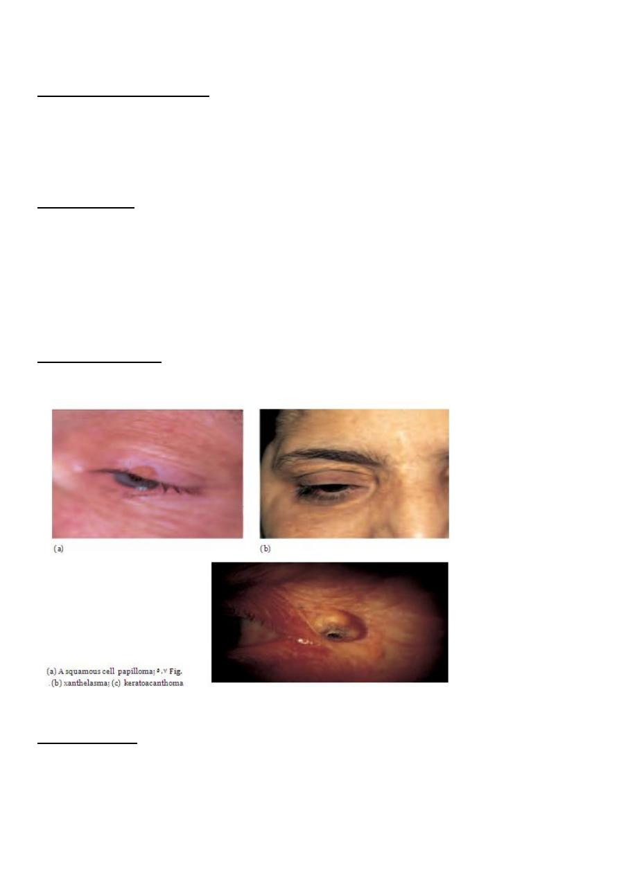
Squamous cell papilloma
o This is a common frond-like lid lesion with a fibrovascular core and thickened squamous
epithelium.
o Symptoms:-usually asymptomatic
o It can be excised for cosmetic reasons with cautery to the base.
Xanthelasmas
These are lipid-containing bilateral lesions which may be associated with
hypercholesterolaemia.
They are excised for cosmetic reasons.
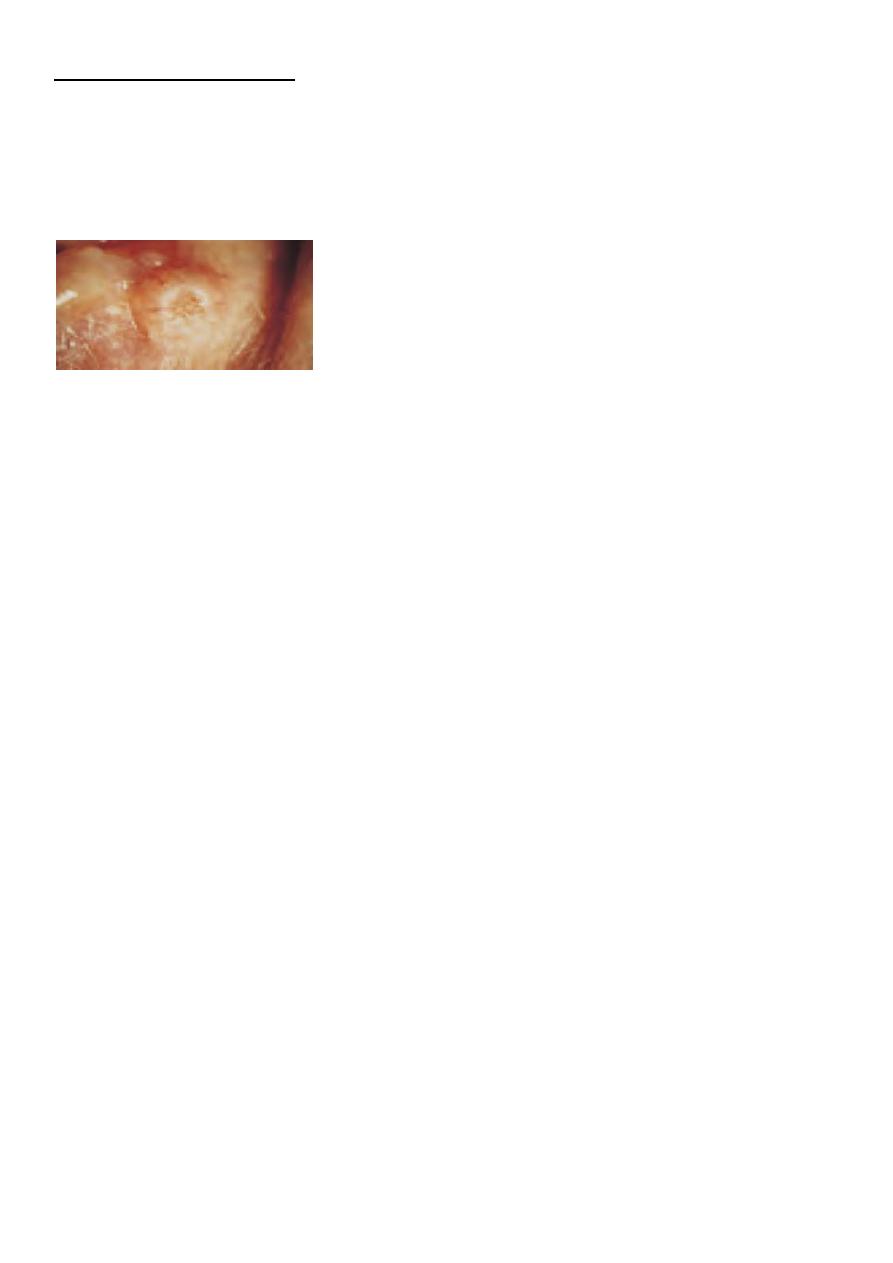
Keratoacanthoma
A brownish pink, fast growing lesion with a central crater filled with keratin
Treatment:- if required, by excision.
Naevus (mole)
These lesions are derived from naevus cells (altered melanocytes) and can be
pigmented or non-pigmented.
No treatment is necessary.
38
MALIGNANT TUMOURS :-
includes
a.Basal cell carcinoma :-
the most common form of malignant tumour(90%).
The tumour is:-
1. slow growing.
2. locally invasive.
3. non-metastasizing.
Features:-
painless lesion on the eyelid which may be
1-nodular
2-sclerosing
3-ulcerative (the so-called rodent ulcer).
*It may have a typical, pale, pearly margin. A high index of suspicion is required.
Treatment :-
1. Excision biopsy with a margin of normal tissue surrounding the lesion. Excision may
also be controlled with frozen sections when
serial histological assessment is used to
determine the need for additional tissue removal (Moh?s surgery). This minimizes
destruction of normal tissue.
2. Cryotherapy.
3. Radiotherapy.
prognosis
:-very good but deep invasion of the tumour can be difficult to treat.
b.Squamous cell carcinoma:-less common but more malignant tumour which can
metastasize to the lymph nodes.
*It can arise de novo or from pre-malignant lesions.
*It may present as a hard nodule or a scaly patch.
Treatment :-excisional biopsy with a margin of healthy tissue.
**UV exposure is an important risk factor for both basal cell and squa- mous cell
carcinoma.

39
ABNORMALITIES OF THE LASHES
--Trichiasis: aberrant eyelashes are directed backwards towards the globe, The lashes rub
against the cornea and cause irritation and abrasion.
*common condition.
Etiology:-
In developing countries trachoma is an important cause and trichiasis is an important basis
for the associated blindness.
*It is distinct from entropion.
*It may result from any cicatricial process.
Treatment :-
epilation of the offending lashes.
Recurrence can be treated with cryotherapy or electrolysis.
Any underlying abnormality of lid position needs surgical correction.
https://www.muhadharaty.com/lecture/13894